 Movie
Movie Controller
 Structure viewers
 About EMN search


-Search query
-Search result
Showing all 24 items for (author: fuller & sd)
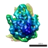
EMDB-1451: 
Human T-lymphotropic virus-1 visualized at the virological synapse by electron tomography.
Method: electron tomography / : Majorovits E, Nejmeddine M, Tanaka Y, Taylor GP, Fuller SD, Bangham CRM
EMDB-1450:
Human T-lymphotropic virus-1 visualized at the virological synapse by electron tomography.
Method: electron tomography / : Majorovits E, Nejmeddine M, Tanaka Y, Taylor GP, Fuller SD, Bangham CRM
EMDB-1452:
Human T-lymphotropic virus-1 visualized at the virological synapse by electron tomography.
Method: electron tomography / : Majorovits E, Nejmeddine M, Tanaka Y, Taylor GP, Fuller SD, Bangham CRM
EMDB-1453:
Human T-lymphotropic virus-1 visualized at the virological synapse by electron tomography.
Method: electron tomography / : Majorovits E, Nejmeddine M, Tanaka Y, Taylor GP, Fuller SD, Bangham CRM
EMDB-1454:
Human T-lymphotropic virus-1 visualized at the virological synapse by electron tomography.
Method: electron tomography / : Majorovits E, Nejmeddine M, Tanaka Y, Taylor GP, Fuller SD, Bangham CRM
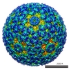
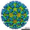
EMDB-1255:
Structure of a hexameric RNA packaging motor in a viral polymerase complex.
Method: single particle / : Huiskonen JT, Jaalinoja HT, Briggs JAG
EMDB-1256:
Structure of a hexameric RNA packaging motor in a viral polymerase complex.
Method: single particle / : Huiskonen JT, Jaalinoja HT, Briggs JAG

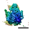
EMDB-1273:
Centrosome polarization delivers secretory granules to the immunological synapse.
Method: electron tomography / : Stinchcombe JC, Majorovits E, Bossi G, Fuller SD, Griffiths GM
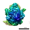
EMDB-1216:
Cryo-electron tomographic structure of an immunodeficiency virus envelope complex in situ.
Method: subtomogram averaging / : Zanetti G, Briggs JAG, Gruenewald K, Sattentau Q, Fuller SD
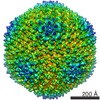
EMDB-1206:
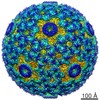
Structure of the bacteriophage phi6 nucleocapsid suggests a mechanism for sequential RNA packaging.
Method: single particle / : Huiskonen JT, de Haas F, Bubeck D, Bamford DH, Fuller SD, Butcher SJ
EMDB-1207:
Structure of the bacteriophage phi6 nucleocapsid suggests a mechanism for sequential RNA packaging.
Method: single particle / : Huiskonen JT, de Haas F, Bubeck D, Bamford DH, Fuller SD, Butcher SJ

EMDB-1155:
The mechanism of HIV-1 core assembly: insights from three-dimensional reconstructions of authentic virions.
Method: electron tomography / : Briggs JAG, Grunewald K, Glass B, Forster F

EMDB-1068:
Three-dimensional structures of translating ribosomes by Cryo-EM.
Method: single particle / : Gilbert RJC, Fucini P, Connell S, Fuller SD, Nierhaus KH, Robinson CV, Dobson CM, Stuart DI
EMDB-1070:
Three-dimensional structures of translating ribosomes by Cryo-EM.
Method: single particle / : Gilbert RJC, Fucini P, Connell S, Fuller SD, Nierhaus KH, Robinson CV, Dobson CM, Stuart DI
EMDB-1071:
Three-dimensional structures of translating ribosomes by Cryo-EM.
Method: single particle / : Gilbert RJC, Fucini P, Connell S, Fuller SD, Nierhaus KH, Robinson CV, Dobson CM, Stuart DI
EMDB-1072:
Three-dimensional structures of translating ribosomes by Cryo-EM.
Method: single particle / : Gilbert RJC, Fucini P, Connell S, Fuller SD, Nierhaus KH, Robinson CV, Dobson CM, Stuart DI

EMDB-1073:
Three-dimensional structures of translating ribosomes by Cryo-EM.
Method: single particle / : Gilbert RJC, Fucini P, Connell S, Fuller SD, Nierhaus KH, Robinson CV, Dobson CM, Stuart DI

EMDB-1018:
The first step: activation of the Semliki Forest virus spike protein precursor causes a localized conformational change in the trimeric spike.
Method: single particle / : Fuller SD

EMDB-1016:
Difference imaging of adenovirus: bridging the resolution gap between X-ray crystallography and electron microscopy.
Method: single particle / : Stewart PL, Fuller SD, Burnett RM
EMDB-1015:
Cryo-electron microscopy reveals the functional organization of an enveloped virus, Semliki Forest virus.
Method: single particle / : Mancini EJ, Clarke M, Gowen B, Rutten T, Fuller SD

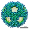
EMDB-1012:
Minor proteins, mobile arms and membrane-capsid interactions in the bacteriophage PRD1 capsid.
Method: single particle / : San Martin C, Huiskonen JT, Bamford JK, Butcher SJ, Fuller SD, Bamford DH, Burnett RM
EMDB-1013:
Combined EM/X-ray imaging yields a quasi-atomic model of the adenovirus-related bacteriophage PRD1 and shows key capsid and membrane interactions.
Method: single particle / : San Martin C, Burnett RM, de Haas F, Heinkel R, Rutten T, Fuller SD, Butcher SJ, Bamford DH
EMDB-1014:
Combined EM/X-ray imaging yields a quasi-atomic model of the adenovirus-related bacteriophage PRD1 and shows key capsid and membrane interactions.
Method: single particle / : San Martin C, Burnett RM, de Haas F, Heinkel R, Rutten T, Fuller SD, Butcher SJ, Bamford DH
EMDB-1011:
Minor proteins, mobile arms and membrane-capsid interactions in the bacteriophage PRD1 capsid.
Method: single particle / : San Martin C, Huiskonen JT, Bamford JK, Butcher SJ, Fuller SD, Bamford DH, Burnett RM
 wwPDB to switch to version 3 of the EMDB data model
