Movie
Movie Controller

[English] 日本語
 Yorodumi
- EMDB-1255: Structure of a hexameric RNA packaging motor in a viral polymeras... -
+ Open data

- Basic information
| Entry | Database: EMDB / ID: EMD-1255 | |||||||||
|---|---|---|---|---|---|---|---|---|---|---|
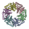
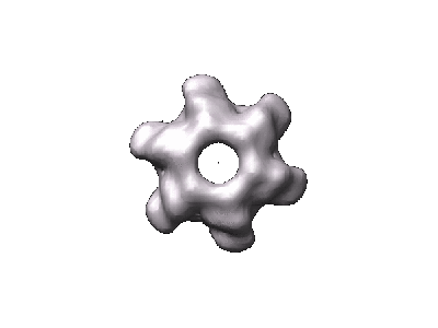
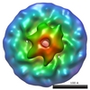
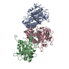
| Title | Structure of a hexameric RNA packaging motor in a viral polymerase complex. | |||||||||
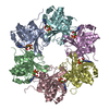
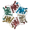
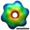
 Map data Map data | Bacteriophage phi8 hexameric packaging motor P4 reconstructed within the viral cores (icosahedral contribution of the core subtracted in the images), applying C6 symmetry | |||||||||
 Sample Sample |
| |||||||||
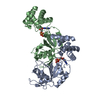
| Biological species |  Pseudomonas phage phi8 (virus) Pseudomonas phage phi8 (virus) | |||||||||
| Method | single particle reconstruction / cryo EM / Resolution: 15.0 Å | |||||||||
 Authors Authors | Huiskonen JT / Jaalinoja HT / Briggs JAG | |||||||||
 Citation Citation | Journal: J Struct Biol / Year: 2007 Title: Structure of a hexameric RNA packaging motor in a viral polymerase complex. Authors: Juha T Huiskonen / Harri T Jäälinoja / John A G Briggs / Stephen D Fuller / Sarah J Butcher /  Abstract: Packaging of the Cystovirus varphi8 genome into the polymerase complex is catalysed by the hexameric P4 packaging motor. The motor is located at the fivefold vertices of the icosahedrally symmetric ...Packaging of the Cystovirus varphi8 genome into the polymerase complex is catalysed by the hexameric P4 packaging motor. The motor is located at the fivefold vertices of the icosahedrally symmetric polymerase complex, and the symmetry mismatch between them may be critical for function. We have developed a novel image-processing approach for the analysis of symmetry-mismatched structures and applied it to cryo-electron microscopy images of P4 bound to the polymerase complex. This approach allowed us to solve the three-dimensional structure of the P4 in situ to 15-A resolution. The C-terminal face of P4 was observed to interact with the polymerase complex, supporting the current view on RNA translocation. We suggest that the symmetry mismatch between the two components may facilitate the ring opening required for RNA loading prior to its translocation. | |||||||||
| History |
|
- Structure visualization
| Movie |
 Movie viewer Movie viewer |
|---|---|
| Structure viewer | EM map: SurfViewMolmilJmol/JSmol |
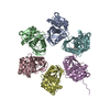
| Supplemental images |
- Downloads & links
-EMDB archive
| Map data | emd_1255.map.gz | 3.3 MB | EMDB map data format | |
|---|---|---|---|---|
| Header (meta data) | emd-1255-v30.xmlemd-1255.xml | 8.9 KB 8.9 KB | Display Display | EMDB header |
| Images |  1255.gif 1255.gif | 10.9 KB | ||
| Archive directory |  http://ftp.pdbj.org/pub/emdb/structures/EMD-1255ftp://ftp.pdbj.org/pub/emdb/structures/EMD-1255 http://ftp.pdbj.org/pub/emdb/structures/EMD-1255ftp://ftp.pdbj.org/pub/emdb/structures/EMD-1255 | HTTPS FTP |
-Related structure data
-Links
| EMDB pages | EMDB (EBI/PDBe) / EMDataResource |
|---|
-Map
| File | Download / File: emd_1255.map.gz / Format: CCP4 / Size: 3.7 MB / Type: IMAGE STORED AS FLOATING POINT NUMBER (4 BYTES) | ||||||||||||||||||||||||||||||||||||||||||||||||||||||||||||||||||||
|---|---|---|---|---|---|---|---|---|---|---|---|---|---|---|---|---|---|---|---|---|---|---|---|---|---|---|---|---|---|---|---|---|---|---|---|---|---|---|---|---|---|---|---|---|---|---|---|---|---|---|---|---|---|---|---|---|---|---|---|---|---|---|---|---|---|---|---|---|---|
| Annotation | Bacteriophage phi8 hexameric packaging motor P4 reconstructed within the viral cores (icosahedral contribution of the core subtracted in the images), applying C6 symmetry | ||||||||||||||||||||||||||||||||||||||||||||||||||||||||||||||||||||
| Projections & slices | Image control
Images are generated by Spider. | ||||||||||||||||||||||||||||||||||||||||||||||||||||||||||||||||||||
| Voxel size | X=Y=Z: 2.8 Å | ||||||||||||||||||||||||||||||||||||||||||||||||||||||||||||||||||||
| Density |
| ||||||||||||||||||||||||||||||||||||||||||||||||||||||||||||||||||||
| Symmetry | Space group: 1 | ||||||||||||||||||||||||||||||||||||||||||||||||||||||||||||||||||||
| Details | EMDB XML:
CCP4 map header:
| ||||||||||||||||||||||||||||||||||||||||||||||||||||||||||||||||||||
 Z (Sec.)
 Y (Row.)
 X (Col.)


-Supplemental data
- Sample components
-Entire : Bacteriophage phi8 core
| Entire | Name: Bacteriophage phi8 core |
|---|---|
| Components |
|
-Supramolecule #1000: Bacteriophage phi8 core
| Supramolecule | Name: Bacteriophage phi8 core / type: sample / ID: 1000 / Number unique components: 1 |
|---|---|
| Molecular weight | Theoretical: 33 MDa |
-Supramolecule #1: Pseudomonas phage phi8
| Supramolecule | Name: Pseudomonas phage phi8 / type: virus / ID: 1 / Name.synonym: Bacteriophage phi8 / NCBI-ID: 120086 / Sci species name: Pseudomonas phage phi8 / Virus type: VIRION / Virus isolate: SPECIES / Virus enveloped: Yes / Virus empty: No / Syn species name: Bacteriophage phi8 |
|---|---|
| Host (natural) | Organism:  Pseudomonas syringae pv. phaseolicola (bacteria) / synonym: BACTERIA(EUBACTERIA) Pseudomonas syringae pv. phaseolicola (bacteria) / synonym: BACTERIA(EUBACTERIA) |
| Molecular weight | Experimental: 33 MDa |
| Virus shell | Shell ID: 1 / T number (triangulation number): 1 |
-Experimental details
-Structure determination
| Method | cryo EM |
|---|---|
 Processing Processing | single particle reconstruction |
| Aggregation state | particle |
-Sample preparation
| Buffer | pH: 7.5 Details: 10 mM potassium phosphate pH 7.5, 1 mM MgCl2, 50 mM NaCl |
|---|---|
| Grid | Details: 200 mesh holey carbon coated copper grid |
| Vitrification | Cryogen name: ETHANE |
- Electron microscopy
| Microscope | FEI TECNAI F20 |
|---|---|
| Image recording | Category: FILM / Film or detector model: KODAK SO-163 FILM / Digitization - Scanner: ZEISS SCAI / Digitization - Sampling interval: 7 µm / Number real images: 66 / Bits/pixel: 12 |
| Electron beam | Acceleration voltage: 200 kV / Electron source:  FIELD EMISSION GUN FIELD EMISSION GUN |
| Electron optics | Illumination mode: FLOOD BEAM / Imaging mode: BRIGHT FIELD / Cs: 2.0 mm / Nominal defocus max: 3.3 µm / Nominal defocus min: 0.7 µm / Nominal magnification: 50000 |
| Sample stage | Specimen holder: Side entry liquid nitrogen-cooled cryo specimen holder Specimen holder model: OTHER |
| Experimental equipment |  Model: Tecnai F20 / Image courtesy: FEI Company |
-Image processing
| CTF correction | Details: Each particle, phases flipped |
|---|---|
| Final reconstruction | Applied symmetry - Point group: C6 (6 fold cyclic) / Algorithm: OTHER / Resolution.type: BY AUTHOR / Resolution: 15.0 Å / Resolution method: FSC 3 SIGMA CUT-OFF / Software - Name: Imagic5 |
| Final two d classification | Number classes: 352 |
-Atomic model buiding 1
| Initial model | PDB ID: |
|---|---|
| Software | Name: colores |
| Details | Protocol: rigid body |
| Refinement | Space: RECIPROCAL / Protocol: RIGID BODY FIT / Target criteria: correlation coefficient |