 Movie
Movie Controller

[English] 日本語
 Yorodumi
- EMDB-1206: Structure of the bacteriophage phi6 nucleocapsid suggests a mecha... -
+ Open data

- Basic information
| Entry | Database: EMDB / ID: EMD-1206 | |||||||||
|---|---|---|---|---|---|---|---|---|---|---|
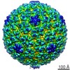
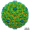
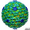
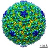
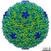
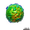
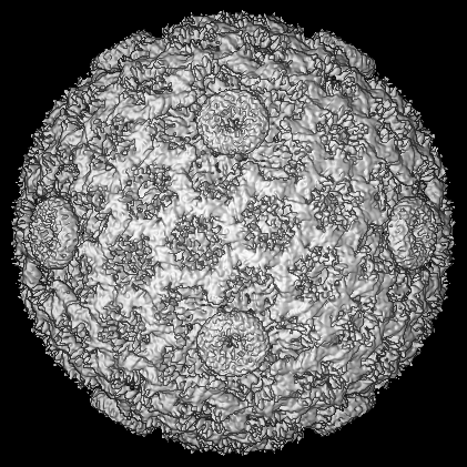
| Title | Structure of the bacteriophage phi6 nucleocapsid suggests a mechanism for sequential RNA packaging. | |||||||||
 Map data Map data | Reconstruction of phi6 nucleocapsid to 7.5-A resolution | |||||||||
 Sample Sample |
| |||||||||
| Function / homology | : / Major inner capsid protein P1 / T=2 icosahedral viral capsid / viral inner capsid / viral nucleocapsid / RNA binding / identical protein binding / Major inner protein P1 Function and homology information Function and homology information | |||||||||
| Biological species |  Pseudomonas phage phi6 (virus) Pseudomonas phage phi6 (virus) | |||||||||
| Method | single particle reconstruction / cryo EM / Resolution: 7.5 Å | |||||||||
 Authors Authors | Huiskonen JT / de Haas F / Bubeck D / Bamford DH / Fuller SD / Butcher SJ | |||||||||
 Citation Citation | Journal: Structure / Year: 2006 Title: Structure of the bacteriophage phi6 nucleocapsid suggests a mechanism for sequential RNA packaging. Authors: Juha T Huiskonen / Felix de Haas / Doryen Bubeck / Dennis H Bamford / Stephen D Fuller / Sarah J Butcher /  Abstract: Bacteriophage phi6 is an enveloped dsRNA virus with a segmented genome. Phi6 specifically packages one copy of each of its three genome segments into a preassembled polymerase complex. This leads to ...Bacteriophage phi6 is an enveloped dsRNA virus with a segmented genome. Phi6 specifically packages one copy of each of its three genome segments into a preassembled polymerase complex. This leads to expansion of the polymerase complex, minus and plus strand RNA synthesis, and assembly of the nucleocapsid. The phi6 in vitro assembly and packaging system is a valuable model for dsRNA virus replication. The structure of the nucleocapsid at 7.5 A resolution presented here reveals the secondary structure of the two major capsid proteins. Asymmetric P1 dimers organize as an inner T = 1 shell, and P8 trimers organize as an outer T = 13 laevo shell. The organization of the P1 molecules in the unexpanded and expanded polymerase complex suggests that the expansion is accomplished by rigid body movements of the P1 monomers. This leads to exposure of new potential RNA binding surfaces to control the sequential packaging of the genome segments. | |||||||||
| History |
|
- Structure visualization
| Movie |
Movie viewer |
|---|---|
| Structure viewer | EM map: SurfViewMolmilJmol/JSmol |
| Supplemental images |


- Downloads & links
-EMDB archive
| Map data | emd_1206.map.gz | 123.6 MB | EMDB map data format | |
|---|---|---|---|---|
| Header (meta data) | emd-1206-v30.xmlemd-1206.xml | 22 KB 22 KB | Display Display | EMDB header |
| Images |  1206.gif 1206.gif | 35.8 KB | ||
| Masks | emd_1206_msk_1.mapemd_1206_msk_2.mapemd_1206_msk_3.mapemd_1206_msk_4.mapemd_1206_msk_5.mapemd_1206_msk_6.mapemd_1206_msk_7.map | 142.3 MB 60.3 MB 142.3 MB 142.3 MB 142.3 MB 142.3 MB 17.7 MB | Mask map | |
| Others | MASKnames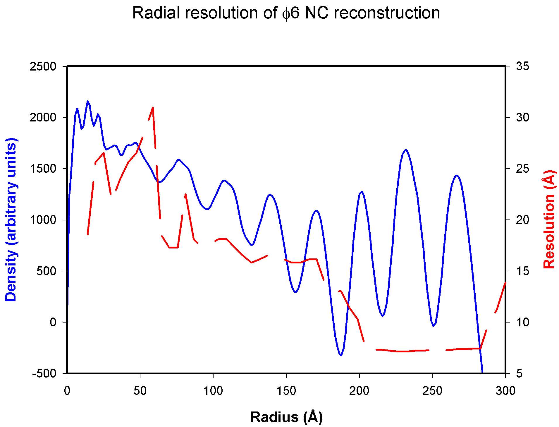 emd_1206_fsc.jpg emd_1206_fsc.jpg | 304 B 131.9 KB | ||
| Archive directory |  http://ftp.pdbj.org/pub/emdb/structures/EMD-1206ftp://ftp.pdbj.org/pub/emdb/structures/EMD-1206 http://ftp.pdbj.org/pub/emdb/structures/EMD-1206ftp://ftp.pdbj.org/pub/emdb/structures/EMD-1206 | HTTPS FTP |
-Related structure data
| Related structure data |  4btqM 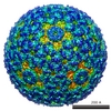 1207C M: atomic model generated by this map C: citing same article ( |
|---|---|
| Similar structure data |
-Links
| EMDB pages | EMDB (EBI/PDBe) / EMDataResource |
|---|---|
| Related items in Molecule of the Month |

-Map
| File | Download / File: emd_1206.map.gz / Format: CCP4 / Size: 278 MB / Type: IMAGE STORED AS FLOATING POINT NUMBER (4 BYTES) | ||||||||||||||||||||||||||||||||||||||||||||||||||||||||||||||||||||
|---|---|---|---|---|---|---|---|---|---|---|---|---|---|---|---|---|---|---|---|---|---|---|---|---|---|---|---|---|---|---|---|---|---|---|---|---|---|---|---|---|---|---|---|---|---|---|---|---|---|---|---|---|---|---|---|---|---|---|---|---|---|---|---|---|---|---|---|---|---|
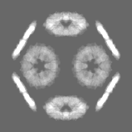
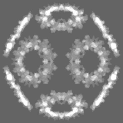
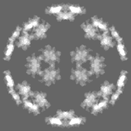
| Annotation | Reconstruction of phi6 nucleocapsid to 7.5-A resolution | ||||||||||||||||||||||||||||||||||||||||||||||||||||||||||||||||||||
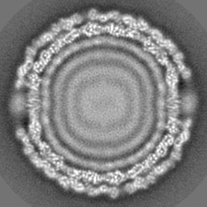
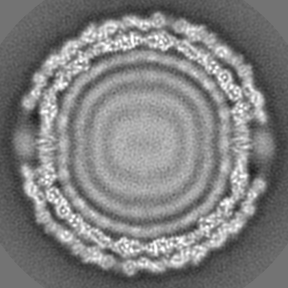
| Projections & slices | Image control
Images are generated by Spider. | ||||||||||||||||||||||||||||||||||||||||||||||||||||||||||||||||||||
| Voxel size | X=Y=Z: 1.4 Å | ||||||||||||||||||||||||||||||||||||||||||||||||||||||||||||||||||||
| Density |
| ||||||||||||||||||||||||||||||||||||||||||||||||||||||||||||||||||||
| Symmetry | Space group: 1 | ||||||||||||||||||||||||||||||||||||||||||||||||||||||||||||||||||||
| Details | EMDB XML:
CCP4 map header:
| ||||||||||||||||||||||||||||||||||||||||||||||||||||||||||||||||||||
 Z (Sec.)
 Y (Row.)
 X (Col.)


-Supplemental data
-Segmentation: Alpha subunit
| Annotation | Alpha subunit | ||||||||||||
|---|---|---|---|---|---|---|---|---|---|---|---|---|---|
| File | emd_1206_msk_1.map | ||||||||||||
| Projections & Slices |
| ||||||||||||
| Density Histograms |


-Segmentation: Beta subunit
| Annotation | Beta subunit | ||||||||||||
|---|---|---|---|---|---|---|---|---|---|---|---|---|---|
| File | emd_1206_msk_2.map | ||||||||||||
| Projections & Slices |
| ||||||||||||
| Density Histograms |


-Segmentation: Q subunit
| Annotation | Q subunit | ||||||||||||
|---|---|---|---|---|---|---|---|---|---|---|---|---|---|
| File | emd_1206_msk_3.map | ||||||||||||
| Projections & Slices |
| ||||||||||||
| Density Histograms |


-Segmentation: R subunit
| Annotation | R subunit | ||||||||||||
|---|---|---|---|---|---|---|---|---|---|---|---|---|---|
| File | emd_1206_msk_4.map | ||||||||||||
| Projections & Slices |
| ||||||||||||
| Density Histograms |


-Segmentation: S subunit
| Annotation | S subunit | ||||||||||||
|---|---|---|---|---|---|---|---|---|---|---|---|---|---|
| File | emd_1206_msk_5.map | ||||||||||||
| Projections & Slices |
| ||||||||||||
| Density Histograms |


-Segmentation: T subunit
| Annotation | T subunit | ||||||||||||
|---|---|---|---|---|---|---|---|---|---|---|---|---|---|
| File | emd_1206_msk_6.map | ||||||||||||
| Projections & Slices |
| ||||||||||||
| Density Histograms |


-Segmentation: tc B
| Annotation | tc_B | ||||||||||||
|---|---|---|---|---|---|---|---|---|---|---|---|---|---|
| File | emd_1206_msk_7.map | ||||||||||||
| Projections & Slices |
| ||||||||||||
| Density Histograms |


-Others
| Details | [MASKnames] emd_1206_msk_1.map > phi6nc_210_tc_B_mask.map emd_1206_msk_2.map > phi6nc_421_A_mask.map emd_1206_msk_3.map > phi6nc_421_B_mask.map emd_1206_msk_4.map > phi6nc_421_Q_mask.map emd_1206_msk_5.map > phi6nc_421_R_mask.map emd_1206_msk_6.map > phi6nc_421_S_mask.map emd_1206_msk_7.map > phi6nc_421_T_mask.ma |
|---|---|
| Image |
- Sample components
-Entire : Bacteriophage phi6 nucleocapsid
| Entire | Name: Bacteriophage phi6 nucleocapsid |
|---|---|
| Components |
|
-Supramolecule #1000: Bacteriophage phi6 nucleocapsid
| Supramolecule | Name: Bacteriophage phi6 nucleocapsid / type: sample / ID: 1000 / Number unique components: 1 |
|---|---|
| Molecular weight | Theoretical: 22 MDa |
-Supramolecule #1: Pseudomonas phage phi6
| Supramolecule | Name: Pseudomonas phage phi6 / type: virus / ID: 1 / Name.synonym: Bacteriophage phi6 / NCBI-ID: 10879 / Sci species name: Pseudomonas phage phi6 / Virus type: VIRION / Virus isolate: SPECIES / Virus enveloped: Yes / Virus empty: No / Syn species name: Bacteriophage phi6 |
|---|---|
| Host (natural) | Organism: Pseudomonas syringae pv phaesolicola HB10Y / synonym: BACTERIA(EUBACTERIA) |
| Virus shell | Shell ID: 1 / Name: PC shell / Diameter: 485 Å / T number (triangulation number): 1 |
| Virus shell | Shell ID: 2 / Name: NC shell / Diameter: 555 Å / T number (triangulation number): 13 |
-Experimental details
-Structure determination
| Method | cryo EM |
|---|---|
 Processing Processing | single particle reconstruction |
| Aggregation state | particle |
-Sample preparation
| Vitrification | Cryogen name: ETHANE |
|---|
- Electron microscopy
| Microscope | FEI TECNAI F20 |
|---|---|
| Temperature | Average: 93 K |
| Alignment procedure | Legacy - Astigmatism: objective lens astigmatism was corrected at |
| Image recording | Category: FILM / Film or detector model: KODAK SO-163 FILM / Digitization - Scanner: ZEISS SCAI / Digitization - Sampling interval: 7 µm / Number real images: 52 / Bits/pixel: 12 |
| Tilt angle min | 0 |
| Tilt angle max | 0 |
| Electron beam | Acceleration voltage: 200 kV / Electron source:  FIELD EMISSION GUN FIELD EMISSION GUN |
| Electron optics | Illumination mode: FLOOD BEAM / Imaging mode: BRIGHT FIELD / Cs: 2.0 mm / Nominal defocus max: 2.5 µm / Nominal defocus min: 0.4 µm / Nominal magnification: 50000 |
| Sample stage | Specimen holder: Side entry liquid nitrogen-cooled cryo specimen holder Specimen holder model: OTHER |
| Experimental equipment |  Model: Tecnai F20 / Image courtesy: FEI Company |
-Image processing
| CTF correction | Details: each particle, full CTF correction |
|---|---|
| Final reconstruction | Applied symmetry - Point group: I (icosahedral) / Algorithm: OTHER / Resolution.type: BY AUTHOR / Resolution: 7.5 Å / Resolution method: FSC 0.5 CUT-OFF / Software - Name: P3DR / Number images used: 10463 |
