 Movie
Movie Controller

[English] 日本語
 Yorodumi
- EMDB-1321: Quasi-atomic model of bacteriophage t7 procapsid shell: insights ... -
+ Open data

- Basic information
| Entry | Database: EMDB / ID: EMD-1321 | |||||||||
|---|---|---|---|---|---|---|---|---|---|---|
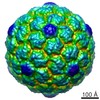
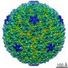
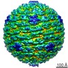
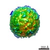
| Title | Quasi-atomic model of bacteriophage t7 procapsid shell: insights into the structure and evolution of a basic fold. | |||||||||
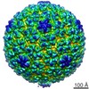
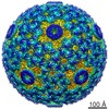
 Map data Map data | phage T7 prohead icosahedral map | |||||||||
 Sample Sample |
| |||||||||
| Function / homology | Capsid Gp10A/Gp10B / : / Major capsid protein / viral capsid / viral translational frameshifting / identical protein binding / Major capsid protein Function and homology information Function and homology information | |||||||||
| Biological species |   Enterobacteria phage T7 (virus) Enterobacteria phage T7 (virus) | |||||||||
| Method | single particle reconstruction / cryo EM / Resolution: 10.9 Å | |||||||||
 Authors Authors | Agirrezabala X / Velazquez-Muriel J / Gomez-Puertas P / Scheres S / Carazo JM / Carrascosa JL | |||||||||
 Citation Citation | Journal: Structure / Year: 2007 Title: Quasi-atomic model of bacteriophage t7 procapsid shell: insights into the structure and evolution of a basic fold. Authors: Xabier Agirrezabala / Javier A Velázquez-Muriel / Paulino Gómez-Puertas / Sjors H W Scheres / José M Carazo / José L Carrascosa /  Abstract: The existence of similar folds among major structural subunits of viral capsids has shown unexpected evolutionary relationships suggesting common origins irrespective of the capsids' host life domain. ...The existence of similar folds among major structural subunits of viral capsids has shown unexpected evolutionary relationships suggesting common origins irrespective of the capsids' host life domain. Tailed bacteriophages are emerging as one such family, and we have studied the possible existence of the HK97-like fold in bacteriophage T7. The procapsid structure at approximately 10 A resolution was used to obtain a quasi-atomic model by fitting a homology model of the T7 capsid protein gp10 that was based on the atomic structure of the HK97 capsid protein. A number of fold similarities, such as the fitting of domains A and P into the L-shaped procapsid subunit, are evident between both viral systems. A different feature is related to the presence of the amino-terminal domain of gp10 found at the inner surface of the capsid that might play an important role in the interaction of capsid and scaffolding proteins. | |||||||||
| History |
|
- Structure visualization
| Movie |
Movie viewer |
|---|---|
| Structure viewer | EM map: SurfViewMolmilJmol/JSmol |
| Supplemental images |
- Downloads & links
-EMDB archive
| Map data | emd_1321.map.gz | 6.8 MB | EMDB map data format | |
|---|---|---|---|---|
| Header (meta data) | emd-1321-v30.xmlemd-1321.xml | 8.4 KB 8.4 KB | Display Display | EMDB header |
| Images |  1321.gif 1321.gif | 85.7 KB | ||
| Archive directory |  http://ftp.pdbj.org/pub/emdb/structures/EMD-1321ftp://ftp.pdbj.org/pub/emdb/structures/EMD-1321 http://ftp.pdbj.org/pub/emdb/structures/EMD-1321ftp://ftp.pdbj.org/pub/emdb/structures/EMD-1321 | HTTPS FTP |
-Related structure data
| Related structure data | 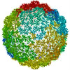 3izgM M: atomic model generated by this map |
|---|---|
| Similar structure data |
-Links
| EMDB pages | EMDB (EBI/PDBe) / EMDataResource |
|---|
-Map
| File | Download / File: emd_1321.map.gz / Format: CCP4 / Size: 65.5 MB / Type: IMAGE STORED AS FLOATING POINT NUMBER (4 BYTES) | ||||||||||||||||||||||||||||||||||||||||||||||||||||||||||||||||||||
|---|---|---|---|---|---|---|---|---|---|---|---|---|---|---|---|---|---|---|---|---|---|---|---|---|---|---|---|---|---|---|---|---|---|---|---|---|---|---|---|---|---|---|---|---|---|---|---|---|---|---|---|---|---|---|---|---|---|---|---|---|---|---|---|---|---|---|---|---|---|
| Annotation | phage T7 prohead icosahedral map | ||||||||||||||||||||||||||||||||||||||||||||||||||||||||||||||||||||
| Projections & slices | Image control
Images are generated by Spider. | ||||||||||||||||||||||||||||||||||||||||||||||||||||||||||||||||||||
| Voxel size | X=Y=Z: 2.72 Å | ||||||||||||||||||||||||||||||||||||||||||||||||||||||||||||||||||||
| Density |
| ||||||||||||||||||||||||||||||||||||||||||||||||||||||||||||||||||||
| Symmetry | Space group: 1 | ||||||||||||||||||||||||||||||||||||||||||||||||||||||||||||||||||||
| Details | EMDB XML:
CCP4 map header:
| ||||||||||||||||||||||||||||||||||||||||||||||||||||||||||||||||||||
 Z (Sec.)
 Y (Row.)
 X (Col.)


-Supplemental data
- Sample components
-Entire : phage T7 prohead
| Entire | Name: phage T7 prohead |
|---|---|
| Components |
|
-Supramolecule #1000: phage T7 prohead
| Supramolecule | Name: phage T7 prohead / type: sample / ID: 1000 / Number unique components: 1 |
|---|
-Macromolecule #1: gp10A
| Macromolecule | Name: gp10A / type: protein_or_peptide / ID: 1 / Name.synonym: major capsid protein / Number of copies: 420 / Oligomeric state: icosahedral / Recombinant expression: Yes |
|---|---|
| Source (natural) | Organism: Enterobacteria phage T7 (virus) / synonym: phage T7 |
| Molecular weight | Theoretical: 37 MDa |
| Recombinant expression | Organism:  |
-Experimental details
-Structure determination
| Method | cryo EM |
|---|---|
 Processing Processing | single particle reconstruction |
| Aggregation state | particle |
-Sample preparation
| Buffer | pH: 7.7 / Details: 50mM Tris-HCl pH:7.7 10mM MgCl2 100mM NaCl |
|---|---|
| Grid | Details: Quantifoil grids 2/2 |
| Vitrification | Cryogen name: ETHANE |
- Electron microscopy
| Microscope | FEI TECNAI 20 |
|---|---|
| Alignment procedure | Legacy - Astigmatism: 100k |
| Image recording | Category: FILM / Film or detector model: KODAK SO-163 FILM / Digitization - Scanner: ZEISS SCAI / Digitization - Sampling interval: 7 µm / Average electron dose: 10 e/Å2 / Bits/pixel: 8 |
| Tilt angle min | 0 |
| Tilt angle max | 0 |
| Electron beam | Acceleration voltage: 200 kV / Electron source:  FIELD EMISSION GUN FIELD EMISSION GUN |
| Electron optics | Calibrated magnification: 51600 / Illumination mode: SPOT SCAN / Imaging mode: BRIGHT FIELD / Cs: 2.26 mm / Nominal defocus max: 3.0 µm / Nominal defocus min: 1.0 µm / Nominal magnification: 50000 |
| Sample stage | Specimen holder: Side entry liquid nitrogen-cooled cryo specimen holder. GATAN. Eucentric Specimen holder model: GATAN LIQUID NITROGEN |
-Image processing
| CTF correction | Details: Wiener filter, defocus groups |
|---|---|
| Final reconstruction | Applied symmetry - Point group: I (icosahedral) / Algorithm: OTHER / Resolution.type: BY AUTHOR / Resolution: 10.9 Å / Resolution method: FSC 0.5 CUT-OFF / Software - Name: spider / Number images used: 4460 |
