+ Open data
- Basic information
| Entry | Database: PDB / ID: 3izg | ||||||
|---|---|---|---|---|---|---|---|
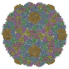
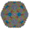
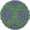
| Title | Bacteriophage T7 prohead shell EM-derived atomic model | ||||||
 Components Components | Major capsid protein 10A | ||||||
 Keywords Keywords | VIRUS / bacteriophage / capsid maturation / cryoelectron microscopy / morphogenetic intermediate / icosahedral | ||||||
| Function / homology | Capsid Gp10A/Gp10B / : / Major capsid protein / viral capsid / viral translational frameshifting / identical protein binding / Major capsid protein Function and homology information Function and homology information | ||||||
| Biological species |   Enterobacteria phage T7 (virus) Enterobacteria phage T7 (virus) | ||||||
| Method | ELECTRON MICROSCOPY / single particle reconstruction / cryo EM / Resolution: 10.9 Å | ||||||
 Authors Authors | Ionel, A. / Velazquez-Muriel, J.A. / Agirrezabala, X. / Luque, D. / Cuervo, A. / Caston, J.R. / Valpuesta, J.M. / Martin-Benito, J. / Carrascosa, J.L. | ||||||
 Citation Citation |  Journal: J Biol Chem / Year: 2011 Journal: J Biol Chem / Year: 2011Title: Molecular rearrangements involved in the capsid shell maturation of bacteriophage T7. Authors: Alina Ionel / Javier A Velázquez-Muriel / Daniel Luque / Ana Cuervo / José R Castón / José M Valpuesta / Jaime Martín-Benito / José L Carrascosa /  Abstract: Maturation of dsDNA bacteriophages involves assembling the virus prohead from a limited set of structural components followed by rearrangements required for the stability that is necessary for ...Maturation of dsDNA bacteriophages involves assembling the virus prohead from a limited set of structural components followed by rearrangements required for the stability that is necessary for infecting a host under challenging environmental conditions. Here, we determine the mature capsid structure of T7 at 1 nm resolution by cryo-electron microscopy and compare it with the prohead to reveal the molecular basis of T7 shell maturation. The mature capsid presents an expanded and thinner shell, with a drastic rearrangement of the major protein monomers that increases in their interacting surfaces, in turn resulting in a new bonding lattice. The rearrangements include tilting, in-plane rotation, and radial expansion of the subunits, as well as a relative bending of the A- and P-domains of each subunit. The unique features of this shell transformation, which does not employ the accessory proteins, inserted domains, or molecular interactions observed in other phages, suggest a simple capsid assembling strategy that may have appeared early in the evolution of these viruses. #1:  Journal: Structure / Year: 2007 Journal: Structure / Year: 2007Title: Quasi-atomic model of bacteriophage t7 procapsid shell: insights into the structure and evolution of a basic fold. Authors: Xabier Agirrezabala / Javier A Velázquez-Muriel / Paulino Gómez-Puertas / Sjors H W Scheres / José M Carazo / José L Carrascosa /  Abstract: The existence of similar folds among major structural subunits of viral capsids has shown unexpected evolutionary relationships suggesting common origins irrespective of the capsids' host life domain. ...The existence of similar folds among major structural subunits of viral capsids has shown unexpected evolutionary relationships suggesting common origins irrespective of the capsids' host life domain. Tailed bacteriophages are emerging as one such family, and we have studied the possible existence of the HK97-like fold in bacteriophage T7. The procapsid structure at approximately 10 A resolution was used to obtain a quasi-atomic model by fitting a homology model of the T7 capsid protein gp10 that was based on the atomic structure of the HK97 capsid protein. A number of fold similarities, such as the fitting of domains A and P into the L-shaped procapsid subunit, are evident between both viral systems. A different feature is related to the presence of the amino-terminal domain of gp10 found at the inner surface of the capsid that might play an important role in the interaction of capsid and scaffolding proteins. | ||||||
| History |
|
- Structure visualization
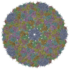
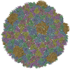
| Movie |
 Movie viewer Movie viewer |
|---|---|
| Structure viewer | Molecule:  Molmil Molmil Jmol/JSmol Jmol/JSmol |
- Downloads & links
- Download
| PDBx/mmCIF format |  3izg.cif.gz 3izg.cif.gz | 296.7 KB | Display |  PDBx/mmCIF format PDBx/mmCIF format |
|---|---|---|---|---|
| PDB format |  pdb3izg.ent.gz pdb3izg.ent.gz | 240.2 KB | Display |  PDB format PDB format |
| PDBx/mmJSON format |  3izg.json.gz 3izg.json.gz | Tree view |  PDBx/mmJSON format PDBx/mmJSON format | |
| Others |  Other downloads Other downloads |
-Validation report
| Arichive directory |  https://data.pdbj.org/pub/pdb/validation_reports/iz/3izg https://data.pdbj.org/pub/pdb/validation_reports/iz/3izg ftp://data.pdbj.org/pub/pdb/validation_reports/iz/3izg ftp://data.pdbj.org/pub/pdb/validation_reports/iz/3izg | HTTPS FTP |
|---|
-Related structure data
| Related structure data |  1321M  1810C  2xvrC M: map data used to model this data C: citing same article ( |
|---|---|
| Similar structure data |
- Links
Links
- Assembly
| Deposited unit | 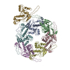
|
|---|---|
| 1 | x 60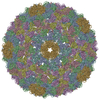
|
| 2 |
|
| 3 | x 5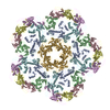
|
| 4 | x 6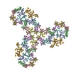
|
| 5 | 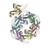
|
| Symmetry | Point symmetry: (Schoenflies symbol: I (icosahedral)) |
- Components
| #1: Protein | Mass: 36589.625 Da / Num. of mol.: 7 / Source method: isolated from a natural source / Source: (natural)   Enterobacteria phage T7 (virus) / References: UniProt: P19726 Enterobacteria phage T7 (virus) / References: UniProt: P19726 |
|---|
-Experimental details
-Experiment
| Experiment | Method: ELECTRON MICROSCOPY |
|---|---|
| EM experiment | Aggregation state: PARTICLE / 3D reconstruction method: single particle reconstruction |
- Sample preparation
| Component | Name: bacteriophage T7 prohead / Type: VIRUS / Details: gp10A |
|---|---|
| Details of virus | Host category: BACTERIA / Type: VIRION |
| Natural host | Organism: Escherichia coli |
| Buffer solution | Name: 50mM Tris-HCl pH7.7 10mM MgCl2 100mM NaCl / pH: 7.7 / Details: 50mM Tris-HCl pH7.7 10mM MgCl2 100mM NaCl |
| Specimen | Embedding applied: NO / Shadowing applied: NO / Staining applied: NO / Vitrification applied: YES |
| Specimen support | Details: Quantifoil R2/2 carbon grids |
| Vitrification | Cryogen name: ETHANE |
- Electron microscopy imaging
| Microscopy | Model: FEI TECNAI 20 |
|---|---|
| Electron gun | Electron source:  FIELD EMISSION GUN / Accelerating voltage: 200 kV / Illumination mode: SPOT SCAN FIELD EMISSION GUN / Accelerating voltage: 200 kV / Illumination mode: SPOT SCAN |
| Electron lens | Mode: BRIGHT FIELD / Nominal magnification: 50000 X / Calibrated magnification: 51600 X / Nominal defocus max: 1000 nm / Nominal defocus min: 3000 nm / Cs: 2.26 mm |
| Specimen holder | Specimen holder model: GATAN LIQUID NITROGEN / Tilt angle max: 0 ° / Tilt angle min: 0 ° |
| Image recording | Electron dose: 10 e/Å2 / Film or detector model: KODAK SO-163 FILM |
- Processing
| EM software | Name: SPIDER / Category: 3D reconstruction | ||||||||||||
|---|---|---|---|---|---|---|---|---|---|---|---|---|---|
| CTF correction | Details: Wiener filter, defocus groups | ||||||||||||
| Symmetry | Point symmetry: I (icosahedral) | ||||||||||||
| 3D reconstruction | Method: Spider / Resolution: 10.9 Å / Num. of particles: 4460 / Actual pixel size: 2.72 Å / Symmetry type: POINT | ||||||||||||
| Atomic model building | PDB-ID: 3E8K Accession code: 3E8K / Source name: PDB / Type: experimental model | ||||||||||||
| Refinement step | Cycle: LAST
|
 Movie
Movie Controller






 PDBj
PDBj