Movie
Movie Controller


+ Open data

- Basic information
| Entry | Database: PDB / ID: 2xvr | ||||||
|---|---|---|---|---|---|---|---|
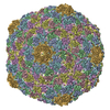
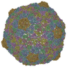
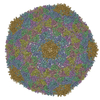
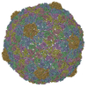
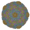
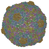
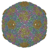
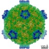
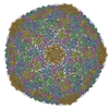
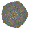
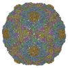
| Title | Phage T7 empty mature head shell | ||||||
 Components Components | MAJOR CAPSID PROTEIN 10A | ||||||
 Keywords Keywords | VIRUS / CAPSID MATURATION / MORPHOGENETIC INTERMEDIATE | ||||||
| Function / homology | Capsid Gp10A/Gp10B / : / Major capsid protein / viral capsid / viral translational frameshifting / identical protein binding / Major capsid protein Function and homology information Function and homology information | ||||||
| Biological species |   ENTEROBACTERIA PHAGE T7 (virus) ENTEROBACTERIA PHAGE T7 (virus) | ||||||
| Method | ELECTRON MICROSCOPY / single particle reconstruction / cryo EM / Resolution: 10.8 Å | ||||||
 Authors Authors | Ionel, A. / Velazquez-Muriel, J.A. / Luque, D. / Cuervo, A. / Caston, J.R. / Valpuesta, J.M. / Martin-Benito, J. / Carrascosa, J.L. | ||||||
 Citation Citation | Journal: J Biol Chem / Year: 2011 Title: Molecular rearrangements involved in the capsid shell maturation of bacteriophage T7. Authors: Alina Ionel / Javier A Velázquez-Muriel / Daniel Luque / Ana Cuervo / José R Castón / José M Valpuesta / Jaime Martín-Benito / José L Carrascosa /  Abstract: Maturation of dsDNA bacteriophages involves assembling the virus prohead from a limited set of structural components followed by rearrangements required for the stability that is necessary for ...Maturation of dsDNA bacteriophages involves assembling the virus prohead from a limited set of structural components followed by rearrangements required for the stability that is necessary for infecting a host under challenging environmental conditions. Here, we determine the mature capsid structure of T7 at 1 nm resolution by cryo-electron microscopy and compare it with the prohead to reveal the molecular basis of T7 shell maturation. The mature capsid presents an expanded and thinner shell, with a drastic rearrangement of the major protein monomers that increases in their interacting surfaces, in turn resulting in a new bonding lattice. The rearrangements include tilting, in-plane rotation, and radial expansion of the subunits, as well as a relative bending of the A- and P-domains of each subunit. The unique features of this shell transformation, which does not employ the accessory proteins, inserted domains, or molecular interactions observed in other phages, suggest a simple capsid assembling strategy that may have appeared early in the evolution of these viruses. | ||||||
| History |
|
- Structure visualization
| Movie |
Movie viewer |
|---|---|
| Structure viewer | Molecule: MolmilJmol/JSmol |
- Downloads & links
-Download
| PDBx/mmCIF format | 2xvr.cif.gz | 288.3 KB | Display | PDBx/mmCIF format |
|---|---|---|---|---|
| PDB format | pdb2xvr.ent.gz | 225.4 KB | Display | PDB format |
| PDBx/mmJSON format | 2xvr.json.gz | Tree view | PDBx/mmJSON format | |
| Others |  Other downloads Other downloads |
-Validation report
| Arichive directory | https://data.pdbj.org/pub/pdb/validation_reports/xv/2xvrftp://data.pdbj.org/pub/pdb/validation_reports/xv/2xvr | HTTPS FTP |
|---|
-Related structure data
| Related structure data | 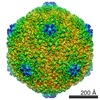 1810MC 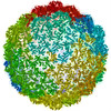 3izgC M: map data used to model this data C: citing same article ( |
|---|---|
| Similar structure data |
-Links
 PDBj
PDBj- Assembly
| Deposited unit | 
|
|---|---|
| 1 | x 60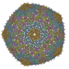
|
| 2 |
|
| 3 | x 5
|
| 4 | x 6
|
| 5 | 
|
| Symmetry | Point symmetry: (Schoenflies symbol: I (icosahedral)) |
-Components
| #1: Protein | Mass: 36589.625 Da / Num. of mol.: 7 / Source method: isolated from a natural source / Source: (natural) ENTEROBACTERIA PHAGE T7 (virus) / References: UniProt: P19726 |
|---|
-Experimental details
-Experiment
| Experiment | Method: ELECTRON MICROSCOPY |
|---|---|
| EM experiment | Aggregation state: PARTICLE / 3D reconstruction method: single particle reconstruction |
- Sample preparation
| Component | Name: PHAGE T7 EMPTY HEAD / Type: VIRUS |
|---|---|
| Buffer solution | Name: 50 MM TRIS-HCL, PH 7.8, 10 MM MGCL2, 0.1 M NACL / pH: 7.8 / Details: 50 MM TRIS-HCL, PH 7.8, 10 MM MGCL2, 0.1 M NACL |
| Specimen | Embedding applied: NO / Shadowing applied: NO / Staining applied: NO / Vitrification applied: YES |
| Specimen support | Details: HOLEY CARBON |
| Vitrification | Cryogen name: ETHANE / Details: VITRIFICATION 1 -- CRYOGEN- ETHANE, |
- Electron microscopy imaging
| Experimental equipment |  Model: Tecnai F20 / Image courtesy: FEI Company |
|---|---|
| Microscopy | Model: FEI TECNAI F20 |
| Electron gun | Electron source:  FIELD EMISSION GUN / Accelerating voltage: 200 kV / Illumination mode: FLOOD BEAM FIELD EMISSION GUN / Accelerating voltage: 200 kV / Illumination mode: FLOOD BEAM |
| Electron lens | Mode: BRIGHT FIELD / Nominal magnification: 50000 X |
| Image recording | Film or detector model: KODAK SO-163 FILM |
- Processing
| EM software | Name: Xmipp / Category: 3D reconstruction | ||||||||||||
|---|---|---|---|---|---|---|---|---|---|---|---|---|---|
| Symmetry | Point symmetry: I (icosahedral) | ||||||||||||
| 3D reconstruction | Resolution: 10.8 Å / Num. of particles: 5100 / Actual pixel size: 1.4 Å Details: SUBMISSION BASED ON EXPERIMENTAL DATA FROM EMDB EMD-1810. (DEPOSITION ID: 7638). Symmetry type: POINT | ||||||||||||
| Atomic model building | Space: REAL | ||||||||||||
| Atomic model building | PDB-ID: 1OHG Accession code: 1OHG / Source name: PDB / Type: experimental model | ||||||||||||
| Refinement | Highest resolution: 10.8 Å | ||||||||||||
| Refinement step | Cycle: LAST / Highest resolution: 10.8 Å
|