 Movie
Movie Controller

[English] 日本語
 Yorodumi
- PDB-3jb5: Capsid Structure of the Propionibacterium acnes Bacteriophage ATC... -
+ Open data

- Basic information
| Entry | Database: PDB / ID: 3jb5 | ||||||
|---|---|---|---|---|---|---|---|
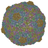
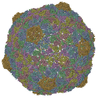
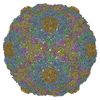
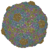
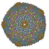
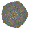
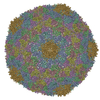
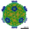
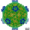
| Title | Capsid Structure of the Propionibacterium acnes Bacteriophage ATCC_Clear | ||||||
 Components Components | major capsid protein | ||||||
 Keywords Keywords | VIRUS / acne / bacteriophage / HK97-like | ||||||
| Function / homology | Gp6 Function and homology information Function and homology information | ||||||
| Biological species |  Propionibacterium phage PA6 (virus) Propionibacterium phage PA6 (virus) | ||||||
| Method | ELECTRON MICROSCOPY / single particle reconstruction / cryo EM / Resolution: 3.7 Å | ||||||
 Authors Authors | Chiou, J. / Zhang, X. / Marinelli, L.J. / Modlin, R.L. / Zhou, Z.H. | ||||||
 Citation Citation | Journal: To be Published Title: Capsid Structure of the Propionibacterium acnes Bacteriophage ATCC_Clear Authors: Chiou, J. / Zhang, X. / Marinelli, L.J. / Modlin, R.L. / Zhou, Z.H. | ||||||
| History |
|
- Structure visualization
| Movie |
Movie viewer |
|---|---|
| Structure viewer | Molecule: MolmilJmol/JSmol |
- Downloads & links
-Download
| PDBx/mmCIF format | 3jb5.cif.gz | 342.1 KB | Display | PDBx/mmCIF format |
|---|---|---|---|---|
| PDB format | pdb3jb5.ent.gz | 287.6 KB | Display | PDB format |
| PDBx/mmJSON format | 3jb5.json.gz | Tree view | PDBx/mmJSON format | |
| Others |  Other downloads Other downloads |
-Validation report
| Arichive directory | https://data.pdbj.org/pub/pdb/validation_reports/jb/3jb5ftp://data.pdbj.org/pub/pdb/validation_reports/jb/3jb5 | HTTPS FTP |
|---|
-Related structure data
| Related structure data | 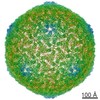 6398MC M: map data used to model this data C: citing same article ( |
|---|---|
| Similar structure data |
-Links
 PDBj
PDBj- Assembly
| Deposited unit | 
|
|---|---|
| 1 | x 60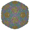
|
| 2 |
|
| 3 | x 5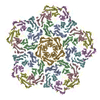
|
| 4 | x 6
|
| 5 | 
|
| Symmetry | Point symmetry: (Schoenflies symbol: I (icosahedral)) |
-Components
| #1: Protein | Mass: 32765.082 Da / Num. of mol.: 7 / Source method: isolated from a natural source / Source: (natural) Propionibacterium phage PA6 (virus) / References: UniProt: A4K473 |
|---|
-Experimental details
-Experiment
| Experiment | Method: ELECTRON MICROSCOPY |
|---|---|
| EM experiment | Aggregation state: PARTICLE / 3D reconstruction method: single particle reconstruction |
- Sample preparation
| Component | Name: Propionibacterium acnes bacteriophage ATCC_Clear / Type: VIRUS |
|---|---|
| Molecular weight | Value: 33 MDa / Experimental value: YES |
| Details of virus | Empty: NO / Enveloped: NO / Host category: BACTERIA(EUBACTERIA) / Isolate: STRAIN / Type: VIRION |
| Natural host | Organism: Propionibacterium acnes / Strain: 6919 |
| Specimen | Embedding applied: NO / Shadowing applied: NO / Staining applied: NO / Vitrification applied: YES |
| Specimen support | Details: Purified sample was applied to a Quantifoil grid. |
| Vitrification | Instrument: FEI VITROBOT MARK IV / Cryogen name: ETHANE / Humidity: 100 % Details: Blot for 15 seconds before plunging into liquid ethane (FEI VITROBOT MARK IV). Method: Blot for 15 seconds before plunging |
- Electron microscopy imaging
| Experimental equipment |  Model: Titan Krios / Image courtesy: FEI Company |
|---|---|
| Microscopy | Model: FEI TITAN KRIOS / Date: May 16, 2014 |
| Electron gun | Electron source:  FIELD EMISSION GUN / Accelerating voltage: 300 kV / Illumination mode: SPOT SCAN FIELD EMISSION GUN / Accelerating voltage: 300 kV / Illumination mode: SPOT SCAN |
| Electron lens | Mode: BRIGHT FIELD / Nominal magnification: 38462 X / Calibrated magnification: 38462 X / Nominal defocus max: 2270 nm / Nominal defocus min: 800 nm |
| Specimen holder | Specimen holder model: FEI TITAN KRIOS AUTOGRID HOLDER |
| Image recording | Electron dose: 25 e/Å2 / Film or detector model: GATAN K2 (4k x 4k) |
| Image scans | Num. digital images: 3168 |
| Radiation | Protocol: SINGLE WAVELENGTH / Monochromatic (M) / Laue (L): M / Scattering type: x-ray |
| Radiation wavelength | Relative weight: 1 |
- Processing
| EM software | Name: FREALIGN / Category: 3D reconstruction | ||||||||||||
|---|---|---|---|---|---|---|---|---|---|---|---|---|---|
| Symmetry | Point symmetry: I (icosahedral) | ||||||||||||
| 3D reconstruction | Resolution: 3.7 Å / Resolution method: FSC 0.143 CUT-OFF / Num. of particles: 27504 / Nominal pixel size: 1.3 Å / Actual pixel size: 1.3 Å Details: (Single particle details: The particles were selected using an in-house selection procedure.) (Single particle--Applied symmetry: I) Symmetry type: POINT | ||||||||||||
| Refinement step | Cycle: LAST
|
