 Movie
Movie Controller


+ Open data

- Basic information
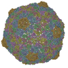
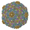
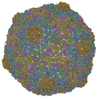
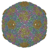
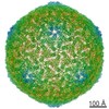
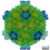
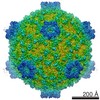
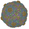
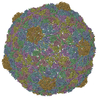
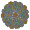
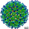
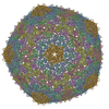
| Entry | Database: PDB / ID: 2fte | ||||||
|---|---|---|---|---|---|---|---|
| Title | Bacteriophage HK97 Expansion Intermediate IV | ||||||
 Components Components | major capsid protein | ||||||
 Keywords Keywords | VIRUS/VIRAL PROTEIN / Bacteriophage / HK97 / capsid protein / expansion intermediate / VIRUS-VIRAL PROTEIN COMPLEX | ||||||
| Function / homology | : / Phage capsid / Phage capsid family / viral procapsid maturation / T=7 icosahedral viral capsid / viral capsid / identical protein binding / Major capsid protein Function and homology information Function and homology information | ||||||
| Biological species |  Enterobacteria phage HK97 (virus) Enterobacteria phage HK97 (virus) | ||||||
| Method | ELECTRON MICROSCOPY / single particle reconstruction / cryo EM / Resolution: 12 Å | ||||||
 Authors Authors | Gan, L. / Speir, J.A. / Conway, J.F. / Lander, G. / Cheng, N. / Firek, B.A. / Hendrix, R.W. / Duda, R.L. / Liljas, L. / Johnson, J.E. | ||||||
 Citation Citation | Journal: Structure / Year: 2006 Title: Capsid conformational sampling in HK97 maturation visualized by X-ray crystallography and cryo-EM. Authors: Lu Gan / Jeffrey A Speir / James F Conway / Gabriel Lander / Naiqian Cheng / Brian A Firek / Roger W Hendrix / Robert L Duda / Lars Liljas / John E Johnson /  Abstract: Maturation of the bacteriophage HK97 capsid from a precursor (Prohead II) to the mature state (Head II) involves a 60 A radial expansion. The mature particle is formed by 420 copies of the major ...Maturation of the bacteriophage HK97 capsid from a precursor (Prohead II) to the mature state (Head II) involves a 60 A radial expansion. The mature particle is formed by 420 copies of the major capsid protein organized on a T = 7 laevo lattice with each subunit covalently crosslinked to two neighbors. Well-characterized pH 4 expansion intermediates make HK97 valuable for investigating quaternary structural dynamics. Here, we use X-ray crystallography and cryo-EM to demonstrate that in the final transition in maturation (requiring neutral pH), pentons in Expansion Intermediate IV (EI-IV) reversibly sample 14 A translations and 6 degrees rotations relative to a fixed hexon lattice. The limit of this trajectory corresponds to the Head II conformation that is secured at this extent only by the formation of the final class of covalent crosslinks. Mutants that cannot crosslink or EI-IV particles that have been rendered incapable of forming the final crosslink remain in the EI-IV state. #1: Journal: Mol Cell / Year: 2004 Title: Control of crosslinking by quaternary structure changes during bacteriophage HK97 maturation. Authors: Lu Gan / James F Conway / Brian A Firek / Naiqian Cheng / Roger W Hendrix / Alasdair C Steven / John E Johnson / Robert L Duda / Abstract: Radical structural changes drive the maturation of the capsid of HK97, a lambda-like, dsDNA bacteriophage of Escherichia coli. These include expansion from approximately 560 to approximately 660 A in ...Radical structural changes drive the maturation of the capsid of HK97, a lambda-like, dsDNA bacteriophage of Escherichia coli. These include expansion from approximately 560 to approximately 660 A in diameter, metamorphosis from a round to an angular shape, and formation of covalent crosslinks between adjacent capsomers. Analogous transformations also occur in unrelated viruses and protein complexes. We find that expansion and crosslinking happen concurrently during maturation at low pH. Expansion causes residues on three different subunits to move up to 35 A to form 420 active sites that each catalyze the formation of a lysine-asparagine crosslink between adjacent subunits, making crosslink formation an indirect reporter of structural change. Intermediate crosslinking patterns support a previously proposed model of expansion, while hydrophobic properties aid in distinguishing discrete intermediates. A structure derived from cryo-EM images reveals the free intermediate conformation of penton arms, supporting our model for coordinated movement of hexons and pentons on the capsid lattice. | ||||||
| History |
|
- Structure visualization
| Movie |
Movie viewer |
|---|---|
| Structure viewer | Molecule: MolmilJmol/JSmol |
- Downloads & links
-Download
| PDBx/mmCIF format | 2fte.cif.gz | 258.2 KB | Display | PDBx/mmCIF format |
|---|---|---|---|---|
| PDB format | pdb2fte.ent.gz | 166.2 KB | Display | PDB format |
| PDBx/mmJSON format | 2fte.json.gz | Tree view | PDBx/mmJSON format | |
| Others |  Other downloads Other downloads |
-Validation report
| Arichive directory | https://data.pdbj.org/pub/pdb/validation_reports/ft/2fteftp://data.pdbj.org/pub/pdb/validation_reports/ft/2fte | HTTPS FTP |
|---|
-Related structure data


-Links
 PDBj
PDBj

- Assembly
| Deposited unit | 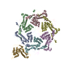
|
|---|---|
| 1 | x 60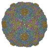
|
| 2 |
|
| 3 | x 5
|
| 4 | x 6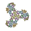
|
| 5 | 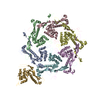
|
| Symmetry | Point symmetry: (Hermann–Mauguin notation: 532 / Schoenflies symbol: I (icosahedral)) |
-Components
| #1: Protein | Mass: 30804.607 Da / Num. of mol.: 7 Source method: isolated from a genetically manipulated source Source: (gene. exp.) Enterobacteria phage HK97 (virus) / Genus: Lambda-like viruses / Gene: 5 / Plasmid: pT7-Hd2.9 / Species (production host): Escherichia coli / Production host:  Has protein modification | N | |
|---|
-Experimental details
-Experiment
| Experiment | Method: ELECTRON MICROSCOPY |
|---|---|
| EM experiment | Aggregation state: PARTICLE / 3D reconstruction method: single particle reconstruction |
- Sample preparation
| Component |
| |||||||||||||||
|---|---|---|---|---|---|---|---|---|---|---|---|---|---|---|---|---|
| Details of virus | Host category: BACTERIA(EUBACTERIA) / Isolate: SPECIES / Type: VIRUS-LIKE PARTICLE | |||||||||||||||
| Natural host | Organism: Escherichia coli | |||||||||||||||
| Buffer solution | Name: 50mM Citric Acid pH 4.0, 200mM KCl / pH: 4 / Details: 50mM Citric Acid pH 4.0, 200mM KCl | |||||||||||||||
| Specimen | Embedding applied: NO / Shadowing applied: NO / Staining applied: NO / Vitrification applied: YES |
- Electron microscopy imaging
| Microscopy | Model: FEI/PHILIPS CM200FEG |
|---|---|
| Electron gun | Electron source:  FIELD EMISSION GUN / Accelerating voltage: 120 kV / Illumination mode: FLOOD BEAM FIELD EMISSION GUN / Accelerating voltage: 120 kV / Illumination mode: FLOOD BEAM |
| Electron lens | Mode: BRIGHT FIELD / Nominal magnification: 50000 X |
| Image recording | Electron dose: 15 e/Å2 |
- Processing
| EM software |
| ||||||||||||
|---|---|---|---|---|---|---|---|---|---|---|---|---|---|
| Symmetry | Point symmetry: I (icosahedral) | ||||||||||||
| 3D reconstruction | Resolution: 12 Å / Nominal pixel size: 1.4 Å Details: THE ENTIRE STRUCTURE WAS MODELED AS ALA AND GLY. THE MISSING ATOMS ARE SUPPRESSED DUE TO TOO MANY TO LIST. RESIDUES 159 to 172 IN CHAIN G WERE DELETED DUE TO DISORDER. Symmetry type: POINT | ||||||||||||
| Atomic model building | B value: 374 / Protocol: RIGID BODY FIT / Space: RECIPROCAL Target criteria: vector reciprocal space target and manual correction with O Details: METHOD--Rigid body and manual fitting REFINEMENT PROTOCOL--rigid body, magnification optimized using van der Waals constraints and real-space correlation coefficient | ||||||||||||
| Atomic model building | PDB-ID: 2FSY Accession code: 2FSY / Source name: PDB / Type: experimental model | ||||||||||||
| Refinement step | Cycle: LAST
|
