 Movie
Movie Controller
 Structure viewers
 About EMN search


-Search query
-Search result
Showing 1 - 50 of 111 items for (author: casper & g)

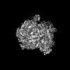
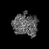
EMDB-55036: 
Human TRPM4 ion channel in MSP2N2 lipid nanodisc in a calcium-bound state
Method: single particle / : Pugh CF, Feilen LP, Zivkovic D, Praestegaard KF, Sideris C, Borthwick NJ, de Lichtenberg C, Bolla JR, Autzen AAA, Autzen HE

PDB-9smk:
Human TRPM4 ion channel in MSP2N2 lipid nanodisc in a calcium-bound state
Method: single particle / : Pugh CF, Feilen LP, Zivkovic D, Praestegaard KF, Sideris C, Borthwick NJ, de Lichtenberg C, Bolla JR, Autzen AAA, Autzen HE

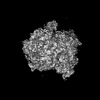
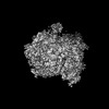
EMDB-52611:
Human TRPM4 ion channel in MAASTY copolymer lipid nanodisc in a calcium-bound state
Method: single particle / : Pugh CF, Feilen LP, Zivkovic D, Praestegaard KF, Sideris C, Borthwick NJ, De Lichtenberg C, Bolla JR, Autzen AAA, Autzen HE

PDB-9i3r:
Human TRPM4 ion channel in MAASTY copolymer lipid nanodisc in a calcium-bound state
Method: single particle / : Pugh CF, Feilen LP, Zivkovic D, Praestegaard KF, Sideris C, Borthwick NJ, De Lichtenberg C, Bolla JR, Autzen AAA, Autzen HE

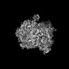
EMDB-53511:
SpCas9 with computationally designed SpCas9_b10 binder
Method: single particle / : Pacesa M, Nickel L, Correia BE
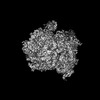
EMDB-53510:
SpCas9 with computationally designed SpCas9_b3 binder
Method: single particle / : Pacesa M, Nickel L, Correia BE
EMDB-50675:
Escherichia coli 70S ribosome in situ structure
Method: subtomogram averaging / : Watson H, Berger C, Grange M
EMDB-50676:
E. coli 70S ribosome in situ structure for xenon damage layer determination: 5 - 10 nm
Method: subtomogram averaging / : Watson H, Berger C, Grange M
EMDB-50678:
E. coli 70S ribosome in situ structure for xenon damage layer determination: 10 - 15 nm
Method: subtomogram averaging / : Watson H, Berger C, Grange M
EMDB-50679:
E. coli 70S ribosome in situ structure for xenon damage layer determination: 15 - 20 nm
Method: subtomogram averaging / : Watson H, Berger C, Grange M

EMDB-50680:
E. coli 70S ribosome in situ structure for xenon damage layer determination: 20 - 25 nm
Method: subtomogram averaging / : Watson H, Berger C, Grange M

EMDB-50681:
E. coli 70S ribosome in situ structure for xenon damage layer determination: 25 - 30 nm
Method: subtomogram averaging / : Watson H, Berger C, Grange M
EMDB-50682:
E. coli 70S ribosome in situ structure for xenon damage layer determination: 30 - 35 nm
Method: subtomogram averaging / : Watson H, Berger C, Grange M
EMDB-50683:
E. coli 70S ribosome in situ structure for xenon damage layer determination: 35 - 40 nm
Method: subtomogram averaging / : Watson H, Berger C, Grange M
EMDB-50684:
E. coli 70S ribosome in situ structure for xenon damage layer determination: 40 - 45 nm
Method: subtomogram averaging / : Watson H, Berger C, Grange M
EMDB-50685:
E. coli 70S ribosome in situ structure for xenon damage layer determination: 45 - 50 nm
Method: subtomogram averaging / : Watson H, Berger C, Grange M
EMDB-50686:
E. coli 70S ribosome in situ structure for xenon damage layer determination: 50 - 55 nm
Method: subtomogram averaging / : Watson H, Berger C, Grange M

EMDB-50687:
E. coli 70S ribosome in situ structure for xenon damage layer determination: 55 - 60 nm
Method: subtomogram averaging / : Watson H, Berger C, Grange M
EMDB-50688:
E. coli 70S ribosome in situ structure for xenon damage layer determination: >10nm matched control for 5 - 10 nm
Method: subtomogram averaging / : Watson H, Berger C, Grange M

EMDB-50689:
E. coli 70S ribosome in situ structure for xenon damage layer determination: >15 nm matched control for 10 - 15 nm
Method: subtomogram averaging / : Watson H, Berger C, Grange M

EMDB-50690:
E. coli 70S ribosome in situ structure for xenon damage layer determination: >20 nm matched control for 15 - 20 nm
Method: subtomogram averaging / : Watson H, Berger C, Grange M

EMDB-50691:
E. coli 70S ribosome in situ structure for xenon damage layer determination: >25 nm matched control for 20 - 25 nm
Method: subtomogram averaging / : Watson H, Berger C, Grange M

EMDB-50692:
E. coli 70S ribosome in situ structure for xenon damage layer determination: >30 nm matched control for 25 - 30 nm
Method: subtomogram averaging / : Watson H, Berger C, Grange M

EMDB-50693:
E. coli 70S ribosome in situ structure for xenon damage layer determination: >35 nm matched control for 30 - 35 nm
Method: subtomogram averaging / : Watson H, Berger C, Grange M

EMDB-50694:
E. coli 70S ribosome in situ structure for xenon damage layer determination: >40 nm matched control for 35 - 40 nm
Method: subtomogram averaging / : Watson H, Berger C, Grange M

EMDB-50695:
E. coli 70S ribosome in situ structure for xenon damage layer determination: >45 nm matched control for 40 - 45 nm
Method: subtomogram averaging / : Watson H, Berger C, Grange M

EMDB-50696:
E. coli 70S ribosome in situ structure for xenon damage layer determination: >50 nm matched control for 45 - 50 nm
Method: subtomogram averaging / : Watson H, Berger C, Grange M

EMDB-50697:
E. coli 70S ribosome in situ structure for xenon damage layer determination: >55 nm matched control for 50 - 55 nm
Method: subtomogram averaging / : Watson H, Berger C, Grange M

EMDB-50698:
E. coli 70S ribosome in situ structure for xenon damage layer determination: >60 nm matched control for 55 - 60 nm
Method: subtomogram averaging / : Watson H, Berger C, Grange M

EMDB-50699:
E. coli 70S ribosome in situ structure for lamellae backside damage analysis: 0 - 1 micron from lamellae backside
Method: subtomogram averaging / : Watson H, Berger C, Grange M

EMDB-50700:
E. coli 70S ribosome in situ structure for lamellae backside damage analysis: 1 - 2 microns from lamellae backside
Method: subtomogram averaging / : Watson H, Berger C, Grange M

EMDB-50701:
E. coli 70S ribosome in situ structure for lamellae backside damage analysis: 2 - 3 microns from lamellae backside
Method: subtomogram averaging / : Watson H, Berger C, Grange M

EMDB-50702:
E. coli 70S ribosome in situ structure for lamellae backside damage analysis: 3 - 4 microns from lamellae backside
Method: subtomogram averaging / : Watson H, Berger C, Grange M

EMDB-50703:
E. coli 70S ribosome in situ structure for lamellae backside damage analysis: 4 - 5 microns from lamellae backside
Method: subtomogram averaging / : Watson H, Berger C, Grange M

EMDB-50704:
E. coli 70S ribosome in situ structure for xenon damage layer determination: 10,000 particles (no depth constraint)
Method: subtomogram averaging / : Watson H, Berger C, Grange M

EMDB-50705:
E. coli 70S ribosome in situ structure for xenon damage layer determination: 10,000 particles (>45 nm)
Method: subtomogram averaging / : Watson H, Berger C, Grange M

EMDB-50706:
E. coli 70S ribosome in situ structure for xenon damage layer determination: 10,000 particles (<=45 nm)
Method: subtomogram averaging / : Watson H, Berger C, Grange M

EMDB-52177:
E. coli 70S ribosome in situ structure for xenon damage layer determination: 10,000 particles (no depth constraint)
Method: subtomogram averaging / : Watson H, Berger C, Grange M

EMDB-52178:
E. coli 70S ribosome in situ structure for xenon damage layer determination: 10,000 particles (<30 nm)
Method: subtomogram averaging / : Watson H, Berger C, Grange M

EMDB-52179:
E. coli 70S ribosome in situ structure for xenon damage layer determination: 10,000 particles (>30 nm)
Method: subtomogram averaging / : Watson H, Berger C, Grange M

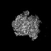
EMDB-51500:
Partial (48mer) encapsulin shell assembly from Mycobacterium tuberculosis
Method: single particle / : Lewis CJ, Berger C, Ravelli RBG

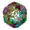
EMDB-52340:
Partial (52mer) encapsulin shell assembly from Mycobacterium tuberculosis
Method: single particle / : Lewis CJ, Berger C, Ravelli RBG

EMDB-52341:
Partial (54mer) encapsulin shell assembly from Mycobacterium tuberculosis
Method: single particle / : Lewis CJ, Berger C, Ravelli RBG

PDB-9got:
Partial (48mer) encapsulin shell assembly from Mycobacterium tuberculosis
Method: single particle / : Lewis CJ, Berger C, Ravelli RBG

PDB-9hq7:
Partial (52mer) encapsulin shell assembly from Mycobacterium tuberculosis
Method: single particle / : Lewis CJ, Berger C, Ravelli RBG
PDB-9hqc:
Partial (54mer) encapsulin shell assembly from Mycobacterium tuberculosis
Method: single particle / : Lewis CJ, Berger C, Ravelli RBG

EMDB-50529:
Cryo-EM map of a Gorilla Foamy Virus fusion glycoprotein in the postfusion conformation (C1 symmetry)
Method: single particle / : Fernandez I, Bontems F, Backovic M
EMDB-50530:
Cryo-EM map of a Gorilla Foamy Virus fusion glycoprotein stabilized in the prefusion conformation (C1 symmetry)
Method: single particle / : Fernandez I, Backovic M

EMDB-19347:
Cryo-EM structure of a Foamy Virus fusion glycoprotein stabilized in the prefusion conformation
Method: single particle / : Fernandez I, Backovic M

EMDB-19348:
Cryo-EM structure of a Foamy Virus fusion glycoprotein in the postfusion conformation
Method: single particle / : Fernandez I, Backovic M
Pages:
 wwPDB to switch to version 3 of the EMDB data model
