Movie
Movie Controller

[English] 日本語
 Yorodumi
- EMDB-19348: Cryo-EM structure of a Foamy Virus fusion glycoprotein in the pos... -
+ Open data

- Basic information
| Entry |  | |||||||||
|---|---|---|---|---|---|---|---|---|---|---|
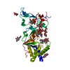
| Title | Cryo-EM structure of a Foamy Virus fusion glycoprotein in the postfusion conformation | |||||||||
 Map data Map data | ||||||||||
 Sample Sample |
| |||||||||
 Keywords Keywords | Envelope / Fusion / Spumavirus / VIRUS | |||||||||
| Function / homology | Foamy virus envelope protein / Foamy virus envelope protein / host cell endoplasmic reticulum membrane / viral envelope / virion membrane / Envelope glycoprotein gp130 Function and homology information Function and homology information | |||||||||
| Biological species |  Spumavirus / Simian foamy virus Spumavirus / Simian foamy virus | |||||||||
| Method | single particle reconstruction / cryo EM / Resolution: 3.1 Å | |||||||||
 Authors Authors | Fernandez I / Backovic M | |||||||||
| Funding support |  France, 1 items France, 1 items
| |||||||||
 Citation Citation | Journal: Sci Adv / Year: 2024 Title: Structures of the Foamy virus fusion protein reveal an unexpected link with the F protein of paramyxo- and pneumoviruses. Authors: Ignacio Fernández / François Bontems / Delphine Brun / Youna Coquin / Casper A Goverde / Bruno E Correia / Antoine Gessain / Florence Buseyne / Felix A Rey / Marija Backovic /  Abstract: Foamy viruses (FVs) constitute a subfamily of retroviruses. Their envelope (Env) glycoprotein drives the merger of viral and cellular membranes during entry into cells. The only available structures ...Foamy viruses (FVs) constitute a subfamily of retroviruses. Their envelope (Env) glycoprotein drives the merger of viral and cellular membranes during entry into cells. The only available structures of retroviral Envs are those from human and simian immunodeficiency viruses from the subfamily of orthoretroviruses, which are only distantly related to the FVs. We report the cryo-electron microscopy structures of the FV Env ectodomain in the pre- and post-fusion states, which unexpectedly demonstrate structural similarity with the fusion protein (F) of paramyxo- and pneumoviruses, implying an evolutionary link between the viral fusogens. We describe the structural features that are unique to the FV Env and propose a mechanistic model for its conformational change, highlighting how the interplay of its structural elements could drive membrane fusion and viral entry. The structural knowledge on the FV Env now provides a framework for functional investigations, which can benefit the design of FV Env variants with improved features for use as gene therapy vectors. | |||||||||
| History |
|
- Structure visualization
| Supplemental images |
|---|

- Downloads & links
-EMDB archive
| Map data | emd_19348.map.gz | 155.9 MB | EMDB map data format | |
|---|---|---|---|---|
| Header (meta data) | emd-19348-v30.xmlemd-19348.xml | 28.3 KB 28.3 KB | Display Display | EMDB header |
| Images |  emd_19348.png emd_19348.png | 35.4 KB | ||
| Filedesc metadata | emd-19348.cif.gz | 8.1 KB | ||
| Others | emd_19348_half_map_1.map.gzemd_19348_half_map_2.map.gz | 131.6 MB 131.7 MB | ||
| Archive directory |  http://ftp.pdbj.org/pub/emdb/structures/EMD-19348ftp://ftp.pdbj.org/pub/emdb/structures/EMD-19348 http://ftp.pdbj.org/pub/emdb/structures/EMD-19348ftp://ftp.pdbj.org/pub/emdb/structures/EMD-19348 | HTTPS FTP |
-Related structure data
| Related structure data |  8rm1MC  8rm0C M: atomic model generated by this map C: citing same article ( |
|---|---|
| Similar structure data |


-Links
| EMDB pages | EMDB (EBI/PDBe) / EMDataResource |
|---|
-Map
| File | Download / File: emd_19348.map.gz / Format: CCP4 / Size: 166.4 MB / Type: IMAGE STORED AS FLOATING POINT NUMBER (4 BYTES) | ||||||||||||||||||||||||||||||||||||
|---|---|---|---|---|---|---|---|---|---|---|---|---|---|---|---|---|---|---|---|---|---|---|---|---|---|---|---|---|---|---|---|---|---|---|---|---|---|
| Projections & slices | Image control
Images are generated by Spider. | ||||||||||||||||||||||||||||||||||||
| Voxel size | X=Y=Z: 0.86 Å | ||||||||||||||||||||||||||||||||||||
| Density |
| ||||||||||||||||||||||||||||||||||||
| Symmetry | Space group: 1 | ||||||||||||||||||||||||||||||||||||
| Details | EMDB XML:
|
 Z (Sec.)
 Y (Row.)
 X (Col.)


-Supplemental data
-Half map: #2
| File | emd_19348_half_map_1.map | ||||||||||||
|---|---|---|---|---|---|---|---|---|---|---|---|---|---|
| Projections & Slices |
| ||||||||||||
| Density Histograms |


-Half map: #1
| File | emd_19348_half_map_2.map | ||||||||||||
|---|---|---|---|---|---|---|---|---|---|---|---|---|---|
| Projections & Slices |
| ||||||||||||
| Density Histograms |


- Sample components
-Entire : Gorilla Foamy Virus Envelope glycoprotein
| Entire | Name: Gorilla Foamy Virus Envelope glycoprotein |
|---|---|
| Components |
|
-Supramolecule #1: Gorilla Foamy Virus Envelope glycoprotein
| Supramolecule | Name: Gorilla Foamy Virus Envelope glycoprotein / type: complex / ID: 1 / Parent: 0 / Macromolecule list: #1 |
|---|---|
| Source (natural) | Organism: Spumavirus |
-Macromolecule #1: Envelope glycoprotein gp130
| Macromolecule | Name: Envelope glycoprotein gp130 / type: protein_or_peptide / ID: 1 / Number of copies: 3 / Enantiomer: LEVO |
|---|---|
| Source (natural) | Organism: Simian foamy virus |
| Molecular weight | Theoretical: 52.050996 KDa |
| Recombinant expression | Organism:  |
| Sequence | String: ALRAQHPVPK YVEVNMTSIP QGVFYQPHPE PIIHTERVLG LSQVLMINSE NVANSANLSQ ETKALLTEMV NEEMQGLSDV MIDFEIPLG DPRDQEQYIH RKCYQEFAHC YLVKYKTPQP WPNEGLIADQ CPLPGLADVS FYPYQAIWDY YAKIENIRPA N WTSSKLYG ...String: ALRAQHPVPK YVEVNMTSIP QGVFYQPHPE PIIHTERVLG LSQVLMINSE NVANSANLSQ ETKALLTEMV NEEMQGLSDV MIDFEIPLG DPRDQEQYIH RKCYQEFAHC YLVKYKTPQP WPNEGLIADQ CPLPGLADVS FYPYQAIWDY YAKIENIRPA N WTSSKLYG KARMGSYYIP KRLRNINNTH ILFCSDVLYS KWYNLQNSIL QNENELTKRL SNLTIGNKLK NRALPYEWAK GG LNRLFRN ISVLDVCSRP EMVLLLNKTY YTFSLWEGDC NITRYNVNET VPECKDFPHR RFNDHPYSCR LWRYREGKEE VKC LTSDHT RCLYYPEYSN PEALFDFGFL SYMRNFPGPQ CIESTSIRQQ DYEVYSIYQE CKLASKTYGI DSVLFSLKNF LNYT GKPVN EMPNARAFVG LIDPKFPPTY PNITRDQYQG CNINQRRKR UniProtKB: Envelope glycoprotein gp130 |
-Macromolecule #2: Envelope glycoprotein gp130
| Macromolecule | Name: Envelope glycoprotein gp130 / type: protein_or_peptide / ID: 2 / Number of copies: 3 / Enantiomer: LEVO |
|---|---|
| Source (natural) | Organism: Simian foamy virus |
| Molecular weight | Theoretical: 42.067801 KDa |
| Recombinant expression | Organism: |
| Sequence | String: EVNNNYSKLR SMGYALTGAV QTLAQISDIN DQNLQQGIYL LRDHIVTLME ATLHDISIME GMFAVQHVHT HLNHLRTMLM ERRIDWTYM SSSWLQTQLQ KSDDEMKVIK RTARSLVYYV KQTYNSLTAT AWEIGLYYEL IIPRHIYLNN WQIVNIGHLI K SAGQLTHV ...String: EVNNNYSKLR SMGYALTGAV QTLAQISDIN DQNLQQGIYL LRDHIVTLME ATLHDISIME GMFAVQHVHT HLNHLRTMLM ERRIDWTYM SSSWLQTQLQ KSDDEMKVIK RTARSLVYYV KQTYNSLTAT AWEIGLYYEL IIPRHIYLNN WQIVNIGHLI K SAGQLTHV TLSHPYEIIN RECSNTLYLH LEECRRLDYV ICDVVKIVQP CGNSSDSSDC PVWAEPVKEP HVQISPLKNG SY LVLASST DCQIPPYVPS VVTVNETTQC FGVTFKKPLV AEEKTSLEPQ LPHLQLRLPH LVGIIAKIKG IKIEVTSSGE SIK DQLERA KAELLRLDIH EDDDDKAGWS HPQFEKGGGS GGGSGGGSWS HPQFEK UniProtKB: Envelope glycoprotein gp130 |
-Macromolecule #5: 2-acetamido-2-deoxy-beta-D-glucopyranose
| Macromolecule | Name: 2-acetamido-2-deoxy-beta-D-glucopyranose / type: ligand / ID: 5 / Number of copies: 3 / Formula: NAG |
|---|---|
| Molecular weight | Theoretical: 221.208 Da |
| Chemical component information |  ChemComp-NAG: |
-Experimental details
-Structure determination
| Method | cryo EM |
|---|---|
 Processing Processing | single particle reconstruction |
| Aggregation state | particle |
-Sample preparation
| Buffer | pH: 8 |
|---|---|
| Grid | Model: Quantifoil R1.2/1.3 / Support film - Material: CARBON / Support film - topology: HOLEY |
| Vitrification | Cryogen name: ETHANE / Chamber humidity: 100 % / Chamber temperature: 281 K / Instrument: FEI VITROBOT MARK IV |
- Electron microscopy
| Microscope | FEI TITAN KRIOS |
|---|---|
| Image recording | Film or detector model: GATAN K3 BIOQUANTUM (6k x 4k) / Average electron dose: 50.0 e/Å2 |
| Electron beam | Acceleration voltage: 300 kV / Electron source:  FIELD EMISSION GUN FIELD EMISSION GUN |
| Electron optics | Illumination mode: FLOOD BEAM / Imaging mode: BRIGHT FIELD / Cs: 2.7 mm / Nominal defocus max: 3.0 µm / Nominal defocus min: 1.0 µm |
| Experimental equipment |  Model: Titan Krios / Image courtesy: FEI Company |