 Movie
Movie Controller

[English] 日本語
 Yorodumi
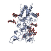
- PDB-8aez: X-ray structure of the deglycosylated receptor binding domain of ... -
+ Open data

- Basic information
| Entry | Database: PDB / ID: 8aez | ||||||
|---|---|---|---|---|---|---|---|
| Title | X-ray structure of the deglycosylated receptor binding domain of Env glycoprotein of Simian Foamy virus | ||||||
 Components Components | Envelope glycoprotein gp130 | ||||||
 Keywords Keywords | VIRAL PROTEIN / Receptor binding domain | ||||||
| Function / homology | Foamy virus envelope protein / Foamy virus envelope protein / host cell endoplasmic reticulum membrane / viral envelope / virion membrane / Envelope glycoprotein gp130 Function and homology information Function and homology information | ||||||
| Biological species |  Simian foamy virus Simian foamy virus | ||||||
| Method |  X-RAY DIFFRACTION / SYNCHROTRON / SAD / Resolution: 2.574 Å X-RAY DIFFRACTION / SYNCHROTRON / SAD / Resolution: 2.574 Å | ||||||
 Authors Authors | Backovic, M. | ||||||
| Funding support |  France, 1items France, 1items
| ||||||
 Citation Citation | Journal: Nat Commun / Year: 2023 Title: The crystal structure of a simian Foamy Virus receptor binding domain provides clues about entry into host cells. Authors: Fernandez, I. / Dynesen, L.T. / Coquin, Y. / Pederzoli, R. / Brun, D. / Haouz, A. / Gessain, A. / Rey, F.A. / Buseyne, F. / Backovic, M. | ||||||
| History |
|
- Structure visualization
| Structure viewer | Molecule: MolmilJmol/JSmol |
|---|
- Downloads & links
-Download
| PDBx/mmCIF format | 8aez.cif.gz | 162.3 KB | Display | PDBx/mmCIF format |
|---|---|---|---|---|
| PDB format | pdb8aez.ent.gz | 128.2 KB | Display | PDB format |
| PDBx/mmJSON format | 8aez.json.gz | Tree view | PDBx/mmJSON format | |
| Others |  Other downloads Other downloads |
-Validation report
| Arichive directory | https://data.pdbj.org/pub/pdb/validation_reports/ae/8aezftp://data.pdbj.org/pub/pdb/validation_reports/ae/8aez | HTTPS FTP |
|---|
-Related structure data
-Links
 PDBj
PDBj- Assembly
| Deposited unit | 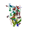
| ||||||||
|---|---|---|---|---|---|---|---|---|---|
| 1 |
| ||||||||
| Unit cell |
|
-Components
| #1: Protein | Mass: 40288.566 Da / Num. of mol.: 1 Source method: isolated from a genetically manipulated source Source: (gene. exp.) Simian foamy virus / Gene: env / Variant: BAK74 / Cell line (production host): 2 / Production host:  | ||||||
|---|---|---|---|---|---|---|---|
| #2: Polysaccharide | beta-D-mannopyranose-(1-4)-2-acetamido-2-deoxy-beta-D-glucopyranose-(1-4)-2-acetamido-2-deoxy-beta- ...beta-D-mannopyranose-(1-4)-2-acetamido-2-deoxy-beta-D-glucopyranose-(1-4)-2-acetamido-2-deoxy-beta-D-glucopyranose Source method: isolated from a genetically manipulated source | ||||||
| #3: Polysaccharide | alpha-D-mannopyranose-(1-2)-alpha-D-mannopyranose-(1-2)-alpha-D-mannopyranose-(1-3)-[alpha-D- ...alpha-D-mannopyranose-(1-2)-alpha-D-mannopyranose-(1-2)-alpha-D-mannopyranose-(1-3)-[alpha-D-mannopyranose-(1-3)-alpha-D-mannopyranose-(1-6)]beta-D-mannopyranose-(1-4)-2-acetamido-2-deoxy-beta-D-glucopyranose-(1-4)-2-acetamido-2-deoxy-beta-D-glucopyranose Source method: isolated from a genetically manipulated source | ||||||
| #4: Sugar | ChemComp-NAG /   Type: D-saccharide, beta linking / Mass: 221.208 Da / Num. of mol.: 5 / Source method: obtained synthetically / Formula: C8H15NO6 Type: D-saccharide, beta linking / Mass: 221.208 Da / Num. of mol.: 5 / Source method: obtained synthetically / Formula: C8H15NO6#5: Water | ChemComp-HOH / |  Mass: 18.015 Da / Num. of mol.: 31 / Source method: isolated from a natural source / Formula: H2O Mass: 18.015 Da / Num. of mol.: 31 / Source method: isolated from a natural source / Formula: H2OHas ligand of interest | N | Has protein modification | Y | |
-Experimental details
-Experiment
| Experiment | Method: X-RAY DIFFRACTION / Number of used crystals: 1 |
|---|
- Sample preparation
| Crystal | Density Matthews: 3.97 Å3/Da / Density % sol: 69.05 % |
|---|---|
| Crystal grow | Temperature: 295 K / Method: vapor diffusion, sitting drop / pH: 8.5 / Details: 0.1M Tris pH 8.5, 3.5M sodium formate |
-Data collection
| Diffraction | Mean temperature: 100 K / Serial crystal experiment: N |
|---|---|
| Diffraction source | Source: SYNCHROTRON / Site: SOLEIL / Beamline: PROXIMA 1 / Wavelength: 0.9786 Å |
| Detector | Type: DECTRIS EIGER X 16M / Detector: PIXEL / Date: Jun 19, 2020 |
| Radiation | Protocol: SINGLE WAVELENGTH / Monochromatic (M) / Laue (L): M / Scattering type: x-ray |
| Radiation wavelength | Wavelength: 0.9786 Å / Relative weight: 1 |
| Reflection | Resolution: 2.574→49.73 Å / Num. obs: 22363 / % possible obs: 99.91 % / Redundancy: 20.3 % / CC1/2: 0.986 / Rmerge(I) obs: 0.2051 / Rpim(I) all: 0.04701 / Net I/σ(I): 10.1 |
| Reflection shell | Resolution: 2.574→2.666 Å / Rmerge(I) obs: 0.9362 / Mean I/σ(I) obs: 1.85 / Num. unique obs: 2101 / CC1/2: 0.846 / Rpim(I) all: 0.2117 |
- Processing
| Software |
| ||||||||||||||||||||||||||||||||||||||||||||||||||||||||||||
|---|---|---|---|---|---|---|---|---|---|---|---|---|---|---|---|---|---|---|---|---|---|---|---|---|---|---|---|---|---|---|---|---|---|---|---|---|---|---|---|---|---|---|---|---|---|---|---|---|---|---|---|---|---|---|---|---|---|---|---|---|---|
| Refinement | Method to determine structure: SAD / Resolution: 2.574→49.73 Å / Cor.coef. Fo:Fc: 0.942 / Cor.coef. Fo:Fc free: 0.909 / SU R Cruickshank DPI: 0.289 / Cross valid method: THROUGHOUT / SU R Blow DPI: 0.285 / SU Rfree Blow DPI: 0.232 / SU Rfree Cruickshank DPI: 0.236
| ||||||||||||||||||||||||||||||||||||||||||||||||||||||||||||
| Displacement parameters | Biso mean: 75.14 Å2
| ||||||||||||||||||||||||||||||||||||||||||||||||||||||||||||
| Refine analyze | Luzzati coordinate error obs: 0.36 Å | ||||||||||||||||||||||||||||||||||||||||||||||||||||||||||||
| Refinement step | Cycle: LAST / Resolution: 2.574→49.73 Å
| ||||||||||||||||||||||||||||||||||||||||||||||||||||||||||||
| Refine LS restraints |
| ||||||||||||||||||||||||||||||||||||||||||||||||||||||||||||
| LS refinement shell | Resolution: 2.574→49.73 Å
| ||||||||||||||||||||||||||||||||||||||||||||||||||||||||||||
| Refinement TLS params. | Origin x: -22.1361 Å / Origin y: -36.3317 Å / Origin z: 18.3941 Å
| ||||||||||||||||||||||||||||||||||||||||||||||||||||||||||||
| Refinement TLS group | Selection details: { A|* } |
