 ムービー
ムービー コントローラー
 構造ビューア
 EMN検索について


-検索条件
-検索結果
検索 (著者・登録者: burg & m)の結果613件中、1から50件目までを表示しています

EMDBエントリ 画像なし
EMDB-43296: 
Constituent EM map: Focused refinement S100A1 of mouse RyR1 in complex with S100A1 (EGTA-only dataset)
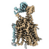
EMDB-50358:
In vitro-induced genome-releasing intermediate of Rhodobacter microvirus Ebor computed with C5 symmetry
EMDB-43712:
Human EBP complexed with compound 1
EMDB-43713:
Human EBP complexed with compound 3a
PDB-8w0r:
Human EBP complexed with compound 1
PDB-8w0s:
Human EBP complexed with compound 3a
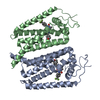
EMDB-17377:
Structure of human SIT1 (focussed map / refinement)

EMDB-17378:
Structure of human SIT1:ACE2 complex (open PD conformation)

EMDB-17379:
Structure of human SIT1:ACE2 complex (closed PD conformation)
EMDB-17380:
Structure of human SIT1 bound to L-pipecolate (focussed map / refinement)
EMDB-17381:
Structure of human SIT1:ACE2 complex (open PD conformation) bound to L-pipecolate
EMDB-17382:
Structure of human SIT1:ACE2 complex (closed PD conformation) bound to L-pipecolate

PDB-8p2w:
Structure of human SIT1 (focussed map / refinement)

PDB-8p2x:
Structure of human SIT1:ACE2 complex (open PD conformation)

PDB-8p2y:
Structure of human SIT1:ACE2 complex (closed PD conformation)

PDB-8p2z:
Structure of human SIT1 bound to L-pipecolate (focussed map / refinement)
PDB-8p30:
Structure of human SIT1:ACE2 complex (open PD conformation) bound to L-pipecolate

PDB-8p31:
Structure of human SIT1:ACE2 complex (closed PD conformation) bound to L-pipecolate
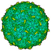
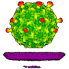
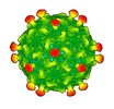
EMDB-50356:
Empty capsid of Rhodobacter microvirus Ebor computed with I4 symmetry
EMDB-50357:
Native capsid of Rhodobacter microvirus Ebor computed with I4 symmetry
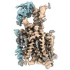
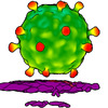
EMDB-50359:
Rhodobacter microvirus Ebor attached to B10 host cell reconstructed by single particle analysis with applied C5 symmetry
EMDB-50360:
Rhodobacter microvirus Ebor attached to the outer membrane vesicle
EMDB-50361:
Rhodobacter microvirus Ebor attached to the host cell reconstructed by subtomogram averaging

PDB-9ffg:
Empty capsid of Rhodobacter microvirus Ebor computed with I4 symmetry

PDB-9ffh:
Native capsid of Rhodobacter microvirus Ebor computed with I4 symmetry
EMDB-17197:
Human TPC2 in Complex with Antagonist (S)-SG-094
EMDB-19108:
Human TPC2 in Complex withAntagonist (R)-SG-094

PDB-8ouo:
Human TPC2 in Complex with Antagonist (S)-SG-094
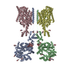
EMDB-19986:
Structure of recombinant alpha-synuclein fibrils 1B capable of seeding GCIs in vivo

PDB-9euu:
Structure of recombinant alpha-synuclein fibrils 1B capable of seeding GCIs in vivo

EMDB-41271:
Consensus map of 96nm repeat of human respiratory doublet microtubule, RS1-2 region

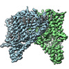
EMDB-19408:
Cryo-EM structure of CDK2-cyclin A in complex with CDC25A

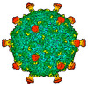
EMDB-41152:
Cryo-EM Structure of Spike Glycoprotein from Civet Coronavirus SZ3 in Closed Conformation

PDB-8tc5:
Cryo-EM Structure of Spike Glycoprotein from Civet Coronavirus SZ3 in Closed Conformation

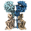
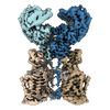
EMDB-18729:
Cryo-EM structure of tetrameric human SAMHD1 with dApNHpp

EMDB-18730:
Cryo-EM structure of tetrameric human SAMHD1 State I - Tense

EMDB-18731:
Cryo-EM structure of tetrameric human SAMHD1 State II - Hemi-relaxed

EMDB-18732:
Cryo-EM structure of tetrameric human SAMHD1 State III - Relaxed

EMDB-18733:
Cryo-EM structure of tetrameric human SAMHD1 State IV - Depleted relaxed

EMDB-18734:
Cryo-EM structure of tetrameric human SAMHD1 State V - Depleted relaxed
PDB-8qxj:
Cryo-EM structure of tetrameric human SAMHD1 with dApNHpp
PDB-8qxk:
Cryo-EM structure of tetrameric human SAMHD1 State I - Tense
PDB-8qxl:
Cryo-EM structure of tetrameric human SAMHD1 State II - Hemi-relaxed
PDB-8qxm:
Cryo-EM structure of tetrameric human SAMHD1 State III - Relaxed
PDB-8qxn:
Cryo-EM structure of tetrameric human SAMHD1 State IV - Depleted relaxed
PDB-8qxo:
Cryo-EM structure of tetrameric human SAMHD1 State V - Depleted relaxed
EMDB-41149:
Cryo-EM Structure of Spike Glycoprotein from Bat Coronavirus WIV1 in Closed Conformation
EMDB-41150:
Cryo-EM Structure of Spike Glycoprotein from Civet Coronavirus 007 in Closed Conformation
PDB-8tc0:
Cryo-EM Structure of Spike Glycoprotein from Bat Coronavirus WIV1 in Closed Conformation

PDB-8tc1:
Cryo-EM Structure of Spike Glycoprotein from Civet Coronavirus 007 in Closed Conformation
ページ:
 wwPDBはEMDBデータモデルのバージョン3へ移行します
