 Movie
Movie Controller

[English] 日本語
 Yorodumi
- PDB-6fuw: Cryo-EM structure of the human CPSF160-WDR33-CPSF30 complex bound... -
+ Open data

- Basic information
| Entry | Database: PDB / ID: 6fuw | |||||||||||||||
|---|---|---|---|---|---|---|---|---|---|---|---|---|---|---|---|---|
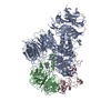
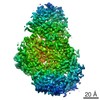
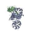
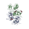
| Title | Cryo-EM structure of the human CPSF160-WDR33-CPSF30 complex bound to the PAS AAUAAA motif at 3.1 Angstrom resolution | |||||||||||||||
 Components Components |
| |||||||||||||||
 Keywords Keywords | RNA BINDING PROTEIN / 3' pre-mRNA processing / polyadenylation / CPSF / beta propeller / AAUAAA | |||||||||||||||
| Function / homology |  Function and homology information Function and homology informationco-transcriptional RNA 3'-end processing, cleavage and polyadenylation pathway / Inhibition of Host mRNA Processing and RNA Silencing / Processing of Intronless Pre-mRNAs / mRNA cleavage and polyadenylation specificity factor complex / collagen trimer / mRNA 3'-UTR AU-rich region binding / Transport of Mature mRNA Derived from an Intronless Transcript / mRNA 3'-end processing / mRNA 3'-end processing / postreplication repair ...co-transcriptional RNA 3'-end processing, cleavage and polyadenylation pathway / Inhibition of Host mRNA Processing and RNA Silencing / Processing of Intronless Pre-mRNAs / mRNA cleavage and polyadenylation specificity factor complex / collagen trimer / mRNA 3'-UTR AU-rich region binding / Transport of Mature mRNA Derived from an Intronless Transcript / mRNA 3'-end processing / mRNA 3'-end processing / postreplication repair / tRNA processing in the nucleus / RNA Polymerase II Transcription Termination / Processing of Capped Intron-Containing Pre-mRNA / fibrillar center / mRNA processing / sequence-specific double-stranded DNA binding / spermatogenesis / intracellular membrane-bounded organelle / enzyme binding / RNA binding / zinc ion binding / nucleoplasm / nucleus Similarity search - Function | |||||||||||||||
| Biological species |  Homo sapiens (human) Homo sapiens (human) unidentified adenovirus unidentified adenovirus | |||||||||||||||
| Method | ELECTRON MICROSCOPY / single particle reconstruction / cryo EM / Resolution: 3.07 Å | |||||||||||||||
 Authors Authors | Clerici, M. / Faini, M. / Jinek, M. | |||||||||||||||
| Funding support |  Belgium, Belgium,  Germany, 4items Germany, 4items
| |||||||||||||||
 Citation Citation | Journal: Nat Struct Mol Biol / Year: 2018 Title: Structural basis of AAUAAA polyadenylation signal recognition by the human CPSF complex. Authors: Marcello Clerici / Marco Faini / Lena M Muckenfuss / Ruedi Aebersold / Martin Jinek /  Abstract: Mammalian mRNA biogenesis requires specific recognition of a hexanucleotide AAUAAA motif in the polyadenylation signals (PAS) of precursor mRNA (pre-mRNA) transcripts by the cleavage and ...Mammalian mRNA biogenesis requires specific recognition of a hexanucleotide AAUAAA motif in the polyadenylation signals (PAS) of precursor mRNA (pre-mRNA) transcripts by the cleavage and polyadenylation specificity factor (CPSF) complex. Here we present a 3.1-Å-resolution cryo-EM structure of a core CPSF module bound to the PAS hexamer motif. The structure reveals the molecular interactions responsible for base-specific recognition, providing a rationale for mechanistic differences between mammalian and yeast 3' polyadenylation. | |||||||||||||||
| History |
|
- Structure visualization
| Movie |
Movie viewer |
|---|---|
| Structure viewer | Molecule: MolmilJmol/JSmol |
- Downloads & links
-Download
| PDBx/mmCIF format | 6fuw.cif.gz | 324.7 KB | Display | PDBx/mmCIF format |
|---|---|---|---|---|
| PDB format | pdb6fuw.ent.gz | 245.5 KB | Display | PDB format |
| PDBx/mmJSON format | 6fuw.json.gz | Tree view | PDBx/mmJSON format | |
| Others |  Other downloads Other downloads |
-Validation report
| Summary document | 6fuw_validation.pdf.gz | 1 MB | Display | wwPDB validaton report |
|---|---|---|---|---|
| Full document | 6fuw_full_validation.pdf.gz | 1 MB | Display | |
| Data in XML | 6fuw_validation.xml.gz | 53.1 KB | Display | |
| Data in CIF | 6fuw_validation.cif.gz | 81.2 KB | Display | |
| Arichive directory | https://data.pdbj.org/pub/pdb/validation_reports/fu/6fuwftp://data.pdbj.org/pub/pdb/validation_reports/fu/6fuw | HTTPS FTP |
-Related structure data
| Related structure data | 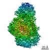 4225MC M: map data used to model this data C: citing same article ( |
|---|---|
| Similar structure data |


-Links
 PDBj
PDBj


- Assembly
| Deposited unit | 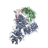
|
|---|---|
| 1 |
|
-Components
| #1: Protein | Mass: 161346.484 Da / Num. of mol.: 1 Source method: isolated from a genetically manipulated source Source: (gene. exp.) Homo sapiens (human) / Gene: CPSF1, CPSF160Production host:  Spodoptera aff. frugiperda 2 RZ-2014 (butterflies/moths) Spodoptera aff. frugiperda 2 RZ-2014 (butterflies/moths)References: UniProt: Q10570 |
|---|---|
| #2: Protein | Mass: 47448.910 Da / Num. of mol.: 1 Source method: isolated from a genetically manipulated source Source: (gene. exp.) Homo sapiens (human) / Gene: WDR33, WDC146Production host: Spodoptera aff. frugiperda 2 RZ-2014 (butterflies/moths)References: UniProt: Q9C0J8 |
| #3: Protein | Mass: 20422.855 Da / Num. of mol.: 1 Source method: isolated from a genetically manipulated source Source: (gene. exp.) Homo sapiens (human) / Gene: CPSF4, CPSF30, NAR, NEB1Production host: Spodoptera aff. frugiperda 2 RZ-2014 (butterflies/moths)References: UniProt: O95639 |
| #4: RNA chain | Mass: 3232.036 Da / Num. of mol.: 1 / Source method: obtained synthetically / Source: (synth.) unidentified adenovirus |
| #5: Chemical |   Mass: 65.409 Da / Num. of mol.: 3 / Source method: obtained synthetically / Formula: Zn Mass: 65.409 Da / Num. of mol.: 3 / Source method: obtained synthetically / Formula: Zn |
-Experimental details
-Experiment
| Experiment | Method: ELECTRON MICROSCOPY |
|---|---|
| EM experiment | Aggregation state: PARTICLE / 3D reconstruction method: single particle reconstruction |
- Sample preparation
| Component |
| ||||||||||||||||||||||||
|---|---|---|---|---|---|---|---|---|---|---|---|---|---|---|---|---|---|---|---|---|---|---|---|---|---|
| Molecular weight | Value: 0.22 MDa / Experimental value: NO | ||||||||||||||||||||||||
| Source (natural) |
| ||||||||||||||||||||||||
| Source (recombinant) |
| ||||||||||||||||||||||||
| Buffer solution | pH: 7.5 | ||||||||||||||||||||||||
| Buffer component |
| ||||||||||||||||||||||||
| Specimen | Conc.: 0.3 mg/ml / Embedding applied: NO / Shadowing applied: NO / Staining applied: NO / Vitrification applied: YES | ||||||||||||||||||||||||
| Specimen support | Grid material: COPPER / Grid mesh size: 400 divisions/in. / Grid type: Quantifoil R1.2/1.3 | ||||||||||||||||||||||||
| Vitrification | Instrument: FEI VITROBOT MARK IV / Cryogen name: ETHANE-PROPANE / Humidity: 100 % / Chamber temperature: 293 K / Details: 15 seconds wait time prior to blotting |
- Electron microscopy imaging
| Experimental equipment |  Model: Titan Krios / Image courtesy: FEI Company |
|---|---|
| Microscopy | Model: FEI TITAN KRIOS |
| Electron gun | Electron source:  FIELD EMISSION GUN / Accelerating voltage: 300 kV / Illumination mode: FLOOD BEAM FIELD EMISSION GUN / Accelerating voltage: 300 kV / Illumination mode: FLOOD BEAM |
| Electron lens | Mode: BRIGHT FIELD / Calibrated magnification: 47259 X / Nominal defocus max: 2200 nm / Nominal defocus min: 1100 nm / Cs: 2.7 mm |
| Specimen holder | Specimen holder model: FEI TITAN KRIOS AUTOGRID HOLDER |
| Image recording | Average exposure time: 10 sec. / Electron dose: 80 e/Å2 / Detector mode: SUPER-RESOLUTION / Film or detector model: GATAN K2 SUMMIT (4k x 4k) / Num. of grids imaged: 1 / Num. of real images: 1070 |
| EM imaging optics | Energyfilter name: GIF Quantum LS |
| Image scans | Movie frames/image: 50 |
- Processing
| Software | Name: PHENIX / Version: 1.12_2829: / Classification: refinement | ||||||||||||||||||||||||||||||||||||||||
|---|---|---|---|---|---|---|---|---|---|---|---|---|---|---|---|---|---|---|---|---|---|---|---|---|---|---|---|---|---|---|---|---|---|---|---|---|---|---|---|---|---|
| EM software |
| ||||||||||||||||||||||||||||||||||||||||
| CTF correction | Type: PHASE FLIPPING AND AMPLITUDE CORRECTION | ||||||||||||||||||||||||||||||||||||||||
| Particle selection | Num. of particles selected: 263000 | ||||||||||||||||||||||||||||||||||||||||
| Symmetry | Point symmetry: C1 (asymmetric) | ||||||||||||||||||||||||||||||||||||||||
| 3D reconstruction | Resolution: 3.07 Å / Resolution method: FSC 0.143 CUT-OFF / Num. of particles: 137000 / Symmetry type: POINT | ||||||||||||||||||||||||||||||||||||||||
| Atomic model building | Protocol: RIGID BODY FIT / Space: REAL / Details: phenix.real_space_refine | ||||||||||||||||||||||||||||||||||||||||
| Atomic model building | PDB-ID: 6F9N Accession code: 6F9N / Details: fitted manually in Coot / Source name: PDB / Type: experimental model | ||||||||||||||||||||||||||||||||||||||||
| Refinement | Highest resolution: 3.07 Å | ||||||||||||||||||||||||||||||||||||||||
| Refine LS restraints |
|

