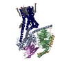
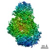
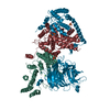
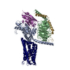
[English] 日本語
Yorodumi
- PDB-5gp4: Lactobacillus brevis CGMCC 1306 Glutamate decarboxylase -

+
Open data


ID or keywords:

Loading...

-
Basic information

Entry
Database: PDB / ID: 5gp4
TitleLactobacillus brevis CGMCC 1306 Glutamate decarboxylase
ComponentsGlutamate decarboxylase
KeywordsLYASE / Glutamate decarboxylase Lactobacillus brevis CGMCC 1306 Gamma-aminobutyric acid pyridoxal phosphate (PLP)
Function / homology
Function and homology information


glutamate decarboxylase / glutamate decarboxylase activity / glutamate metabolic process / pyridoxal phosphate binding
Similarity search - Function
Glutamate decarboxylase / Pyridoxal phosphate-dependent decarboxylase / Pyridoxal-dependent decarboxylase conserved domain / Aspartate Aminotransferase; domain 2 / Type I PLP-dependent aspartate aminotransferase-like (Major domain) / Pyridoxal phosphate-dependent transferase, major domain / Pyridoxal phosphate-dependent transferase / 3-Layer(aba) Sandwich / Alpha Beta
Similarity search - Domain/homology
PYRIDOXAL-5'-PHOSPHATE / Glutamate decarboxylase
Similarity search - Component
Biological speciesLactobacillus brevis (bacteria)
MethodX-RAY DIFFRACTION / SYNCHROTRON / MOLECULAR REPLACEMENT / Resolution: 2.16 Å
AuthorsMei, L. / Huang, J.
Funding support China, 2items
OrganizationGrant numberCountry
National Natural Science Foundation of China21176220 and 31470793 China
the Natural Science Foundation of Zhejiang ProvinceZ13B060008 and LY16B060008 China
CitationJournal: Biochem. Biophys. Res. Commun. / Year: 2018
Title: Lactobacillus brevis CGMCC 1306 glutamate decarboxylase: Crystal structure and functional analysis.
Authors: Huang, J. / Fang, H. / Gai, Z.C. / Mei, J.Q. / Li, J.N. / Hu, S. / Lv, C.J. / Zhao, W.R. / Mei, L.H.
History
DepositionJul 31, 2016Deposition site: PDBJ / Processing site: PDBJ
Revision 1.0Aug 2, 2017Provider: repository / Type: Initial release
Revision 1.1Sep 12, 2018Group: Data collection / Database references / Category: citation / citation_author
Item: _citation.country / _citation.journal_abbrev ..._citation.country / _citation.journal_abbrev / _citation.journal_id_ASTM / _citation.journal_id_CSD / _citation.journal_id_ISSN / _citation.journal_volume / _citation.page_first / _citation.page_last / _citation.pdbx_database_id_DOI / _citation.pdbx_database_id_PubMed / _citation.title / _citation.year
Revision 1.2Nov 8, 2023Group: Data collection / Database references / Refinement description
Category: chem_comp_atom / chem_comp_bond ...chem_comp_atom / chem_comp_bond / database_2 / pdbx_initial_refinement_model / refine_hist
Item: _database_2.pdbx_DOI / _database_2.pdbx_database_accession / _refine_hist.d_res_low

-
Structure visualization

Structure viewerMolecule:
MolmilJmol/JSmol

Downloads & links

-
Assembly

Deposited unit
C: Glutamate decarboxylase
A: Glutamate decarboxylase
B: Glutamate decarboxylase
hetero molecules


Theoretical massNumber of molelcules
Total (without water)161,5796
Polymers160,8383
Non-polymers7413
Water3,333185
1


  • Idetical with deposited unit
  • defined by author
TypeNameSymmetry operationNumber
identity operation1_555x,y,z1
Buried area14520 Å2
ΔGint-51 kcal/mol
Surface area50440 Å2
2
C: Glutamate decarboxylase
A: Glutamate decarboxylase
B: Glutamate decarboxylase
hetero molecules

C: Glutamate decarboxylase
A: Glutamate decarboxylase
B: Glutamate decarboxylase
hetero molecules


Theoretical massNumber of molelcules
Total (without water)323,15912
Polymers321,6766
Non-polymers1,4836
Water1086
TypeNameSymmetry operationNumber
identity operation1_555x,y,z1
crystal symmetry operation3_654-x+1,y,-z-1/21
Buried area45160 Å2
ΔGint-159 kcal/mol
Surface area84740 Å2
MethodPISA
Unit cell
Length a, b, c (Å)114.333, 170.129, 164.567
Angle α, β, γ (deg.)90.00, 90.00, 90.00
Int Tables number20
Space group name H-MC2221

-
Components

#1: Protein Glutamate decarboxylase


Mass: 53612.664 Da / Num. of mol.: 3
Source method: isolated from a genetically manipulated source
Source: (gene. exp.) Lactobacillus brevis (bacteria) / Gene: gad / Plasmid: pet28 / Production host: Escherichia coli BL21 (bacteria) / Strain (production host): BL21 / References: UniProt: D6PXK5, glutamate decarboxylase
#2: Chemical ChemComp-PLP / PYRIDOXAL-5'-PHOSPHATE / VITAMIN B6 Phosphate


Mass: 247.142 Da / Num. of mol.: 3 / Source method: isolated from a natural source / Formula: C8H10NO6P
#3: Water ChemComp-HOH / water


Mass: 18.015 Da / Num. of mol.: 185 / Source method: isolated from a natural source / Formula: H2O

-
Experimental details

-
Experiment

ExperimentMethod: X-RAY DIFFRACTION / Number of used crystals: 1

-
Sample preparation

CrystalDensity Matthews: 2.52 Å3/Da / Density % sol: 51.27 %
Crystal growTemperature: 287 K / Method: vapor diffusion, hanging drop / pH: 5.5
Details: 10% W/V PEG 2000 MME, 100mM Sodium Acetate/Acetic acid, 200mM Ammonium Sulfate

-
Data collection

DiffractionMean temperature: 82 K / Ambient temp details: 82
Diffraction sourceSource: SYNCHROTRON / Site: NFPSS / Beamline: BL18U / Wavelength: 0.97851 Å
DetectorType: ADSC QUANTUM 315r / Detector: CCD / Date: Apr 25, 2016
RadiationProtocol: SINGLE WAVELENGTH / Monochromatic (M) / Laue (L): M / Scattering type: x-ray
Radiation wavelengthWavelength: 0.97851 Å / Relative weight: 1
ReflectionResolution: 2.15→50.01 Å / Num. obs: 85974 / % possible obs: 100 % / Redundancy: 6.7 % / CC1/2: 0.93 / Rmerge(I) obs: 0.176 / Net I/σ(I): 10.88
Reflection shellResolution: 2.15→2.23 Å / Redundancy: 6.7 % / Rmerge(I) obs: 0.7 / CC1/2: 0.84 / % possible all: 100

-
Processing

Software
NameVersionClassification
REFMAC5.8.0135refinement
HKL-3000data collection
SCALEPACKdata scaling
PHASERphasing
MERLOTdata processing
RefinementMethod to determine structure: MOLECULAR REPLACEMENT
Starting model: 1xey
Resolution: 2.16→48.543 Å / Cor.coef. Fo:Fc: 0.945 / Cor.coef. Fo:Fc free: 0.917 / SU B: 5.117 / SU ML: 0.132 / Cross valid method: THROUGHOUT / ESU R: 0.217 / ESU R Free: 0.183 / Stereochemistry target values: MAXIMUM LIKELIHOOD / Details: HYDROGENS HAVE BEEN ADDED IN THE RIDING POSITIONS
RfactorNum. reflection% reflectionSelection details
Rfree0.23005 4290 5 %RANDOM
Rwork0.18694 ---
obs0.18911 81517 99.32 %-
Solvent computationIon probe radii: 0.8 Å / Shrinkage radii: 0.8 Å / VDW probe radii: 1.2 Å / Solvent model: MASK
Displacement parametersBiso mean: 31.556 Å2
Baniso -1Baniso -2Baniso -3
1-0.34 Å2-0 Å2-0 Å2
2---0.35 Å2-0 Å2
3---0.02 Å2
Refinement stepCycle: 1 / Resolution: 2.16→48.543 Å
ProteinNucleic acidLigandSolventTotal
Num. atoms10498 0 45 185 10728
Refine LS restraints
Refine-IDTypeDev idealDev ideal targetNumber
X-RAY DIFFRACTIONr_bond_refined_d0.0190.01910809
X-RAY DIFFRACTIONr_bond_other_d0.0020.0210051
X-RAY DIFFRACTIONr_angle_refined_deg1.9831.95114687
X-RAY DIFFRACTIONr_angle_other_deg1.188323095
X-RAY DIFFRACTIONr_dihedral_angle_1_deg6.61851301
X-RAY DIFFRACTIONr_dihedral_angle_2_deg35.44923.992531
X-RAY DIFFRACTIONr_dihedral_angle_3_deg17.737151792
X-RAY DIFFRACTIONr_dihedral_angle_4_deg23.7051565
X-RAY DIFFRACTIONr_chiral_restr0.1420.21583
X-RAY DIFFRACTIONr_gen_planes_refined0.010.02112239
X-RAY DIFFRACTIONr_gen_planes_other0.0020.022560
X-RAY DIFFRACTIONr_nbd_refined
X-RAY DIFFRACTIONr_nbd_other
X-RAY DIFFRACTIONr_nbtor_refined
X-RAY DIFFRACTIONr_nbtor_other
X-RAY DIFFRACTIONr_xyhbond_nbd_refined
X-RAY DIFFRACTIONr_xyhbond_nbd_other
X-RAY DIFFRACTIONr_metal_ion_refined
X-RAY DIFFRACTIONr_metal_ion_other
X-RAY DIFFRACTIONr_symmetry_vdw_refined
X-RAY DIFFRACTIONr_symmetry_vdw_other
X-RAY DIFFRACTIONr_symmetry_hbond_refined
X-RAY DIFFRACTIONr_symmetry_hbond_other
X-RAY DIFFRACTIONr_symmetry_metal_ion_refined
X-RAY DIFFRACTIONr_symmetry_metal_ion_other
X-RAY DIFFRACTIONr_mcbond_it2.8692.9185213
X-RAY DIFFRACTIONr_mcbond_other2.8672.9185212
X-RAY DIFFRACTIONr_mcangle_it4.2534.3576511
X-RAY DIFFRACTIONr_mcangle_other4.2544.3586512
X-RAY DIFFRACTIONr_scbond_it3.5933.3155594
X-RAY DIFFRACTIONr_scbond_other3.5923.3155595
X-RAY DIFFRACTIONr_scangle_it
X-RAY DIFFRACTIONr_scangle_other5.564.8118177
X-RAY DIFFRACTIONr_long_range_B_refined6.98822.76512283
X-RAY DIFFRACTIONr_long_range_B_other6.98822.76512284
X-RAY DIFFRACTIONr_rigid_bond_restr
X-RAY DIFFRACTIONr_sphericity_free
X-RAY DIFFRACTIONr_sphericity_bonded
LS refinement shellResolution: 2.156→2.212 Å / Total num. of bins used: 20
RfactorNum. reflection% reflection
Rfree0.287 290 -
Rwork0.239 5558 -
obs--92.87 %

+
About Yorodumi

-
News

-
Feb 9, 2022. New format data for meta-information of EMDB entries

New format data for meta-information of EMDB entries

  • Version 3 of the EMDB header file is now the official format.
  • The previous official version 1.9 will be removed from the archive.

Related info.:EMDB header

External links:wwPDB to switch to version 3 of the EMDB data model

-
Aug 12, 2020. Covid-19 info

Covid-19 info

URL: https://pdbjlvh1.pdbj.org/emnavi/covid19.php

New page: Covid-19 featured information page in EM Navigator.

Related info.:Covid-19 info / Mar 5, 2020. Novel coronavirus structure data

+
Mar 5, 2020. Novel coronavirus structure data

Novel coronavirus structure data

Related info.:Yorodumi Speices / Aug 12, 2020. Covid-19 info

External links:COVID-19 featured content - PDBj / Molecule of the Month (242):Coronavirus Proteases

+
Jan 31, 2019. EMDB accession codes are about to change! (news from PDBe EMDB page)

EMDB accession codes are about to change! (news from PDBe EMDB page)

  • The allocation of 4 digits for EMDB accession codes will soon come to an end. Whilst these codes will remain in use, new EMDB accession codes will include an additional digit and will expand incrementally as the available range of codes is exhausted. The current 4-digit format prefixed with “EMD-” (i.e. EMD-XXXX) will advance to a 5-digit format (i.e. EMD-XXXXX), and so on. It is currently estimated that the 4-digit codes will be depleted around Spring 2019, at which point the 5-digit format will come into force.
  • The EM Navigator/Yorodumi systems omit the EMD- prefix.

Related info.:Q: What is EMD? / ID/Accession-code notation in Yorodumi/EM Navigator

External links:EMDB Accession Codes are Changing Soon! / Contact to PDBj

+
Jul 12, 2017. Major update of PDB

Major update of PDB

  • wwPDB released updated PDB data conforming to the new PDBx/mmCIF dictionary.
  • This is a major update changing the version number from 4 to 5, and with Remediation, in which all the entries are updated.
  • In this update, many items about electron microscopy experimental information are reorganized (e.g. em_software).
  • Now, EM Navigator and Yorodumi are based on the updated data.

External links:wwPDB Remediation / Enriched Model Files Conforming to OneDep Data Standards Now Available in the PDB FTP Archive

-
Yorodumi

Thousand views of thousand structures

  • Yorodumi is a browser for structure data from EMDB, PDB, SASBDB, etc.
  • This page is also the successor to EM Navigator detail page, and also detail information page/front-end page for Omokage search.
  • The word "yorodu" (or yorozu) is an old Japanese word meaning "ten thousand". "mi" (miru) is to see.

Related info.:EMDB / PDB / SASBDB / Comparison of 3 databanks / Yorodumi Search / Aug 31, 2016. New EM Navigator & Yorodumi / Yorodumi Papers / Jmol/JSmol / Function and homology information / Changes in new EM Navigator and Yorodumi

Read more