[English] 日本語
 Yorodumi
- EMDB-2529: Single particle electron microscopy of the human mitochondrial tr... -
+ Open data
- Basic information
| Entry | Database: EMDB / ID: EMD-2529 | |||||||||
|---|---|---|---|---|---|---|---|---|---|---|
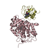
| Title | Single particle electron microscopy of the human mitochondrial transcription initiation complex | |||||||||
 Map data Map data | Reconstruction of the human mitochondrial transcription initiation complex | |||||||||
 Sample Sample |
| |||||||||
 Keywords Keywords | human mitochondrial transcription / POLRMT / TFAM / TFB2M | |||||||||
| Function / homology |  Function and homology information Function and homology informationMitochondrial transcription initiation / mitochondrial promoter sequence-specific DNA binding / mitochondrial transcription factor activity / mitochondrial respiratory chain complex assembly / transcription initiation at mitochondrial promoter / rRNA (adenine-N6,N6-)-dimethyltransferase activity / mitochondrial transcription / rRNA methylation / mitochondrial nucleoid / heat shock protein binding ...Mitochondrial transcription initiation / mitochondrial promoter sequence-specific DNA binding / mitochondrial transcription factor activity / mitochondrial respiratory chain complex assembly / transcription initiation at mitochondrial promoter / rRNA (adenine-N6,N6-)-dimethyltransferase activity / mitochondrial transcription / rRNA methylation / mitochondrial nucleoid / heat shock protein binding / response to nutrient / Mitochondrial protein degradation / Transferases; Transferring one-carbon groups; Methyltransferases / transcription coactivator binding / Transcriptional activation of mitochondrial biogenesis / sequence-specific DNA binding / response to hypoxia / transcription cis-regulatory region binding / mitochondrial matrix / chromatin binding / positive regulation of DNA-templated transcription / protein-containing complex / mitochondrion / RNA binding / nucleus / cytosol Similarity search - Function | |||||||||
| Biological species |  Homo sapiens (human) Homo sapiens (human) | |||||||||
| Method | single particle reconstruction / negative staining / Resolution: 25.0 Å | |||||||||
 Authors Authors | Yakubovskaya E / Guja KE / Eng ET / Choi WS / Mejia E / Beglov B / Lukin M / Kozakov D / Garcia-Diaz M | |||||||||
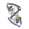
 Citation Citation |  Journal: Nucleic Acids Res / Year: 2014 Journal: Nucleic Acids Res / Year: 2014Title: Organization of the human mitochondrial transcription initiation complex. Authors: Elena Yakubovskaya / Kip E Guja / Edward T Eng / Woo Suk Choi / Edison Mejia / Dmitri Beglov / Mark Lukin / Dima Kozakov / Miguel Garcia-Diaz /  Abstract: Initiation of transcription in human mitochondria involves two factors, TFAM and TFB2M, in addition to the mitochondrial RNA polymerase, POLRMT. We have investigated the organization of the human ...Initiation of transcription in human mitochondria involves two factors, TFAM and TFB2M, in addition to the mitochondrial RNA polymerase, POLRMT. We have investigated the organization of the human mitochondrial transcription initiation complex on the light-strand promoter (LSP) through solution X-ray scattering, electron microscopy (EM) and biochemical studies. Our EM results demonstrate a compact organization of the initiation complex, suggesting that protein-protein interactions might help mediate initiation. We demonstrate that, in the absence of DNA, only POLRMT and TFAM form a stable interaction, albeit one with low affinity. This is consistent with the expected transient nature of the interactions necessary for initiation and implies that the promoter DNA acts as a scaffold that enables formation of the full initiation complex. Docking of known crystal structures into our EM maps results in a model for transcriptional initiation that strongly correlates with new and existing biochemical observations. Our results reveal the organization of TFAM, POLRMT and TFB2M around the LSP and represent the first structural characterization of the entire mitochondrial transcriptional initiation complex. | |||||||||
| History |
|
- Structure visualization
| Movie |
 Movie viewer Movie viewer |
|---|---|
| Structure viewer | EM map:  SurfView SurfView Molmil Molmil Jmol/JSmol Jmol/JSmol |
| Supplemental images |
- Downloads & links
-EMDB archive
| Map data |  emd_2529.map.gz emd_2529.map.gz | 198.4 KB |  EMDB map data format EMDB map data format | |
|---|---|---|---|---|
| Header (meta data) |  emd-2529-v30.xml emd-2529-v30.xml emd-2529.xml emd-2529.xml | 14.1 KB 14.1 KB | Display Display |  EMDB header EMDB header |
| Images |  EMD-2529.png EMD-2529.png | 57.6 KB | ||
| Archive directory |  http://ftp.pdbj.org/pub/emdb/structures/EMD-2529 http://ftp.pdbj.org/pub/emdb/structures/EMD-2529 ftp://ftp.pdbj.org/pub/emdb/structures/EMD-2529 ftp://ftp.pdbj.org/pub/emdb/structures/EMD-2529 | HTTPS FTP |
-Validation report
| Summary document |  emd_2529_validation.pdf.gz emd_2529_validation.pdf.gz | 211.5 KB | Display |  EMDB validaton report EMDB validaton report |
|---|---|---|---|---|
| Full document |  emd_2529_full_validation.pdf.gz emd_2529_full_validation.pdf.gz | 210.6 KB | Display | |
| Data in XML |  emd_2529_validation.xml.gz emd_2529_validation.xml.gz | 4.8 KB | Display | |
| Arichive directory |  https://ftp.pdbj.org/pub/emdb/validation_reports/EMD-2529 https://ftp.pdbj.org/pub/emdb/validation_reports/EMD-2529 ftp://ftp.pdbj.org/pub/emdb/validation_reports/EMD-2529 ftp://ftp.pdbj.org/pub/emdb/validation_reports/EMD-2529 | HTTPS FTP |
-Related structure data
| Similar structure data |
|---|
- Links
Links
| EMDB pages |  EMDB (EBI/PDBe) / EMDB (EBI/PDBe) /  EMDataResource EMDataResource |
|---|---|
| Related items in Molecule of the Month |
- Map
Map
| File |  Download / File: emd_2529.map.gz / Format: CCP4 / Size: 284.2 KB / Type: IMAGE STORED AS FLOATING POINT NUMBER (4 BYTES) Download / File: emd_2529.map.gz / Format: CCP4 / Size: 284.2 KB / Type: IMAGE STORED AS FLOATING POINT NUMBER (4 BYTES) | ||||||||||||||||||||||||||||||||||||||||||||||||||||||||||||||||||||
|---|---|---|---|---|---|---|---|---|---|---|---|---|---|---|---|---|---|---|---|---|---|---|---|---|---|---|---|---|---|---|---|---|---|---|---|---|---|---|---|---|---|---|---|---|---|---|---|---|---|---|---|---|---|---|---|---|---|---|---|---|---|---|---|---|---|---|---|---|---|
| Annotation | Reconstruction of the human mitochondrial transcription initiation complex | ||||||||||||||||||||||||||||||||||||||||||||||||||||||||||||||||||||
| Projections & slices | Image control
Images are generated by Spider. | ||||||||||||||||||||||||||||||||||||||||||||||||||||||||||||||||||||
| Voxel size | X=Y=Z: 3.4 Å | ||||||||||||||||||||||||||||||||||||||||||||||||||||||||||||||||||||
| Density |
| ||||||||||||||||||||||||||||||||||||||||||||||||||||||||||||||||||||
| Symmetry | Space group: 1 | ||||||||||||||||||||||||||||||||||||||||||||||||||||||||||||||||||||
| Details | EMDB XML:
CCP4 map header:
| ||||||||||||||||||||||||||||||||||||||||||||||||||||||||||||||||||||
-Supplemental data
- Sample components
-Entire : Quaternary complex of POLRMT, TFAM, TFB2M and LSP DNA
| Entire | Name: Quaternary complex of POLRMT, TFAM, TFB2M and LSP DNA |
|---|---|
| Components |
|
-Supramolecule #1000: Quaternary complex of POLRMT, TFAM, TFB2M and LSP DNA
| Supramolecule | Name: Quaternary complex of POLRMT, TFAM, TFB2M and LSP DNA / type: sample / ID: 1000 Details: The complex also has a DNA, that has a mitochondrial light strand promoter sequence Oligomeric state: one POLRMT one TFB2M and one TFAM TFAM) / Number unique components: 3 |
|---|---|
| Molecular weight | Experimental: 250 KDa / Theoretical: 250 KDa / Method: Gel filtration, electrophoresis |
-Macromolecule #1: Dimethyladenosine transferase 2, mitochondrial
| Macromolecule | Name: Dimethyladenosine transferase 2, mitochondrial / type: protein_or_peptide / ID: 1 / Name.synonym: TFB2M / Number of copies: 1 / Oligomeric state: 1 / Recombinant expression: Yes |
|---|---|
| Source (natural) | Organism:  Homo sapiens (human) / synonym: human Homo sapiens (human) / synonym: human |
| Molecular weight | Experimental: 39 KDa / Theoretical: 39 KDa |
| Recombinant expression | Organism:  |
| Sequence | UniProtKB: Dimethyladenosine transferase 2, mitochondrial |
-Macromolecule #2: Dimethyladenosine transferase 2, mitochondrial
| Macromolecule | Name: Dimethyladenosine transferase 2, mitochondrial / type: protein_or_peptide / ID: 2 / Name.synonym: TFB2M / Number of copies: 1 / Oligomeric state: 1 / Recombinant expression: Yes |
|---|---|
| Source (natural) | Organism:  Homo sapiens (human) / synonym: human Homo sapiens (human) / synonym: human |
| Molecular weight | Experimental: 39 KDa / Theoretical: 39 KDa |
| Recombinant expression | Organism:  |
| Sequence | UniProtKB: Dimethyladenosine transferase 2, mitochondrial |
-Macromolecule #3: Transcription factor A, mitochondrial
| Macromolecule | Name: Transcription factor A, mitochondrial / type: protein_or_peptide / ID: 3 / Name.synonym: TFAM / Number of copies: 1 / Oligomeric state: 1 / Recombinant expression: Yes |
|---|---|
| Source (natural) | Organism:  Homo sapiens (human) / synonym: human Homo sapiens (human) / synonym: human |
| Molecular weight | Experimental: 29 KDa / Theoretical: 29 KDa |
| Recombinant expression | Organism:  |
| Sequence | UniProtKB: Transcription factor A, mitochondrial |
-Experimental details
-Structure determination
| Method | negative staining |
|---|---|
 Processing Processing | single particle reconstruction |
| Aggregation state | particle |
- Sample preparation
| Concentration | 0.2 mg/mL |
|---|---|
| Buffer | pH: 8 / Details: 50mM KCl,20mM,HEPES, 10mM MgCl2 |
| Staining | Type: NEGATIVE Details: Grids with adsorbed complex were washed 2 times in buffer and then floated on 1% w/v uranyl acetate for 30 seconds. |
| Grid | Details: 200 mesh gold grid with thin carbon support, glow discharged in amylamine atmosphere |
| Vitrification | Cryogen name: NONE / Instrument: OTHER |
- Electron microscopy
| Microscope | FEI TECNAI F20 |
|---|---|
| Alignment procedure | Legacy - Astigmatism: Objective lens astigmatism was corrected at 50,000 times magnification at each new area of the grid |
| Specialist optics | Energy filter - Name: FEI |
| Date | Oct 21, 2013 |
| Image recording | Category: CCD / Film or detector model: OTHER / Digitization - Sampling interval: 10 µm / Number real images: 200 |
| Electron beam | Acceleration voltage: 200 kV / Electron source:  FIELD EMISSION GUN FIELD EMISSION GUN |
| Electron optics | Calibrated magnification: 88249 / Illumination mode: SPOT SCAN / Imaging mode: BRIGHT FIELD / Nominal defocus max: -0.5 µm / Nominal defocus min: -1.5 µm / Nominal magnification: 50000 |
| Sample stage | Specimen holder: liquid nitrogen cooled / Specimen holder model: GATAN LIQUID NITROGEN |
| Experimental equipment |  Model: Tecnai F20 / Image courtesy: FEI Company |
- Image processing
| Details | Reference free classes were divided into two subsets, based on particle size, and two initial models were generated from each subset. One model had a visibly elongated shape, whereas the other was more compact. We utilized both models as starting points for parallel refinement, as implemented in EMAN MULTIREFINE routine |
|---|---|
| CTF correction | Details: EMAN2 protocol each particle |
| Final reconstruction | Applied symmetry - Point group: C1 (asymmetric) / Algorithm: OTHER / Resolution.type: BY AUTHOR / Resolution: 25.0 Å / Resolution method: OTHER / Software - Name: EMAN2 / Number images used: 12453 |
| Final two d classification | Number classes: 15 |
-Atomic model buiding 1
| Initial model | PDB ID: Chain - Chain ID: A |
|---|---|
| Software | Name: Chimera, SITUS |
| Details | 3 proteins were fitted separately by manual docking and then refine with SITUS and in-home program. |
| Refinement | Space: REAL / Protocol: RIGID BODY FIT |
-Atomic model buiding 2
| Initial model | PDB ID: Chain - Chain ID: A |
|---|---|
| Software | Name: Chimera, SITUS |
| Details | 3 proteins were fitted separately by manual docking and then refine with SITUS and in-home program. |
| Refinement | Space: REAL / Protocol: RIGID BODY FIT |
-Atomic model buiding 3
| Initial model | PDB ID: Chain - Chain ID: A |
|---|---|
| Software | Name: Chimera, SITUS |
| Details | 3 proteins were fitted separately by manual docking and then refine with SITUS and in-home program. |
| Refinement | Space: REAL / Protocol: RIGID BODY FIT |
 Movie
Movie Controller







 Z (Sec.)
 Y (Row.)
 X (Col.)


