[English] 日本語
Yorodumi
- PDB-3tq6: Crystal structure of human mitochondrial transcription factor A, ... -

+
Open data


ID or keywords:

Loading...

-
Basic information

Entry
Database: PDB / ID: 3tq6
TitleCrystal structure of human mitochondrial transcription factor A, TFAM or mtTFA, bound to the light strand promoter LSP
Components
  • DNA (5'-D(*GP*TP*TP*AP*GP*TP*TP*GP*GP*GP*GP*GP*GP*TP*GP*AP*CP*TP*GP*TP*TP*A)-3')
  • DNA (5'-D(*TP*AP*AP*CP*AP*GP*TP*CP*AP*CP*CP*CP*CP*CP*CP*AP*AP*CP*(BRU)P*AP*AP*C)-3')
  • Transcription factor A, mitochondrial
KeywordsTRANSCRIPTION/DNA / Transcription / Transcription regulation / Mitochondrion / DNA-binding / HMGB-UBF_HMG-box / minor groove / tandem HMG boxes / TRANSCRIPTION-DNA complex / mitochondrial nucleoid / light-strand mitochondrial promoter / LSP
Function / homology
Function and homology information


Mitochondrial transcription initiation / mitochondrial promoter sequence-specific DNA binding / mitochondrial respiratory chain complex assembly / mitochondrial transcription factor activity / transcription initiation at mitochondrial promoter / mitochondrial transcription / mitochondrial nucleoid / heat shock protein binding / response to nutrient / Transcriptional activation of mitochondrial biogenesis ...Mitochondrial transcription initiation / mitochondrial promoter sequence-specific DNA binding / mitochondrial respiratory chain complex assembly / mitochondrial transcription factor activity / transcription initiation at mitochondrial promoter / mitochondrial transcription / mitochondrial nucleoid / heat shock protein binding / response to nutrient / Transcriptional activation of mitochondrial biogenesis / transcription coactivator binding / sequence-specific DNA binding / response to hypoxia / transcription cis-regulatory region binding / mitochondrial matrix / chromatin binding / positive regulation of DNA-templated transcription / protein-containing complex / mitochondrion / RNA binding / nucleus / cytosol
Similarity search - Function
High mobility group box domain / DNA Binding (I), subunit A / HMG-box domain / HMG (high mobility group) box / HMG boxes A and B DNA-binding domains profile. / high mobility group / High mobility group box domain / High mobility group box domain superfamily / Orthogonal Bundle / Mainly Alpha
Similarity search - Domain/homology
DI(HYDROXYETHYL)ETHER / PHOSPHATE ION / DNA / DNA (> 10) / Transcription factor A, mitochondrial
Similarity search - Component
Biological speciesHomo sapiens (human)
MethodX-RAY DIFFRACTION / SYNCHROTRON / MOLECULAR REPLACEMENT / Resolution: 2.45 Å
AuthorsRubio-Cosials, A. / Sydow, J.F. / Jimenez-Menendez, N. / Fernandez-Millan, P. / Montoya, J. / Jacobs, H.T. / Coll, M. / Bernado, P. / Sola, M.
CitationJournal: Nat.Struct.Mol.Biol. / Year: 2011
Title: Human mitochondrial transcription factor A induces a U-turn structure in the light strand promoter.
Authors: Rubio-Cosials, A. / Sidow, J.F. / Jimenez-Menendez, N. / Fernandez-Millan, P. / Montoya, J. / Jacobs, H.T. / Coll, M. / Bernado, P. / Sola, M.
History
DepositionSep 9, 2011Deposition site: RCSB / Processing site: RCSB
Revision 1.0Nov 2, 2011Provider: repository / Type: Initial release
Revision 1.1Nov 9, 2011Group: Database references
Revision 1.2Nov 16, 2011Group: Database references
Revision 1.3Jan 11, 2017Group: Other
Revision 1.4Feb 28, 2024Group: Data collection / Database references / Derived calculations
Category: chem_comp_atom / chem_comp_bond ...chem_comp_atom / chem_comp_bond / database_2 / struct_conn / struct_ref_seq_dif / struct_site
Item: _database_2.pdbx_DOI / _database_2.pdbx_database_accession ..._database_2.pdbx_DOI / _database_2.pdbx_database_accession / _struct_conn.pdbx_leaving_atom_flag / _struct_ref_seq_dif.details / _struct_site.pdbx_auth_asym_id / _struct_site.pdbx_auth_comp_id / _struct_site.pdbx_auth_seq_id

-
Structure visualization

Structure viewerMolecule:
MolmilJmol/JSmol

Downloads & links

-
Assembly

Deposited unit
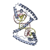
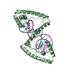
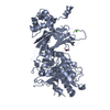
A: Transcription factor A, mitochondrial
B: Transcription factor A, mitochondrial
C: DNA (5'-D(*TP*AP*AP*CP*AP*GP*TP*CP*AP*CP*CP*CP*CP*CP*CP*AP*AP*CP*(BRU)P*AP*AP*C)-3')
D: DNA (5'-D(*GP*TP*TP*AP*GP*TP*TP*GP*GP*GP*GP*GP*GP*TP*GP*AP*CP*TP*GP*TP*TP*A)-3')
E: DNA (5'-D(*TP*AP*AP*CP*AP*GP*TP*CP*AP*CP*CP*CP*CP*CP*CP*AP*AP*CP*(BRU)P*AP*AP*C)-3')
F: DNA (5'-D(*GP*TP*TP*AP*GP*TP*TP*GP*GP*GP*GP*GP*GP*TP*GP*AP*CP*TP*GP*TP*TP*A)-3')
hetero molecules


Theoretical massNumber of molelcules
Total (without water)78,9949
Polymers78,5546
Non-polymers4393
Water3,351186
1
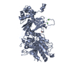
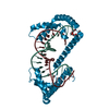
A: Transcription factor A, mitochondrial
C: DNA (5'-D(*TP*AP*AP*CP*AP*GP*TP*CP*AP*CP*CP*CP*CP*CP*CP*AP*AP*CP*(BRU)P*AP*AP*C)-3')
D: DNA (5'-D(*GP*TP*TP*AP*GP*TP*TP*GP*GP*GP*GP*GP*GP*TP*GP*AP*CP*TP*GP*TP*TP*A)-3')
hetero molecules


Theoretical massNumber of molelcules
Total (without water)39,3724
Polymers39,2773
Non-polymers951
Water543
TypeNameSymmetry operationNumber
identity operation1_555x,y,z1
Buried area8590 Å2
ΔGint-41 kcal/mol
Surface area17840 Å2
MethodPISA
2
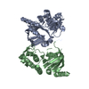
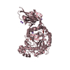
B: Transcription factor A, mitochondrial
E: DNA (5'-D(*TP*AP*AP*CP*AP*GP*TP*CP*AP*CP*CP*CP*CP*CP*CP*AP*AP*CP*(BRU)P*AP*AP*C)-3')
F: DNA (5'-D(*GP*TP*TP*AP*GP*TP*TP*GP*GP*GP*GP*GP*GP*TP*GP*AP*CP*TP*GP*TP*TP*A)-3')
hetero molecules


Theoretical massNumber of molelcules
Total (without water)39,6225
Polymers39,2773
Non-polymers3442
Water543
TypeNameSymmetry operationNumber
identity operation1_555x,y,z1
Buried area8960 Å2
ΔGint-34 kcal/mol
Surface area17510 Å2
MethodPISA
Unit cell
Length a, b, c (Å)113.900, 117.200, 56.530
Angle α, β, γ (deg.)90.00, 90.00, 90.00
Int Tables number18
Space group name H-MP21212

-
Components

-
Protein , 1 types, 2 molecules AB

#1: Protein Transcription factor A, mitochondrial / mtTFA / Mitochondrial transcription factor 1 / MtTF1 / Transcription factor 6 / TCF-6 / ...mtTFA / Mitochondrial transcription factor 1 / MtTF1 / Transcription factor 6 / TCF-6 / Transcription factor 6-like 2


Mass: 25708.611 Da / Num. of mol.: 2
Source method: isolated from a genetically manipulated source
Source: (gene. exp.) Homo sapiens (human) / Gene: CR407653.1, TCF6, TCF6L2, TFAM / Plasmid: pET28b(+) / Production host: Escherichia coli (E. coli) / Strain (production host): BL21(DE3)pLysS / References: UniProt: Q00059

-
DNA chain , 2 types, 4 molecules CEDF

#2: DNA chain DNA (5'-D(*TP*AP*AP*CP*AP*GP*TP*CP*AP*CP*CP*CP*CP*CP*CP*AP*AP*CP*(BRU)P*AP*AP*C)-3')


Mass: 6659.169 Da / Num. of mol.: 2 / Source method: obtained synthetically
Details: This sequence occurs naturally in humans. DNA nt 424-445 LSP
Source: (synth.) Homo sapiens (human)
#3: DNA chain DNA (5'-D(*GP*TP*TP*AP*GP*TP*TP*GP*GP*GP*GP*GP*GP*TP*GP*AP*CP*TP*GP*TP*TP*A)-3')


Mass: 6909.445 Da / Num. of mol.: 2 / Source method: obtained synthetically
Details: This sequence occurs naturally in humans. Complementary DNA nt 424-445 LSP
Source: (synth.) Homo sapiens (human)

-
Non-polymers , 4 types, 189 molecules

#4: Chemical ChemComp-1PE / PENTAETHYLENE GLYCOL / PEG400 / Polyethylene glycol


Mass: 238.278 Da / Num. of mol.: 1 / Source method: obtained synthetically / Formula: C10H22O6 / Comment: precipitant*YM
#5: Chemical ChemComp-PO4 / PHOSPHATE ION / Phosphate


Mass: 94.971 Da / Num. of mol.: 1 / Source method: obtained synthetically / Formula: PO4
#6: Chemical ChemComp-PEG / DI(HYDROXYETHYL)ETHER / Diethylene glycol


Mass: 106.120 Da / Num. of mol.: 1 / Source method: obtained synthetically / Formula: C4H10O3
#7: Water ChemComp-HOH / water / Water


Mass: 18.015 Da / Num. of mol.: 186 / Source method: isolated from a natural source / Formula: H2O

-
Experimental details

-
Experiment

ExperimentMethod: X-RAY DIFFRACTION / Number of used crystals: 1

-
Sample preparation

CrystalDensity Matthews: 2.4 Å3/Da / Density % sol: 48.78 %
Crystal growTemperature: 293.15 K / Method: vapor diffusion, sitting drop / pH: 6.2
Details: 37.5% (v/v) PEG1000, 300 mM sodium chloride, 0.1 M Na,K-phosphate, pH 6.2, VAPOR DIFFUSION, SITTING DROP, temperature 293.15K

-
Data collection

DiffractionMean temperature: 100 K
Diffraction sourceSource: SYNCHROTRON / Site: ESRF / Beamline: ID23-2 / Wavelength: 0.8726 Å
DetectorType: MARMOSAIC 225 mm CCD / Detector: CCD / Date: Nov 13, 2010
Details: Focusing mirrors: one pair of (300x40x15) mm3 long Pt coated Si mirror, 260mm usable, in a Kirkpatrick-Baez geometry;
RadiationMonochromator: monochromator (horizontally side diffracting Silicon 111 crystal)
Protocol: SINGLE WAVELENGTH / Monochromatic (M) / Laue (L): M / Scattering type: x-ray
Radiation wavelengthWavelength: 0.8726 Å / Relative weight: 1
ReflectionResolution: 2.45→40.84 Å / Num. obs: 28529 / % possible obs: 99.8 % / Observed criterion σ(F): 1.5 / Observed criterion σ(I): 1.6 / Redundancy: 7 % / Biso Wilson estimate: 59.217 Å2 / Rmerge(I) obs: 0.08 / Rsym value: 0.049 / Net I/σ(I): 8.2
Reflection shell

Diffraction-ID: 1

Resolution (Å)Redundancy (%)Rmerge(I) obsMean I/σ(I) obsNum. unique allRsym value% possible all
2.45-2.577.40.4674.2303500.467100
2.58-2.737.30.3345.9282680.33499.9
2.74-2.927.30.2268.3267530.226100
2.93-3.157.10.1412.6243770.1499.9
3.16-3.456.90.08119.8216130.08199.7
3.46-3.866.70.06224.8192580.06299.9
3.87-4.466.60.04830.7168500.04899.7
4.47-5.476.60.04233.6144760.04299.8
5.48-7.746.50.03835.1113110.038100
7.75-406.10.03140.761620.03198.3

-
Processing

Software
NameVersionClassification
ADSCQuantumdata collection
MOLREPphasing
BUSTER2.8.0refinement
XDSdata reduction
SCALAdata scaling
RefinementMethod to determine structure: MOLECULAR REPLACEMENT / Resolution: 2.45→40.84 Å / Cor.coef. Fo:Fc: 0.9378 / Cor.coef. Fo:Fc free: 0.9137 / Cross valid method: THROUGHOUT / σ(F): 0
RfactorNum. reflection% reflectionSelection details
Rfree0.2283 992 3.48 %RANDOM
Rwork0.1816 ---
obs0.1832 28483 --
all-2954 --
Displacement parametersBiso mean: 41.79 Å2
Baniso -1Baniso -2Baniso -3
1-3.9362 Å20 Å20 Å2
2---8.0574 Å20 Å2
3---4.1212 Å2
Refine analyzeLuzzati coordinate error obs: 0.309 Å
Refinement stepCycle: LAST / Resolution: 2.45→40.84 Å
ProteinNucleic acidLigandSolventTotal
Num. atoms3237 1792 28 186 5243
Refine LS restraints
Refine-IDTypeDev idealNumberRestraint functionWeight
X-RAY DIFFRACTIONt_bond_d0.0125380HARMONIC2
X-RAY DIFFRACTIONt_angle_deg1.47593HARMONIC2
X-RAY DIFFRACTIONt_dihedral_angle_d22293SINUSOIDAL2
X-RAY DIFFRACTIONt_trig_c_planes2108HARMONIC2
X-RAY DIFFRACTIONt_gen_planes5588HARMONIC5
X-RAY DIFFRACTIONt_it205380HARMONIC20
X-RAY DIFFRACTIONt_nbd50SEMIHARMONIC5
X-RAY DIFFRACTIONt_omega_torsion2.83
X-RAY DIFFRACTIONt_other_torsion20.65
X-RAY DIFFRACTIONt_chiral_improper_torsion5686SEMIHARMONIC5
X-RAY DIFFRACTIONt_utility_distance194HARMONIC1
X-RAY DIFFRACTIONt_ideal_dist_contact45465SEMIHARMONIC4
LS refinement shellResolution: 2.45→2.54 Å / Total num. of bins used: 14
RfactorNum. reflection% reflection
Rfree0.2836 103 3.49 %
Rwork0.2084 2851 -
all0.2111 2954 -
obs-28483 3.48 %
Refinement TLS params.

Method: refined / Refine-ID: X-RAY DIFFRACTION

IDL112)L122)L132)L222)L232)L332)S11 (Å °)S12 (Å °)S13 (Å °)S21 (Å °)S22 (Å °)S23 (Å °)S31 (Å °)S32 (Å °)S33 (Å °)T112)T122)T132)T222)T232)T332)Origin x (Å)Origin y (Å)Origin z (Å)
10.8527-0.56120.39340.7703-0.27890.38160.0796-0.03130.0827-0.1221-0.0259-0.0886-0.0674-0.0425-0.0537-0.0330.01020.0543-0.0101-0.0191-0.1198-22.215133.8141-15.0633
20.4205-0.6371-0.23991.23880.1050.60450.03620.0111-0.0172-0.093-0.00430.1290.0077-0.0886-0.0319-0.11130.0174-0.03190.0013-0.0028-0.0847-47.590519.7731-10.5944
32.3396-1.04070.80842.8134-1.338900.02050.0122-0.09670.0312-0.1207-0.25110.0262-0.13270.1003-0.06080.00790.0558-0.0478-0.0011-0.0023-24.013638.4397-14.8951
41.6322-2.76070.87473.3082-0.3390-0.0367-0.0502-0.12930.0442-0.0705-0.06090.1292-0.09870.1072-0.0216-0.00390.056-0.02110.0186-0.0262-22.395736.1384-15.0827
51.6481-2.09840.32042.9159-0.8070-0.00070.0531-0.0409-0.01320.04730.1758-0.07070.0739-0.0467-0.07010.0047-0.0217-0.00840.03740.0073-45.512315.3216-9.2941
63.6447-4.64031.16585.125-1.66260.4432-0.0069-0.0456-0.0108-0.0040.030.2291-0.252-0.0649-0.0231-0.10010.0301-0.0140.04710.0178-0.0992-47.029217.3789-10.2617
Refinement TLS group
IDRefine-IDRefine TLS-IDSelection detailsAuth asym-IDAuth seq-ID
1X-RAY DIFFRACTION1{ A|* }A44 - 237
2X-RAY DIFFRACTION2{ B|* }B44 - 233
3X-RAY DIFFRACTION3{ C|* }C1 - 22
4X-RAY DIFFRACTION4{ D|* }D1 - 22
5X-RAY DIFFRACTION5{ E|* }E1 - 22
6X-RAY DIFFRACTION6{ F|* }F1 - 22

+
About Yorodumi

-
News

-
Feb 9, 2022. New format data for meta-information of EMDB entries

New format data for meta-information of EMDB entries

  • Version 3 of the EMDB header file is now the official format.
  • The previous official version 1.9 will be removed from the archive.

Related info.:EMDB header

External links:wwPDB to switch to version 3 of the EMDB data model

-
Aug 12, 2020. Covid-19 info

Covid-19 info

URL: https://pdbj.org/emnavi/covid19.php

New page: Covid-19 featured information page in EM Navigator.

Related info.:Covid-19 info / Mar 5, 2020. Novel coronavirus structure data

+
Mar 5, 2020. Novel coronavirus structure data

Novel coronavirus structure data

Related info.:Yorodumi Speices / Aug 12, 2020. Covid-19 info

External links:COVID-19 featured content - PDBj / Molecule of the Month (242):Coronavirus Proteases

+
Jan 31, 2019. EMDB accession codes are about to change! (news from PDBe EMDB page)

EMDB accession codes are about to change! (news from PDBe EMDB page)

  • The allocation of 4 digits for EMDB accession codes will soon come to an end. Whilst these codes will remain in use, new EMDB accession codes will include an additional digit and will expand incrementally as the available range of codes is exhausted. The current 4-digit format prefixed with “EMD-” (i.e. EMD-XXXX) will advance to a 5-digit format (i.e. EMD-XXXXX), and so on. It is currently estimated that the 4-digit codes will be depleted around Spring 2019, at which point the 5-digit format will come into force.
  • The EM Navigator/Yorodumi systems omit the EMD- prefix.

Related info.:Q: What is EMD? / ID/Accession-code notation in Yorodumi/EM Navigator

External links:EMDB Accession Codes are Changing Soon! / Contact to PDBj

+
Jul 12, 2017. Major update of PDB

Major update of PDB

  • wwPDB released updated PDB data conforming to the new PDBx/mmCIF dictionary.
  • This is a major update changing the version number from 4 to 5, and with Remediation, in which all the entries are updated.
  • In this update, many items about electron microscopy experimental information are reorganized (e.g. em_software).
  • Now, EM Navigator and Yorodumi are based on the updated data.

External links:wwPDB Remediation / Enriched Model Files Conforming to OneDep Data Standards Now Available in the PDB FTP Archive

-
Yorodumi

Thousand views of thousand structures

  • Yorodumi is a browser for structure data from EMDB, PDB, SASBDB, etc.
  • This page is also the successor to EM Navigator detail page, and also detail information page/front-end page for Omokage search.
  • The word "yorodu" (or yorozu) is an old Japanese word meaning "ten thousand". "mi" (miru) is to see.

Related info.:EMDB / PDB / SASBDB / Comparison of 3 databanks / Yorodumi Search / Aug 31, 2016. New EM Navigator & Yorodumi / Yorodumi Papers / Jmol/JSmol / Function and homology information / Changes in new EM Navigator and Yorodumi

Read more