[English] 日本語
Yorodumi
- EMDB-21544: Cryo-EM structure of MLL1 in complex with RbBP5, WDR5, SET1, and ... -

+
Open data


ID or keywords:

Loading...

-
Basic information

Entry
Database: EMDB / ID: EMD-21544
TitleCryo-EM structure of MLL1 in complex with RbBP5, WDR5, SET1, and ASH2L bound to the nucleosome (Class05)
Map dataMLL1 in complex with RbBP5, WDR5, SET1, and ASH2L bound to the nucleosome (Class05)
Sample
  • Complex: MLL1 in complex with RbBP5, WDR5, SET1, and ASH2L bound to the nucleosome
    • Complex: Retinoblastoma-binding protein 5, WD repeat-containing protein 5, Histone-lysine N-methyltransferase 2A, Set1/Ash2 histone methyltransferase complex subunit ASH2
      • Protein or peptide: Retinoblastoma-binding protein 5
      • Protein or peptide: WD repeat-containing protein 5
      • Protein or peptide: Histone-lysine N-methyltransferase 2A
      • Protein or peptide: Set1/Ash2 histone methyltransferase complex subunit ASH2
    • Complex: Histone H3.2, Histone H4, Histone H2A type 1, Histone H2B 1.1
      • Protein or peptide: Histone H3.2
      • Protein or peptide: Histone H4
      • Protein or peptide: Histone H2A type 1
      • Protein or peptide: Histone H2B 1.1
    • Complex: DNA
      • DNA: DNA (147-MER)
      • DNA: DNA (147-MER)
KeywordsMLL1-NCP / H3K4 methylation / TRANSFERASE / TRANSFERASE-STRUCTURAL PROTEIN-DNA complex
Function / homology
Function and homology information


protein-cysteine methyltransferase activity / [histone H3]-lysine4 N-methyltransferase / histone H3K4 monomethyltransferase activity / response to potassium ion / unmethylated CpG binding / histone H3K4 trimethyltransferase activity / negative regulation of DNA methylation-dependent heterochromatin formation / T-helper 2 cell differentiation / MLL3/4 complex / regulation of short-term neuronal synaptic plasticity ...protein-cysteine methyltransferase activity / [histone H3]-lysine4 N-methyltransferase / histone H3K4 monomethyltransferase activity / response to potassium ion / unmethylated CpG binding / histone H3K4 trimethyltransferase activity / negative regulation of DNA methylation-dependent heterochromatin formation / T-helper 2 cell differentiation / MLL3/4 complex / regulation of short-term neuronal synaptic plasticity / Set1C/COMPASS complex / MLL1/2 complex / ATAC complex / definitive hemopoiesis / NSL complex / histone H3K4 methyltransferase activity / Cardiogenesis / embryonic hemopoiesis / anterior/posterior pattern specification / exploration behavior / histone methyltransferase complex / regulation of tubulin deacetylation / Formation of WDR5-containing histone-modifying complexes / regulation of cell division / minor groove of adenine-thymine-rich DNA binding / regulation of embryonic development / membrane depolarization / MLL1 complex / hemopoiesis / histone acetyltransferase complex / negative regulation of fibroblast proliferation / spleen development / cellular response to transforming growth factor beta stimulus / positive regulation of gluconeogenesis / homeostasis of number of cells within a tissue / methylated histone binding / post-embryonic development / transcription initiation-coupled chromatin remodeling / Transferases; Transferring one-carbon groups; Methyltransferases / skeletal system development / gluconeogenesis / Deactivation of the beta-catenin transactivating complex / lysine-acetylated histone binding / circadian regulation of gene expression / Formation of the beta-catenin:TCF transactivating complex / protein modification process / RUNX1 regulates genes involved in megakaryocyte differentiation and platelet function / visual learning / euchromatin / mitotic spindle / PKMTs methylate histone lysines / beta-catenin binding / RMTs methylate histone arginines / Activation of anterior HOX genes in hindbrain development during early embryogenesis / response to estrogen / Transcriptional regulation of granulopoiesis / structural constituent of chromatin / nucleosome / nucleosome assembly / RUNX1 regulates transcription of genes involved in differentiation of HSCs / Neddylation / HATs acetylate histones / histone binding / fibroblast proliferation / protein-containing complex assembly / methylation / regulation of cell cycle / transcription cis-regulatory region binding / protein heterodimerization activity / DNA damage response / chromatin binding / positive regulation of cell population proliferation / regulation of DNA-templated transcription / regulation of transcription by RNA polymerase II / nucleolus / apoptotic process / positive regulation of DNA-templated transcription / negative regulation of transcription by RNA polymerase II / protein homodimerization activity / positive regulation of transcription by RNA polymerase II / DNA binding / zinc ion binding / nucleoplasm / identical protein binding / nucleus / metal ion binding / cytosol
Similarity search - Function
: / : / Set1/Ash2 histone methyltransferase complex subunit ASH2-like, WH / ASH2, PHD zinc finger / Histone methyltransferase complex subunit ASH2 / Retinoblastoma-binding protein 5/Swd1 / KMT2A, ePHD domain / KMT2A, PHD domain 1 / KMT2A, PHD domain 2 / KMT2A, PHD domain 3 ...: / : / Set1/Ash2 histone methyltransferase complex subunit ASH2-like, WH / ASH2, PHD zinc finger / Histone methyltransferase complex subunit ASH2 / Retinoblastoma-binding protein 5/Swd1 / KMT2A, ePHD domain / KMT2A, PHD domain 1 / KMT2A, PHD domain 2 / KMT2A, PHD domain 3 / Methyltransferase, trithorax / : / FY-rich, N-terminal / F/Y-rich N-terminus / PHD-like zinc-binding domain / FYR domain FYRN motif profile. / "FY-rich" domain, N-terminal region / FY-rich, C-terminal / F/Y rich C-terminus / FYR domain FYRC motif profile. / "FY-rich" domain, C-terminal region / CXXC zinc finger domain / Zinc finger, CXXC-type / Zinc finger CXXC-type profile. / Cysteine-rich motif following a subset of SET domains / Post-SET domain / Post-SET domain profile. / SET (Su(var)3-9, Enhancer-of-zeste, Trithorax) domain / SPRY domain / B30.2/SPRY domain / B30.2/SPRY domain profile. / B30.2/SPRY domain superfamily / Domain in SPla and the RYanodine Receptor. / SPRY domain / SET domain / Extended PHD (ePHD) domain / Extended PHD (ePHD) domain profile. / SET domain superfamily / SET domain profile. / SET domain / PHD-finger / Zinc finger PHD-type signature. / Histone H2B signature. / Histone H2B / Histone H2B / Histone H2A conserved site / Histone H2A signature. / Histone H2A, C-terminal domain / C-terminus of histone H2A / Histone H4, conserved site / Histone H4 signature. / Histone H4 / Histone H4 / Histone H2A / Histone 2A / CENP-T/Histone H4, histone fold / Centromere kinetochore component CENP-T histone fold / TATA box binding protein associated factor / TATA box binding protein associated factor (TAF), histone-like fold domain / Zinc finger PHD-type profile. / Zinc finger, PHD-finger / Zinc finger, PHD-type / PHD zinc finger / Histone H3 signature 1. / Histone H3 signature 2. / Histone H3 / Histone H3/CENP-A / Zinc finger, FYVE/PHD-type / Histone H2A/H2B/H3 / Core histone H2A/H2B/H3/H4 / Histone-fold / Bromodomain profile. / bromo domain / Bromodomain / Bromodomain-like superfamily / Concanavalin A-like lectin/glucanase domain superfamily / G-protein beta WD-40 repeat / Zinc finger, RING/FYVE/PHD-type / WD40 repeat, conserved site / Trp-Asp (WD) repeats signature. / Trp-Asp (WD) repeats profile. / Trp-Asp (WD) repeats circular profile. / WD domain, G-beta repeat / WD40 repeats / WD40 repeat / WD40-repeat-containing domain superfamily / WD40/YVTN repeat-like-containing domain superfamily
Similarity search - Domain/homology
Histone H2B 1.1 / Histone H2A type 1 / WD repeat-containing protein 5 / Histone H4 / Histone H3.2 / Histone-lysine N-methyltransferase 2A / Retinoblastoma-binding protein 5 / Set1/Ash2 histone methyltransferase complex subunit ASH2
Similarity search - Component
Biological speciesHomo sapiens (human) / Xenopus laevis (African clawed frog) / synthetic construct (others)
Methodsingle particle reconstruction / cryo EM / Resolution: 6.0 Å
AuthorsPark SH / Lee YT
Funding support United States, 2 items
OrganizationGrant numberCountry
National Institutes of Health/National Center for Research Resources (NIH/NCRR)DK111465 United States
National Institutes of Health/National Cancer Institute (NIH/NCI)CA250329 United States
CitationJournal: Nat Commun / Year: 2021
Title: Mechanism for DPY30 and ASH2L intrinsically disordered regions to modulate the MLL/SET1 activity on chromatin.
Authors: Young-Tae Lee / Alex Ayoub / Sang-Ho Park / Liang Sha / Jing Xu / Fengbiao Mao / Wei Zheng / Yang Zhang / Uhn-Soo Cho / Yali Dou /
Abstract: Recent cryo-EM structures show the highly dynamic nature of the MLL1-NCP (nucleosome core particle) interaction. Functional implication and regulation of such dynamics remain unclear. Here we show ...Recent cryo-EM structures show the highly dynamic nature of the MLL1-NCP (nucleosome core particle) interaction. Functional implication and regulation of such dynamics remain unclear. Here we show that DPY30 and the intrinsically disordered regions (IDRs) of ASH2L work together in restricting the rotational dynamics of the MLL1 complex on the NCP. We show that DPY30 binding to ASH2L leads to stabilization and integration of ASH2L IDRs into the MLL1 complex and establishes new ASH2L-NCP contacts. The significance of ASH2L-DPY30 interactions is demonstrated by requirement of both ASH2L IDRs and DPY30 for dramatic increase of processivity and activity of the MLL1 complex. This DPY30 and ASH2L-IDR dependent regulation is NCP-specific and applies to all members of the MLL/SET1 family of enzymes. We further show that DPY30 is causal for de novo establishment of H3K4me3 in ESCs. Our study provides a paradigm of how H3K4me3 is regulated on chromatin and how H3K4me3 heterogeneity can be modulated by ASH2L IDR interacting proteins.
History
DepositionMar 13, 2020-
Header (metadata) releaseMar 31, 2021-
Map releaseMar 31, 2021-
UpdateMay 29, 2024-
Current statusMay 29, 2024Processing site: RCSB / Status: Released

-
Structure visualization

Movie
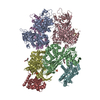
  • Surface view with section colored by density value
  • Surface level: 0.01
  • Imaged by UCSF Chimera
  • Download
  • Surface view colored by cylindrical radius
  • Surface level: 0.01
  • Imaged by UCSF Chimera
  • Download
  • Surface view with fitted model
  • Atomic models: PDB-6w5n
  • Surface level: 0.01
  • Imaged by UCSF Chimera
  • Download
Movie viewer
Structure viewerEM map:
SurfViewMolmilJmol/JSmol
Supplemental images

Downloads & links

-
Map

FileDownload / File: emd_21544.map.gz / Format: CCP4 / Size: 103 MB / Type: IMAGE STORED AS FLOATING POINT NUMBER (4 BYTES)
AnnotationMLL1 in complex with RbBP5, WDR5, SET1, and ASH2L bound to the nucleosome (Class05)
Projections & slices

Image control

Size
Brightness
Contrast
Others
AxesZ (Sec.)Y (Row.)X (Col.)
1 Å/pix.
x 300 pix.
= 300. Å
1 Å/pix.
x 300 pix.
= 300. Å
1 Å/pix.
x 300 pix.
= 300. Å

Surface

Projections

Slices (1/3)

Slices (1/2)

Slices (2/3)

Images are generated by Spider.

Voxel sizeX=Y=Z: 1 Å
Density
Contour LevelBy AUTHOR: 0.0078 / Movie #1: 0.01
Minimum - Maximum-0.017511481 - 0.05429599
Average (Standard dev.)0.00025798933 (±0.0020596227)
SymmetrySpace group: 1
Details

EMDB XML:

Map geometry
Axis orderXYZ
Origin000
Dimensions300300300
Spacing300300300
CellA=B=C: 300.0 Å
α=β=γ: 90.0 °

CCP4 map header:

modeImage stored as Reals
Å/pix. X/Y/Z111
M x/y/z300300300
origin x/y/z0.0000.0000.000
length x/y/z300.000300.000300.000
α/β/γ90.00090.00090.000
MAP C/R/S123
start NC/NR/NS000
NC/NR/NS300300300
D min/max/mean-0.0180.0540.000

-
Supplemental data

-
Sample components

+
Entire : MLL1 in complex with RbBP5, WDR5, SET1, and ASH2L bound to the nu...

EntireName: MLL1 in complex with RbBP5, WDR5, SET1, and ASH2L bound to the nucleosome
Components
  • Complex: MLL1 in complex with RbBP5, WDR5, SET1, and ASH2L bound to the nucleosome
    • Complex: Retinoblastoma-binding protein 5, WD repeat-containing protein 5, Histone-lysine N-methyltransferase 2A, Set1/Ash2 histone methyltransferase complex subunit ASH2
      • Protein or peptide: Retinoblastoma-binding protein 5
      • Protein or peptide: WD repeat-containing protein 5
      • Protein or peptide: Histone-lysine N-methyltransferase 2A
      • Protein or peptide: Set1/Ash2 histone methyltransferase complex subunit ASH2
    • Complex: Histone H3.2, Histone H4, Histone H2A type 1, Histone H2B 1.1
      • Protein or peptide: Histone H3.2
      • Protein or peptide: Histone H4
      • Protein or peptide: Histone H2A type 1
      • Protein or peptide: Histone H2B 1.1
    • Complex: DNA
      • DNA: DNA (147-MER)
      • DNA: DNA (147-MER)

+
Supramolecule #1: MLL1 in complex with RbBP5, WDR5, SET1, and ASH2L bound to the nu...

SupramoleculeName: MLL1 in complex with RbBP5, WDR5, SET1, and ASH2L bound to the nucleosome
type: complex / ID: 1 / Parent: 0 / Macromolecule list: all
Molecular weightTheoretical: 380 KDa

+
Supramolecule #2: Retinoblastoma-binding protein 5, WD repeat-containing protein 5,...

SupramoleculeName: Retinoblastoma-binding protein 5, WD repeat-containing protein 5, Histone-lysine N-methyltransferase 2A, Set1/Ash2 histone methyltransferase complex subunit ASH2
type: complex / ID: 2 / Parent: 1 / Macromolecule list: #1-#4
Source (natural)Organism: Homo sapiens (human)

+
Supramolecule #3: Histone H3.2, Histone H4, Histone H2A type 1, Histone H2B 1.1

SupramoleculeName: Histone H3.2, Histone H4, Histone H2A type 1, Histone H2B 1.1
type: complex / ID: 3 / Parent: 1 / Macromolecule list: #5-#8
Source (natural)Organism: Xenopus laevis (African clawed frog)

+
Supramolecule #4: DNA

SupramoleculeName: DNA / type: complex / ID: 4 / Parent: 1 / Macromolecule list: #9-#10
Source (natural)Organism: synthetic construct (others)

+
Macromolecule #1: Retinoblastoma-binding protein 5

MacromoleculeName: Retinoblastoma-binding protein 5 / type: protein_or_peptide / ID: 1 / Number of copies: 1 / Enantiomer: LEVO
Source (natural)Organism: Homo sapiens (human)
Molecular weightTheoretical: 59.179359 KDa
Recombinant expressionOrganism: Escherichia coli (E. coli)
SequenceString: SNLELLESFG QNYPEEADGT LDCISMALTC TFNRWGTLLA VGCNDGRIVI WDFLTRGIAK IISAHIHPVC SLCWSRDGHK LVSASTDNI VSQWDVLSGD CDQRFRFPSP ILKVQYHPRD QNKVLVCPMK SAPVMLTLSD SKHVVLPVDD DSDLNVVASF D RRGEYIYT ...String:
SNLELLESFG QNYPEEADGT LDCISMALTC TFNRWGTLLA VGCNDGRIVI WDFLTRGIAK IISAHIHPVC SLCWSRDGHK LVSASTDNI VSQWDVLSGD CDQRFRFPSP ILKVQYHPRD QNKVLVCPMK SAPVMLTLSD SKHVVLPVDD DSDLNVVASF D RRGEYIYT GNAKGKILVL KTDSQDLVAS FRVTTGTSNT TAIKSIEFAR KGSCFLINTA DRIIRVYDGR EILTCGRDGE PE PMQKLQD LVNRTPWKKC CFSGDGEYIV AGSARQHALY IWEKSIGNLV KILHGTRGEL LLDVAWHPVR PIIASISSGV VSI WAQNQV ENWSAFAPDF KELDENVEYE ERESEFDIED EDKSEPEQTG ADAAEDEEVD VTSVDPIAAF CSSDEELEDS KALL YLPIA PEVEDPEENP YGPPPDAVQT SLMDEGASSE KKRQSSADGS QPPKKKPKTT NIELQGVPND EVHPLLGVKG DGKSK KKQA GRPKGSKGKE KDSPFKPKLY KGDRGLPLEG SAKGKVQAEL SQPLTAGGAI SELL

UniProtKB: Retinoblastoma-binding protein 5

+
Macromolecule #2: WD repeat-containing protein 5

MacromoleculeName: WD repeat-containing protein 5 / type: protein_or_peptide / ID: 2 / Number of copies: 1 / Enantiomer: LEVO
Source (natural)Organism: Homo sapiens (human)
Molecular weightTheoretical: 34.390992 KDa
Recombinant expressionOrganism: Escherichia coli (E. coli)
SequenceString: SATQSKPTPV KPNYALKFTL AGHTKAVSSV KFSPNGEWLA SSSADKLIKI WGAYDGKFEK TISGHKLGIS DVAWSSDSNL LVSASDDKT LKIWDVSSGK CLKTLKGHSN YVFCCNFNPQ SNLIVSGSFD ESVRIWDVKT GKCLKTLPAH SDPVSAVHFN R DGSLIVSS ...String:
SATQSKPTPV KPNYALKFTL AGHTKAVSSV KFSPNGEWLA SSSADKLIKI WGAYDGKFEK TISGHKLGIS DVAWSSDSNL LVSASDDKT LKIWDVSSGK CLKTLKGHSN YVFCCNFNPQ SNLIVSGSFD ESVRIWDVKT GKCLKTLPAH SDPVSAVHFN R DGSLIVSS SYDGLCRIWD TASGQCLKTL IDDDNPPVSF VKFSPNGKYI LAATLDNTLK LWDYSKGKCL KTYTGHKNEK YC IFANFSV TGGKWIVSGS EDNLVYIWNL QTKEIVQKLQ GHTDVVISTA CHPTENIIAS AALENDKTIK LWKSDC

UniProtKB: WD repeat-containing protein 5

+
Macromolecule #3: Histone-lysine N-methyltransferase 2A

MacromoleculeName: Histone-lysine N-methyltransferase 2A / type: protein_or_peptide / ID: 3 / Number of copies: 1 / Enantiomer: LEVO / EC number: [histone H3]-lysine4 N-trimethyltransferase
Source (natural)Organism: Homo sapiens (human)
Molecular weightTheoretical: 24.141732 KDa
Recombinant expressionOrganism: Escherichia coli (E. coli)
SequenceString: SGSARAEVHL RKSAFDMFNF LASKHRQPPE YNPNDEEEEE VQLKSARRAT SMDLPMPMRF RHLKKTSKEA VGVYRSPIHG RGLFCKRNI DAGEMVIEYA GIVIRSILTD KREKYYDSKG IGCYMFRIDD SEVVDATMHG NAARFINHSC EPNCYSRVIN I DGQKHIVI ...String:
SGSARAEVHL RKSAFDMFNF LASKHRQPPE YNPNDEEEEE VQLKSARRAT SMDLPMPMRF RHLKKTSKEA VGVYRSPIHG RGLFCKRNI DAGEMVIEYA GIVIRSILTD KREKYYDSKG IGCYMFRIDD SEVVDATMHG NAARFINHSC EPNCYSRVIN I DGQKHIVI FAMRKIYRGE ELTYDYKFPI EDASNKLPCN CGAKKCRKFL N

UniProtKB: Histone-lysine N-methyltransferase 2A

+
Macromolecule #4: Set1/Ash2 histone methyltransferase complex subunit ASH2

MacromoleculeName: Set1/Ash2 histone methyltransferase complex subunit ASH2
type: protein_or_peptide / ID: 4 / Number of copies: 1 / Enantiomer: LEVO
Source (natural)Organism: Homo sapiens (human)
Molecular weightTheoretical: 60.244641 KDa
Recombinant expressionOrganism: Escherichia coli (E. coli)
SequenceString: SDTQAGSVDE ENGRQLGEVE LQCGICTKWF TADTFGIDTS SCLPFMTNYS FHCNVCHHSG NTYFLRKQAN LKEMCLSALA NLTWQSRTQ DEHPKTMFSK DKDIIPFIDK YWECMTTRQR PGKMTWPNNI VKTMSKERDV FLVKEHPDPG SKDPEEDYPK F GLLDQDLS ...String:
SDTQAGSVDE ENGRQLGEVE LQCGICTKWF TADTFGIDTS SCLPFMTNYS FHCNVCHHSG NTYFLRKQAN LKEMCLSALA NLTWQSRTQ DEHPKTMFSK DKDIIPFIDK YWECMTTRQR PGKMTWPNNI VKTMSKERDV FLVKEHPDPG SKDPEEDYPK F GLLDQDLS NIGPAYDNQK QSSAVSTSGN LNGGIAAGSS GKGRGAKRKQ QDGGTTGTTK KARSDPLFSA QRLPPHGYPL EH PFNKDGY RYILAEPDPH APDPEKLELD CWAGKPIPGD LYRACLYERV LLALHDRAPQ LKISDDRLTV VGEKGYSMVR ASH GVRKGA WYFEITVDEM PPDTAARLGW SQPLGNLQAP LGYDKFSYSW RSKKGTKFHQ SIGKHYSSGY GQGDVLGFYI NLPE DTETA KSLPDTYKDK ALIKFKSYLY FEEKDFVDKA EKSLKQTPHS EIIFYKNGVN QGVAYKDIFE GVYFPAISLY KSCTV SINF GPCFKYPPKD LTYRPMSDMG WGAVVEHTLA DVLYHVETEV DGRRSPPWEP

UniProtKB: Set1/Ash2 histone methyltransferase complex subunit ASH2

+
Macromolecule #5: Histone H3.2

MacromoleculeName: Histone H3.2 / type: protein_or_peptide / ID: 5 / Number of copies: 2 / Enantiomer: LEVO
Source (natural)Organism: Xenopus laevis (African clawed frog)
Molecular weightTheoretical: 15.435126 KDa
Recombinant expressionOrganism: Escherichia coli (E. coli)
SequenceString:
MARTKQTARK STGGKAPRKQ LATKAARKSA PATGGVKKPH RYRPGTVALR EIRRYQKSTE LLIRKLPFQR LVREIAQDFK TDLRFQSSA VMALQEASEA YLVALFEDTN LCAIHAKRVT IMPKDIQLAR RIRGERA

UniProtKB: Histone H3.2

+
Macromolecule #6: Histone H4

MacromoleculeName: Histone H4 / type: protein_or_peptide / ID: 6 / Number of copies: 2 / Enantiomer: LEVO
Source (natural)Organism: Xenopus laevis (African clawed frog)
Molecular weightTheoretical: 11.394426 KDa
Recombinant expressionOrganism: Escherichia coli (E. coli)
SequenceString:
MSGRGKGGKG LGKGGAKRHR KVLRDNIQGI TKPAIRRLAR RGGVKRISGL IYEETRGVLK VFLENVIRDA VTYTEHAKRK TVTAMDVVY ALKRQGRTLY GFGG

UniProtKB: Histone H4

+
Macromolecule #7: Histone H2A type 1

MacromoleculeName: Histone H2A type 1 / type: protein_or_peptide / ID: 7 / Number of copies: 2 / Enantiomer: LEVO
Source (natural)Organism: Xenopus laevis (African clawed frog)
Molecular weightTheoretical: 13.978241 KDa
Recombinant expressionOrganism: Escherichia coli (E. coli)
SequenceString:
SGRGKQGGKT RAKAKTRSSR AGLQFPVGRV HRLLRKGNYA ERVGAGAPVY LAAVLEYLTA EILELAGNAA RDNKKTRIIP RHLQLAVRN DEELNKLLGR VTIAQGGVLP NIQSVLLPKK TESSKSAKSK

UniProtKB: Histone H2A type 1

+
Macromolecule #8: Histone H2B 1.1

MacromoleculeName: Histone H2B 1.1 / type: protein_or_peptide / ID: 8 / Number of copies: 2 / Enantiomer: LEVO
Source (natural)Organism: Xenopus laevis (African clawed frog)
Molecular weightTheoretical: 13.655948 KDa
Recombinant expressionOrganism: Escherichia coli (E. coli)
SequenceString:
MAKSAPAPKK GSKKAVTKTQ KKDGKKRRKT RKESYAIYVY KVLKQVHPDT GISSKAMSIM NSFVNDVFER IAGEASRLAH YNKRSTITS REIQTAVRLL LPGELAKHAV SEGTKAVTKY TSAK

UniProtKB: Histone H2B 1.1

+
Macromolecule #9: DNA (147-MER)

MacromoleculeName: DNA (147-MER) / type: dna / ID: 9 / Number of copies: 1 / Classification: DNA
Source (natural)Organism: synthetic construct (others)
Molecular weightTheoretical: 45.13877 KDa
SequenceString: (DA)(DT)(DC)(DG)(DA)(DG)(DA)(DA)(DT)(DC) (DC)(DC)(DG)(DG)(DT)(DG)(DC)(DC)(DG)(DA) (DG)(DG)(DC)(DC)(DG)(DC)(DT)(DC)(DA) (DA)(DT)(DT)(DG)(DG)(DT)(DC)(DG)(DT)(DA) (DG) (DA)(DC)(DA)(DG)(DC)(DT) ...String:
(DA)(DT)(DC)(DG)(DA)(DG)(DA)(DA)(DT)(DC) (DC)(DC)(DG)(DG)(DT)(DG)(DC)(DC)(DG)(DA) (DG)(DG)(DC)(DC)(DG)(DC)(DT)(DC)(DA) (DA)(DT)(DT)(DG)(DG)(DT)(DC)(DG)(DT)(DA) (DG) (DA)(DC)(DA)(DG)(DC)(DT)(DC)(DT) (DA)(DG)(DC)(DA)(DC)(DC)(DG)(DC)(DT)(DT) (DA)(DA) (DA)(DC)(DG)(DC)(DA)(DC)(DG) (DT)(DA)(DC)(DG)(DC)(DG)(DC)(DT)(DG)(DT) (DC)(DC)(DC) (DC)(DC)(DG)(DC)(DG)(DT) (DT)(DT)(DT)(DA)(DA)(DC)(DC)(DG)(DC)(DC) (DA)(DA)(DG)(DG) (DG)(DG)(DA)(DT)(DT) (DA)(DC)(DT)(DC)(DC)(DC)(DT)(DA)(DG)(DT) (DC)(DT)(DC)(DC)(DA) (DG)(DG)(DC)(DA) (DC)(DG)(DT)(DG)(DT)(DC)(DA)(DG)(DA)(DT) (DA)(DT)(DA)(DT)(DA)(DC) (DA)(DT)(DC) (DC)(DG)(DA)(DT)

+
Macromolecule #10: DNA (147-MER)

MacromoleculeName: DNA (147-MER) / type: dna / ID: 10 / Number of copies: 1 / Classification: DNA
Source (natural)Organism: synthetic construct (others)
Molecular weightTheoretical: 45.610043 KDa
SequenceString: (DA)(DT)(DC)(DG)(DG)(DA)(DT)(DG)(DT)(DA) (DT)(DA)(DT)(DA)(DT)(DC)(DT)(DG)(DA)(DC) (DA)(DC)(DG)(DT)(DG)(DC)(DC)(DT)(DG) (DG)(DA)(DG)(DA)(DC)(DT)(DA)(DG)(DG)(DG) (DA) (DG)(DT)(DA)(DA)(DT)(DC) ...String:
(DA)(DT)(DC)(DG)(DG)(DA)(DT)(DG)(DT)(DA) (DT)(DA)(DT)(DA)(DT)(DC)(DT)(DG)(DA)(DC) (DA)(DC)(DG)(DT)(DG)(DC)(DC)(DT)(DG) (DG)(DA)(DG)(DA)(DC)(DT)(DA)(DG)(DG)(DG) (DA) (DG)(DT)(DA)(DA)(DT)(DC)(DC)(DC) (DC)(DT)(DT)(DG)(DG)(DC)(DG)(DG)(DT)(DT) (DA)(DA) (DA)(DA)(DC)(DG)(DC)(DG)(DG) (DG)(DG)(DG)(DA)(DC)(DA)(DG)(DC)(DG)(DC) (DG)(DT)(DA) (DC)(DG)(DT)(DG)(DC)(DG) (DT)(DT)(DT)(DA)(DA)(DG)(DC)(DG)(DG)(DT) (DG)(DC)(DT)(DA) (DG)(DA)(DG)(DC)(DT) (DG)(DT)(DC)(DT)(DA)(DC)(DG)(DA)(DC)(DC) (DA)(DA)(DT)(DT)(DG) (DA)(DG)(DC)(DG) (DG)(DC)(DC)(DT)(DC)(DG)(DG)(DC)(DA)(DC) (DC)(DG)(DG)(DG)(DA)(DT) (DT)(DC)(DT) (DC)(DG)(DA)(DT)

-
Experimental details

-
Structure determination

Methodcryo EM
Processingsingle particle reconstruction
Aggregation stateparticle

-
Sample preparation

BufferpH: 7.5
VitrificationCryogen name: ETHANE / Chamber humidity: 100 % / Chamber temperature: 277.15 K

-
Electron microscopy

MicroscopeFEI TITAN KRIOS
Image recordingFilm or detector model: GATAN K2 SUMMIT (4k x 4k) / Average electron dose: 64.0 e/Å2
Electron beamAcceleration voltage: 300 kV / Electron source: FIELD EMISSION GUN
Electron opticsIllumination mode: OTHER / Imaging mode: BRIGHT FIELD
Experimental equipment
Model: Titan Krios / Image courtesy: FEI Company

+
Image processing

Startup modelType of model: EMDB MAP
Final reconstructionApplied symmetry - Point group: C1 (asymmetric) / Resolution.type: BY AUTHOR / Resolution: 6.0 Å / Resolution method: FSC 0.143 CUT-OFF / Number images used: 23236
Initial angle assignmentType: RANDOM ASSIGNMENT
Final angle assignmentType: ANGULAR RECONSTITUTION
FSC plot (resolution estimation)

-
Atomic model buiding 1

RefinementProtocol: RIGID BODY FIT
Output model

PDB-6w5n:
Cryo-EM structure of MLL1 in complex with RbBP5, WDR5, SET1, and ASH2L bound to the nucleosome (Class05)

+
About Yorodumi

-
News

-
Feb 9, 2022. New format data for meta-information of EMDB entries

New format data for meta-information of EMDB entries

  • Version 3 of the EMDB header file is now the official format.
  • The previous official version 1.9 will be removed from the archive.

Related info.:EMDB header

External links:wwPDB to switch to version 3 of the EMDB data model

-
Aug 12, 2020. Covid-19 info

Covid-19 info

URL: https://pdbj.org/emnavi/covid19.php

New page: Covid-19 featured information page in EM Navigator.

Related info.:Covid-19 info / Mar 5, 2020. Novel coronavirus structure data

+
Mar 5, 2020. Novel coronavirus structure data

Novel coronavirus structure data

Related info.:Yorodumi Speices / Aug 12, 2020. Covid-19 info

External links:COVID-19 featured content - PDBj / Molecule of the Month (242):Coronavirus Proteases

+
Jan 31, 2019. EMDB accession codes are about to change! (news from PDBe EMDB page)

EMDB accession codes are about to change! (news from PDBe EMDB page)

  • The allocation of 4 digits for EMDB accession codes will soon come to an end. Whilst these codes will remain in use, new EMDB accession codes will include an additional digit and will expand incrementally as the available range of codes is exhausted. The current 4-digit format prefixed with “EMD-” (i.e. EMD-XXXX) will advance to a 5-digit format (i.e. EMD-XXXXX), and so on. It is currently estimated that the 4-digit codes will be depleted around Spring 2019, at which point the 5-digit format will come into force.
  • The EM Navigator/Yorodumi systems omit the EMD- prefix.

Related info.:Q: What is EMD? / ID/Accession-code notation in Yorodumi/EM Navigator

External links:EMDB Accession Codes are Changing Soon! / Contact to PDBj

+
Jul 12, 2017. Major update of PDB

Major update of PDB

  • wwPDB released updated PDB data conforming to the new PDBx/mmCIF dictionary.
  • This is a major update changing the version number from 4 to 5, and with Remediation, in which all the entries are updated.
  • In this update, many items about electron microscopy experimental information are reorganized (e.g. em_software).
  • Now, EM Navigator and Yorodumi are based on the updated data.

External links:wwPDB Remediation / Enriched Model Files Conforming to OneDep Data Standards Now Available in the PDB FTP Archive

-
Yorodumi

Thousand views of thousand structures

  • Yorodumi is a browser for structure data from EMDB, PDB, SASBDB, etc.
  • This page is also the successor to EM Navigator detail page, and also detail information page/front-end page for Omokage search.
  • The word "yorodu" (or yorozu) is an old Japanese word meaning "ten thousand". "mi" (miru) is to see.

Related info.:EMDB / PDB / SASBDB / Comparison of 3 databanks / Yorodumi Search / Aug 31, 2016. New EM Navigator & Yorodumi / Yorodumi Papers / Jmol/JSmol / Function and homology information / Changes in new EM Navigator and Yorodumi

Read more