 Movie
Movie Controller

[English] 日本語
 Yorodumi
- PDB-6w5i: Cryo-EM structure of MLL1 in complex with RbBP5, WDR5, SET1, and ... -
+ Open data

- Basic information
| Entry | Database: PDB / ID: 6w5i | |||||||||
|---|---|---|---|---|---|---|---|---|---|---|
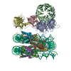
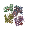
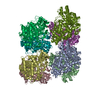
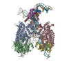
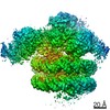
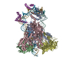
| Title | Cryo-EM structure of MLL1 in complex with RbBP5, WDR5, SET1, and ASH2L bound to the nucleosome (Class01) | |||||||||
 Components Components |
| |||||||||
 Keywords Keywords | TRANSFERASE/STRUCTURAL PROTEIN/DNA / MLL1-NCP / H3K4 methylation / TRANSFERASE / TRANSFERASE-STRUCTURAL PROTEIN-DNA complex | |||||||||
| Function / homology |  Function and homology information Function and homology informationnegative regulation of DNA methylation-dependent heterochromatin formation / protein-cysteine methyltransferase activity / [histone H3]-lysine4 N-methyltransferase / histone H3K4 monomethyltransferase activity / response to potassium ion / histone H3K4 trimethyltransferase activity / unmethylated CpG binding / Phosphorylation of CLOCK, acetylation of BMAL1 (ARNTL) at target gene promoters / histone H3Q5ser reader activity / histone H3K4me1 reader activity ...negative regulation of DNA methylation-dependent heterochromatin formation / protein-cysteine methyltransferase activity / [histone H3]-lysine4 N-methyltransferase / histone H3K4 monomethyltransferase activity / response to potassium ion / histone H3K4 trimethyltransferase activity / unmethylated CpG binding / Phosphorylation of CLOCK, acetylation of BMAL1 (ARNTL) at target gene promoters / histone H3Q5ser reader activity / histone H3K4me1 reader activity / Loss of Function of KMT2D in MLL4 Complex Formation in Kabuki Syndrome / Epigenetic regulation of gene expression by MLL3 and MLL4 complexes / The CRY:PER:kinase complex represses transactivation by the BMAL:CLOCK (ARNTL:CLOCK) complex / MLL3/4 complex / definitive hemopoiesis / Set1C/COMPASS complex / ATAC complex / MLL1/2 complex / NSL complex / regulation of short-term neuronal synaptic plasticity / histone H3K4 methyltransferase activity / anterior/posterior pattern specification / Cardiogenesis / Phosphorylated BMAL1:CLOCK (ARNTL:CLOCK) activates expression of core clock genes / embryonic hemopoiesis / T-helper 2 cell differentiation / Formation of WDR5-containing histone-modifying complexes / histone methyltransferase complex / minor groove of adenine-thymine-rich DNA binding / exploration behavior / MLL1 complex / hemopoiesis / regulation of embryonic development / histone acetyltransferase complex / regulation of cell division / cellular response to transforming growth factor beta stimulus / membrane depolarization / negative regulation of fibroblast proliferation / spleen development / homeostasis of number of cells within a tissue / positive regulation of gluconeogenesis / Transferases; Transferring one-carbon groups; Methyltransferases / transcription initiation-coupled chromatin remodeling / gluconeogenesis / post-embryonic development / skeletal system development / circadian regulation of gene expression / Deactivation of the beta-catenin transactivating complex / Formation of the beta-catenin:TCF transactivating complex / RUNX1 regulates genes involved in megakaryocyte differentiation and platelet function / visual learning / response to estrogen / beta-catenin binding / PKMTs methylate histone lysines / Activation of anterior HOX genes in hindbrain development during early embryogenesis / Transcriptional regulation of granulopoiesis / RMTs methylate histone arginines / structural constituent of chromatin / mitotic spindle / nucleosome / nucleosome assembly / heterochromatin formation / HATs acetylate histones / MLL4 and MLL3 complexes regulate expression of PPARG target genes in adipogenesis and hepatic steatosis / sperm principal piece / RUNX1 regulates transcription of genes involved in differentiation of HSCs / Neddylation / protein-containing complex assembly / fibroblast proliferation / methylation / histone binding / regulation of cell cycle / transcription cis-regulatory region binding / protein heterodimerization activity / apoptotic process / positive regulation of cell population proliferation / chromatin binding / DNA damage response / regulation of transcription by RNA polymerase II / regulation of DNA-templated transcription / positive regulation of DNA-templated transcription / nucleolus / negative regulation of transcription by RNA polymerase II / protein homodimerization activity / positive regulation of transcription by RNA polymerase II / DNA binding / zinc ion binding / nucleoplasm / identical protein binding / nucleus / cytosol Similarity search - Function | |||||||||
| Biological species |  Homo sapiens (human) Homo sapiens (human)synthetic construct (others) | |||||||||
| Method | ELECTRON MICROSCOPY / single particle reconstruction / cryo EM / Resolution: 6.9 Å | |||||||||
 Authors Authors | Park, S.H. / Lee, Y.T. / Ayoub, A. / Dou, Y. / Cho, U. | |||||||||
| Funding support |  United States, 2items United States, 2items
| |||||||||
 Citation Citation | Journal: Nat Commun / Year: 2021 Title: Mechanism for DPY30 and ASH2L intrinsically disordered regions to modulate the MLL/SET1 activity on chromatin. Authors: Young-Tae Lee / Alex Ayoub / Sang-Ho Park / Liang Sha / Jing Xu / Fengbiao Mao / Wei Zheng / Yang Zhang / Uhn-Soo Cho / Yali Dou / Abstract: Recent cryo-EM structures show the highly dynamic nature of the MLL1-NCP (nucleosome core particle) interaction. Functional implication and regulation of such dynamics remain unclear. Here we show ...Recent cryo-EM structures show the highly dynamic nature of the MLL1-NCP (nucleosome core particle) interaction. Functional implication and regulation of such dynamics remain unclear. Here we show that DPY30 and the intrinsically disordered regions (IDRs) of ASH2L work together in restricting the rotational dynamics of the MLL1 complex on the NCP. We show that DPY30 binding to ASH2L leads to stabilization and integration of ASH2L IDRs into the MLL1 complex and establishes new ASH2L-NCP contacts. The significance of ASH2L-DPY30 interactions is demonstrated by requirement of both ASH2L IDRs and DPY30 for dramatic increase of processivity and activity of the MLL1 complex. This DPY30 and ASH2L-IDR dependent regulation is NCP-specific and applies to all members of the MLL/SET1 family of enzymes. We further show that DPY30 is causal for de novo establishment of H3K4me3 in ESCs. Our study provides a paradigm of how H3K4me3 is regulated on chromatin and how H3K4me3 heterogeneity can be modulated by ASH2L IDR interacting proteins. | |||||||||
| History |
|
- Structure visualization
| Movie |
Movie viewer |
|---|---|
| Structure viewer | Molecule: MolmilJmol/JSmol |
- Downloads & links
-Download
| PDBx/mmCIF format | 6w5i.cif.gz | 602.3 KB | Display | PDBx/mmCIF format |
|---|---|---|---|---|
| PDB format | pdb6w5i.ent.gz | 430 KB | Display | PDB format |
| PDBx/mmJSON format | 6w5i.json.gz | Tree view | PDBx/mmJSON format | |
| Others |  Other downloads Other downloads |
-Validation report
| Arichive directory | https://data.pdbj.org/pub/pdb/validation_reports/w5/6w5iftp://data.pdbj.org/pub/pdb/validation_reports/w5/6w5i | HTTPS FTP |
|---|
-Related structure data
| Related structure data |  21542MC 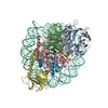 6w5mC  6w5nC M: map data used to model this data C: citing same article ( |
|---|---|
| Similar structure data |


-Links
 PDBj
PDBj


- Assembly
| Deposited unit | 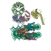
|
|---|---|
| 1 |
|
-Components
-Protein , 8 types, 12 molecules ABCDGKHLIMJN
| #1: Protein | Mass: 59179.359 Da / Num. of mol.: 1 Source method: isolated from a genetically manipulated source Source: (gene. exp.) Homo sapiens (human) / Gene: RBBP5, RBQ3 / Production host:  | ||||||
|---|---|---|---|---|---|---|---|
| #2: Protein | Mass: 34390.992 Da / Num. of mol.: 1 Source method: isolated from a genetically manipulated source Source: (gene. exp.) Homo sapiens (human) / Gene: WDR5, BIG3 / Production host: | ||||||
| #3: Protein | Mass: 24141.732 Da / Num. of mol.: 1 Source method: isolated from a genetically manipulated source Source: (gene. exp.) Homo sapiens (human) / Gene: KMT2A, ALL1, CXXC7, HRX, HTRX, MLL, MLL1, TRX1 / Production host: References: UniProt: Q03164, [histone H3]-lysine4 N-trimethyltransferase | ||||||
| #4: Protein | Mass: 60244.641 Da / Num. of mol.: 1 Source method: isolated from a genetically manipulated source Source: (gene. exp.) Homo sapiens (human) / Gene: ASH2L, ASH2L1 / Production host: | ||||||
| #5: Protein | Mass: 15435.126 Da / Num. of mol.: 2 Source method: isolated from a genetically manipulated source Source: (gene. exp.) #6: Protein | Mass: 11394.426 Da / Num. of mol.: 2 Source method: isolated from a genetically manipulated source Source: (gene. exp.) #7: Protein | Mass: 14250.499 Da / Num. of mol.: 2 Source method: isolated from a genetically manipulated source Source: (gene. exp.) #8: Protein | Mass: 13524.752 Da / Num. of mol.: 2 Source method: isolated from a genetically manipulated source Source: (gene. exp.) |
-DNA chain , 2 types, 2 molecules OP
| #9: DNA chain | Mass: 45138.770 Da / Num. of mol.: 1 / Source method: obtained synthetically / Source: (synth.) synthetic construct (others) |
|---|---|
| #10: DNA chain | Mass: 45610.043 Da / Num. of mol.: 1 / Source method: obtained synthetically / Source: (synth.) synthetic construct (others) |
-Experimental details
-Experiment
| Experiment | Method: ELECTRON MICROSCOPY |
|---|---|
| EM experiment | Aggregation state: PARTICLE / 3D reconstruction method: single particle reconstruction |
- Sample preparation
| Component |
| ||||||||||||||||||||||||||||||
|---|---|---|---|---|---|---|---|---|---|---|---|---|---|---|---|---|---|---|---|---|---|---|---|---|---|---|---|---|---|---|---|
| Molecular weight | Value: 0.38 MDa / Experimental value: NO | ||||||||||||||||||||||||||||||
| Source (natural) |
| ||||||||||||||||||||||||||||||
| Source (recombinant) |
| ||||||||||||||||||||||||||||||
| Buffer solution | pH: 7.5 | ||||||||||||||||||||||||||||||
| Specimen | Embedding applied: NO / Shadowing applied: NO / Staining applied: NO / Vitrification applied: YES | ||||||||||||||||||||||||||||||
| Vitrification | Cryogen name: ETHANE / Humidity: 100 % / Chamber temperature: 277.15 K |
- Electron microscopy imaging
| Experimental equipment |  Model: Titan Krios / Image courtesy: FEI Company |
|---|---|
| Microscopy | Model: FEI TITAN KRIOS |
| Electron gun | Electron source:  FIELD EMISSION GUN / Accelerating voltage: 300 kV / Illumination mode: OTHER FIELD EMISSION GUN / Accelerating voltage: 300 kV / Illumination mode: OTHER |
| Electron lens | Mode: BRIGHT FIELD |
| Image recording | Electron dose: 64 e/Å2 / Film or detector model: GATAN K2 SUMMIT (4k x 4k) |
- Processing
| Software |
| ||||||||||||||||||||||||
|---|---|---|---|---|---|---|---|---|---|---|---|---|---|---|---|---|---|---|---|---|---|---|---|---|---|
| CTF correction | Type: PHASE FLIPPING ONLY | ||||||||||||||||||||||||
| Symmetry | Point symmetry: C1 (asymmetric) | ||||||||||||||||||||||||
| 3D reconstruction | Resolution: 6.9 Å / Resolution method: FSC 0.143 CUT-OFF / Num. of particles: 13086 / Symmetry type: POINT | ||||||||||||||||||||||||
| Atomic model building | Protocol: RIGID BODY FIT | ||||||||||||||||||||||||
| Refinement | Cross valid method: NONE Stereochemistry target values: GeoStd + Monomer Library + CDL v1.2 | ||||||||||||||||||||||||
| Displacement parameters | Biso mean: 355.17 Å2 | ||||||||||||||||||||||||
| Refine LS restraints |
|
