- EMDB-12697: Structure of a Minimal Photosystem I -
+
Open data
ID or keywords:
Loading...
-
Basic information
Entry
Database: EMDB / ID: EMD-12697
Title
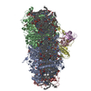
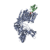
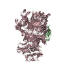
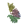
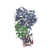
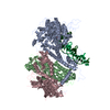
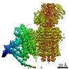
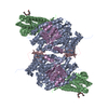
Structure of a Minimal Photosystem I
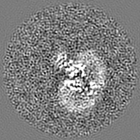
Map data
Sample
Complex: Minimal Photosystem I
Protein or peptide: x 7 types
Ligand: x 8 types
Keywords
Photosystem I / excitation energy transfer / cyanobacteria / minimal structure / PHOTOSYNTHESIS
Function / homology
Function and homology information
photosystem I reaction center / photosystem I / photosynthetic electron transport in photosystem I / photosystem I / chlorophyll binding / plasma membrane-derived thylakoid membrane / photosynthesis / 4 iron, 4 sulfur cluster binding / electron transfer activity / oxidoreductase activity ...photosystem I reaction center / photosystem I / photosynthetic electron transport in photosystem I / photosystem I / chlorophyll binding / plasma membrane-derived thylakoid membrane / photosynthesis / 4 iron, 4 sulfur cluster binding / electron transfer activity / oxidoreductase activity / magnesium ion binding / metal ion binding / plasma membrane Similarity search - Function
Photosystem I reaction center subunit PsaK / Photosystem I reaction centre subunit PsaK / Photosystem I PsaM, reaction centre superfamily / Photosystem I PsaM, reaction centre / Photosystem I protein M (PsaM) / Photosystem I reaction centre subunit PsaK superfamily / Photosystem I psaG and psaK proteins signature. / Photosystem I reaction center subunit V/PsaK / Photosystem I psaG / psaK / Photosystem I PsaD ...Photosystem I reaction center subunit PsaK / Photosystem I reaction centre subunit PsaK / Photosystem I PsaM, reaction centre superfamily / Photosystem I PsaM, reaction centre / Photosystem I protein M (PsaM) / Photosystem I reaction centre subunit PsaK superfamily / Photosystem I psaG and psaK proteins signature. / Photosystem I reaction center subunit V/PsaK / Photosystem I psaG / psaK / Photosystem I PsaD / Photosystem I, reaction centre subunit PsaD superfamily / PsaD / Photosystem I PsaE, reaction centre subunit IV / Photosystem I reaction centre subunit IV / PsaE / : / Photosystem I protein PsaC / Photosystem I PsaA / Photosystem I PsaB / Photosystem I PsaA/PsaB, conserved site / Photosystem I psaA and psaB proteins signature. / Photosystem I PsaA/PsaB / Photosystem I PsaA/PsaB superfamily / Photosystem I psaA/psaB protein / Electron transport accessory-like domain superfamily / 4Fe-4S dicluster domain / 4Fe-4S ferredoxin, iron-sulphur binding, conserved site / 4Fe-4S ferredoxin-type iron-sulfur binding region signature. / 4Fe-4S ferredoxin-type iron-sulfur binding domain profile. / 4Fe-4S ferredoxin-type, iron-sulphur binding domain Similarity search - Domain/homology
Photosystem I reaction center subunit IV / Photosystem I reaction center subunit II / Photosystem I P700 chlorophyll a apoprotein A1 / Photosystem I P700 chlorophyll a apoprotein A2 / Photosystem I iron-sulfur center / Photosystem I reaction center subunit XII / Photosystem I reaction center subunit PsaK 2 Similarity search - Component
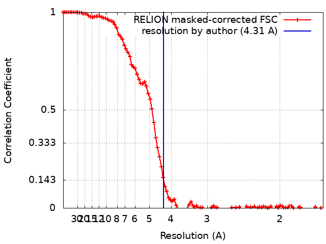
Journal: J Am Chem Soc / Year: 2021 Title: Two-Dimensional Electronic Spectroscopy of a Minimal Photosystem I Complex Reveals the Rate of Primary Charge Separation. Authors: Parveen Akhtar / Ido Caspy / Paweł J Nowakowski / Tirupathi Malavath / Nathan Nelson / Howe-Siang Tan / Petar H Lambrev / Abstract: Photosystem I (PSI), found in all oxygenic photosynthetic organisms, uses solar energy to drive electron transport with nearly 100% quantum efficiency, thanks to fast energy transfer among antenna ...Photosystem I (PSI), found in all oxygenic photosynthetic organisms, uses solar energy to drive electron transport with nearly 100% quantum efficiency, thanks to fast energy transfer among antenna chlorophylls and charge separation in the reaction center. There is no complete consensus regarding the kinetics of the elementary steps involved in the overall trapping, especially the rate of primary charge separation. In this work, we employed two-dimensional coherent electronic spectroscopy to follow the dynamics of energy and electron transfer in a monomeric PSI complex from PCC 6803, containing only subunits A-E, K, and M, at 77 K. We also determined the structure of the complex to 4.3 Å resolution by cryoelectron microscopy with refinements to 2.5 Å. We applied structure-based modeling using a combined Redfield-Förster theory to compute the excitation dynamics. The absorptive 2D electronic spectra revealed fast excitonic/vibronic relaxation on time scales of 50-100 fs from the high-energy side of the absorption spectrum. Antenna excitations were funneled within 1 ps to a small pool of chlorophylls absorbing around 687 nm, thereafter decaying with 4-20 ps lifetimes, independently of excitation wavelength. Redfield-Förster energy transfer computations showed that the kinetics is limited by transfer from these red-shifted pigments. The rate of primary charge separation, upon direct excitation of the reaction center, was determined to be 1.2-1.5 ps. This result implies activationless electron transfer in PSI.
History
Deposition
Mar 30, 2021
-
Header (metadata) release
Sep 1, 2021
-
Map release
Sep 1, 2021
-
Update
Jul 10, 2024
-
Current status
Jul 10, 2024
Processing site: PDBe / Status: Released
-
Structure visualization
Movie
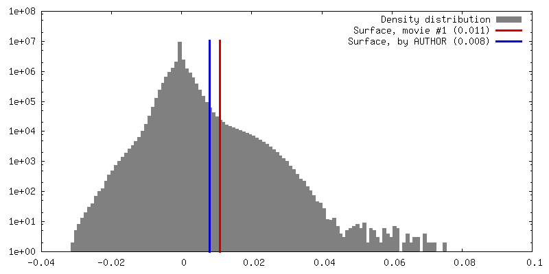
Surface view with section colored by density value
In the structure databanks used in Yorodumi, some data are registered as the other names, "COVID-19 virus" and "2019-nCoV". Here are the details of the virus and the list of structure data.
Jan 31, 2019. EMDB accession codes are about to change! (news from PDBe EMDB page)
EMDB accession codes are about to change! (news from PDBe EMDB page)
The allocation of 4 digits for EMDB accession codes will soon come to an end. Whilst these codes will remain in use, new EMDB accession codes will include an additional digit and will expand incrementally as the available range of codes is exhausted. The current 4-digit format prefixed with “EMD-” (i.e. EMD-XXXX) will advance to a 5-digit format (i.e. EMD-XXXXX), and so on. It is currently estimated that the 4-digit codes will be depleted around Spring 2019, at which point the 5-digit format will come into force.
The EM Navigator/Yorodumi systems omit the EMD- prefix.
Related info.:Q: What is EMD? / ID/Accession-code notation in Yorodumi/EM Navigator
Yorodumi is a browser for structure data from EMDB, PDB, SASBDB, etc.
This page is also the successor to EM Navigator detail page, and also detail information page/front-end page for Omokage search.
The word "yorodu" (or yorozu) is an old Japanese word meaning "ten thousand". "mi" (miru) is to see.
Related info.:EMDB / PDB / SASBDB / Comparison of 3 databanks / Yorodumi Search / Aug 31, 2016. New EM Navigator & Yorodumi / Yorodumi Papers / Jmol/JSmol / Function and homology information / Changes in new EM Navigator and Yorodumi
 Movie
Movie Controller


 Open data
 Basic information
 Map data
 Sample
 Keywords
 Function and homology information
 Authors
 Israel, 1 items
  Citation

 Structure visualization
 Downloads & links
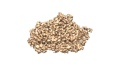 emd_12697.png
 http://ftp.pdbj.org/pub/emdb/structures/EMD-12697


 Z (Sec.)
 Y (Row.)
 X (Col.)


 Sample components


 Processing
 Electron microscopy
 FIELD EMISSION GUN