 Movie
Movie Controller


+ Open data

- Basic information
| Entry | Database: PDB / ID: 7o1v | ||||||
|---|---|---|---|---|---|---|---|
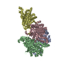
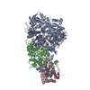
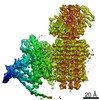
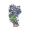
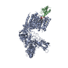
| Title | Structure of a Minimal Photosystem I | ||||||
 Components Components |
| ||||||
 Keywords Keywords | PHOTOSYNTHESIS / Photosystem I / excitation energy transfer / cyanobacteria / minimal structure | ||||||
| Function / homology |  Function and homology information Function and homology informationphotosystem I reaction center / photosystem I / photosynthetic electron transport in photosystem I / photosystem I / plasma membrane-derived thylakoid membrane / chlorophyll binding / photosynthesis / 4 iron, 4 sulfur cluster binding / electron transfer activity / oxidoreductase activity ...photosystem I reaction center / photosystem I / photosynthetic electron transport in photosystem I / photosystem I / plasma membrane-derived thylakoid membrane / chlorophyll binding / photosynthesis / 4 iron, 4 sulfur cluster binding / electron transfer activity / oxidoreductase activity / magnesium ion binding / metal ion binding / plasma membrane Similarity search - Function | ||||||
| Biological species |  | ||||||
| Method | ELECTRON MICROSCOPY / single particle reconstruction / cryo EM / Resolution: 4.31 Å | ||||||
 Authors Authors | Nelson, N. / Caspy, I. / Lambrev, P. | ||||||
| Funding support |  Israel, 1items Israel, 1items
| ||||||
 Citation Citation | Journal: J Am Chem Soc / Year: 2021 Title: Two-Dimensional Electronic Spectroscopy of a Minimal Photosystem I Complex Reveals the Rate of Primary Charge Separation. Authors: Parveen Akhtar / Ido Caspy / Paweł J Nowakowski / Tirupathi Malavath / Nathan Nelson / Howe-Siang Tan / Petar H Lambrev /   Abstract: Photosystem I (PSI), found in all oxygenic photosynthetic organisms, uses solar energy to drive electron transport with nearly 100% quantum efficiency, thanks to fast energy transfer among antenna ...Photosystem I (PSI), found in all oxygenic photosynthetic organisms, uses solar energy to drive electron transport with nearly 100% quantum efficiency, thanks to fast energy transfer among antenna chlorophylls and charge separation in the reaction center. There is no complete consensus regarding the kinetics of the elementary steps involved in the overall trapping, especially the rate of primary charge separation. In this work, we employed two-dimensional coherent electronic spectroscopy to follow the dynamics of energy and electron transfer in a monomeric PSI complex from PCC 6803, containing only subunits A-E, K, and M, at 77 K. We also determined the structure of the complex to 4.3 Å resolution by cryoelectron microscopy with refinements to 2.5 Å. We applied structure-based modeling using a combined Redfield-Förster theory to compute the excitation dynamics. The absorptive 2D electronic spectra revealed fast excitonic/vibronic relaxation on time scales of 50-100 fs from the high-energy side of the absorption spectrum. Antenna excitations were funneled within 1 ps to a small pool of chlorophylls absorbing around 687 nm, thereafter decaying with 4-20 ps lifetimes, independently of excitation wavelength. Redfield-Förster energy transfer computations showed that the kinetics is limited by transfer from these red-shifted pigments. The rate of primary charge separation, upon direct excitation of the reaction center, was determined to be 1.2-1.5 ps. This result implies activationless electron transfer in PSI. | ||||||
| History |
|
- Structure visualization
| Movie |
Movie viewer |
|---|---|
| Structure viewer | Molecule: MolmilJmol/JSmol |
- Downloads & links
-Download
| PDBx/mmCIF format | 7o1v.cif.gz | 597.4 KB | Display | PDBx/mmCIF format |
|---|---|---|---|---|
| PDB format | pdb7o1v.ent.gz | 485.6 KB | Display | PDB format |
| PDBx/mmJSON format | 7o1v.json.gz | Tree view | PDBx/mmJSON format | |
| Others |  Other downloads Other downloads |
-Validation report
| Arichive directory | https://data.pdbj.org/pub/pdb/validation_reports/o1/7o1vftp://data.pdbj.org/pub/pdb/validation_reports/o1/7o1v | HTTPS FTP |
|---|
-Related structure data
| Related structure data | 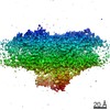 12697MC M: map data used to model this data C: citing same article ( |
|---|---|
| Similar structure data |


-Links
 PDBj
PDBj


- Assembly
| Deposited unit | 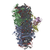
|
|---|---|
| 1 |
|
-Components
-Photosystem I P700 chlorophyll a apoprotein ... , 2 types, 2 molecules AB
| #1: Protein | Mass: 81722.875 Da / Num. of mol.: 1 / Source method: isolated from a natural source Source: (natural) Strain: PCC 6803 / Kazusa / References: UniProt: P29254, photosystem I |
|---|---|
| #2: Protein | Mass: 81167.258 Da / Num. of mol.: 1 / Source method: isolated from a natural source Source: (natural) Strain: PCC 6803 / Kazusa / References: UniProt: P29255, photosystem I |
-Protein , 1 types, 1 molecules C
| #3: Protein | Mass: 8706.064 Da / Num. of mol.: 1 / Source method: isolated from a natural source Source: (natural) Strain: PCC 6803 / Kazusa / References: UniProt: P32422, photosystem I |
|---|
-Photosystem I reaction center subunit ... , 4 types, 4 molecules DEKM
| #4: Protein | Mass: 15663.749 Da / Num. of mol.: 1 / Source method: isolated from a natural source Source: (natural) Strain: PCC 6803 / Kazusa / References: UniProt: P19569 |
|---|---|
| #5: Protein | Mass: 7680.478 Da / Num. of mol.: 1 / Source method: isolated from a natural source Source: (natural) Strain: PCC 6803 / Kazusa / References: UniProt: P12975 |
| #6: Protein | Mass: 8237.783 Da / Num. of mol.: 1 / Source method: isolated from a natural source Source: (natural) Strain: PCC 6803 / Kazusa / References: UniProt: P74564 |
| #7: Protein/peptide | Mass: 3382.063 Da / Num. of mol.: 1 / Source method: isolated from a natural source Source: (natural) Strain: PCC 6803 / Kazusa / References: UniProt: P72986 |
-Non-polymers , 8 types, 120 molecules 


| #8: Chemical | ChemComp-CLA /  Mass: 893.489 Da / Num. of mol.: 88 / Source method: obtained synthetically / Formula: C55H72MgN4O5 Mass: 893.489 Da / Num. of mol.: 88 / Source method: obtained synthetically / Formula: C55H72MgN4O5#9: Chemical |  Mass: 450.696 Da / Num. of mol.: 2 / Source method: obtained synthetically / Formula: C31H46O2 Mass: 450.696 Da / Num. of mol.: 2 / Source method: obtained synthetically / Formula: C31H46O2#10: Chemical | ChemComp-BCR /  Mass: 536.873 Da / Num. of mol.: 15 / Source method: obtained synthetically / Formula: C40H56 Mass: 536.873 Da / Num. of mol.: 15 / Source method: obtained synthetically / Formula: C40H56#11: Chemical | ChemComp-LHG /  Mass: 722.970 Da / Num. of mol.: 5 / Source method: obtained synthetically / Formula: C38H75O10P / Comment: phospholipid*YM Mass: 722.970 Da / Num. of mol.: 5 / Source method: obtained synthetically / Formula: C38H75O10P / Comment: phospholipid*YM#12: Chemical | ChemComp-LMG /  Mass: 787.158 Da / Num. of mol.: 4 / Source method: obtained synthetically / Formula: C45H86O10 Mass: 787.158 Da / Num. of mol.: 4 / Source method: obtained synthetically / Formula: C45H86O10#13: Chemical |  Mass: 351.640 Da / Num. of mol.: 3 / Source method: obtained synthetically / Formula: Fe4S4 Mass: 351.640 Da / Num. of mol.: 3 / Source method: obtained synthetically / Formula: Fe4S4#14: Chemical |  Mass: 550.856 Da / Num. of mol.: 2 / Source method: obtained synthetically / Formula: C40H54O Mass: 550.856 Da / Num. of mol.: 2 / Source method: obtained synthetically / Formula: C40H54O#15: Chemical | ChemComp-SQD / |  Mass: 795.116 Da / Num. of mol.: 1 / Source method: obtained synthetically / Formula: C41H78O12S Mass: 795.116 Da / Num. of mol.: 1 / Source method: obtained synthetically / Formula: C41H78O12S |
|---|
-Details
| Has ligand of interest | N |
|---|
-Experimental details
-Experiment
| Experiment | Method: ELECTRON MICROSCOPY |
|---|---|
| EM experiment | Aggregation state: PARTICLE / 3D reconstruction method: single particle reconstruction |
- Sample preparation
| Component | Name: Minimal Photosystem I / Type: COMPLEX / Entity ID: #1-#7 / Source: NATURAL |
|---|---|
| Molecular weight | Experimental value: NO |
| Source (natural) | Organism: |
| Buffer solution | pH: 6.5 |
| Specimen | Embedding applied: NO / Shadowing applied: NO / Staining applied: NO / Vitrification applied: YES |
| Vitrification | Cryogen name: ETHANE |
- Electron microscopy imaging
| Experimental equipment |  Model: Titan Krios / Image courtesy: FEI Company |
|---|---|
| Microscopy | Model: FEI TITAN KRIOS |
| Electron gun | Electron source:  FIELD EMISSION GUN / Accelerating voltage: 300 kV / Illumination mode: FLOOD BEAM FIELD EMISSION GUN / Accelerating voltage: 300 kV / Illumination mode: FLOOD BEAM |
| Electron lens | Mode: BRIGHT FIELD |
| Image recording | Electron dose: 43.6 e/Å2 / Film or detector model: GATAN K3 (6k x 4k) / Num. of real images: 2493 |
| EM imaging optics | Phase plate: VOLTA PHASE PLATE |
- Processing
| Software |
| ||||||||||||||||||||||||
|---|---|---|---|---|---|---|---|---|---|---|---|---|---|---|---|---|---|---|---|---|---|---|---|---|---|
| EM software | Name: RELION / Category: particle selection | ||||||||||||||||||||||||
| CTF correction | Type: NONE | ||||||||||||||||||||||||
| Particle selection | Num. of particles selected: 286538 | ||||||||||||||||||||||||
| Symmetry | Point symmetry: C1 (asymmetric) | ||||||||||||||||||||||||
| 3D reconstruction | Resolution: 4.31 Å / Resolution method: FSC 0.143 CUT-OFF / Num. of particles: 74303 / Symmetry type: POINT | ||||||||||||||||||||||||
| Refinement | Cross valid method: NONE Stereochemistry target values: GeoStd + Monomer Library + CDL v1.2 | ||||||||||||||||||||||||
| Displacement parameters | Biso mean: 85.65 Å2 | ||||||||||||||||||||||||
| Refine LS restraints |
|
