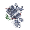
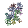
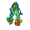
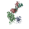
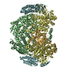
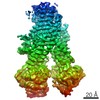
Entry Database : PDB / ID : 6skeTitle Teneurin 2 in complex with Latrophilin 2 Lec domain Adhesion G protein-coupled receptor L2 Teneurin-2 Keywords / / / / / / / / / / Function / homology Function Domain/homology Component
/ / / / / / / / / / / / / / / / / / / / / / / / / / / / / / / / / / / / / / / / / / / / / / / / / / / / / / / / / / / / / / / / / / / / / / / / / / / / / / / / / / / / / / / / / / / / / / / / / / / / / / / / / / / / / / / / / / Biological species Gallus gallus (chicken)Mus musculus (house mouse)Method / / / Resolution : 3.62 Å Authors Shahin, M. / Jackson, V.A. / Carrasquero, M. / Lowe, E. / Seiradake, E. Journal : Cell / Year : 2020Title : Structural Basis of Teneurin-Latrophilin Interaction in Repulsive Guidance of Migrating Neurons.Authors: Del Toro, D. / Carrasquero-Ordaz, M.A. / Chu, A. / Ruff, T. / Shahin, M. / Jackson, V.A. / Chavent, M. / Berbeira-Santana, M. / Seyit-Bremer, G. / Brignani, S. / Kaufmann, R. / Lowe, E. / ... Authors : Del Toro, D. / Carrasquero-Ordaz, M.A. / Chu, A. / Ruff, T. / Shahin, M. / Jackson, V.A. / Chavent, M. / Berbeira-Santana, M. / Seyit-Bremer, G. / Brignani, S. / Kaufmann, R. / Lowe, E. / Klein, R. / Seiradake, E. History Deposition Aug 15, 2019 Deposition site / Processing site Revision 1.0 Feb 12, 2020 Provider / Type Revision 2.0 Jul 29, 2020 Group Atomic model / Data collection ... Atomic model / Data collection / Derived calculations / Structure summary Category atom_site / atom_site_anisotrop ... atom_site / atom_site_anisotrop / chem_comp / entity / pdbx_branch_scheme / pdbx_chem_comp_identifier / pdbx_entity_branch / pdbx_entity_branch_descriptor / pdbx_entity_branch_link / pdbx_entity_branch_list / pdbx_entity_nonpoly / pdbx_nonpoly_scheme / pdbx_struct_assembly_gen / struct_asym / struct_conn / struct_site / struct_site_gen Item _atom_site.B_iso_or_equiv / _atom_site.Cartn_x ... _atom_site.B_iso_or_equiv / _atom_site.Cartn_x / _atom_site.Cartn_y / _atom_site.Cartn_z / _atom_site.auth_asym_id / _atom_site.auth_atom_id / _atom_site.auth_comp_id / _atom_site.auth_seq_id / _atom_site.label_asym_id / _atom_site.label_atom_id / _atom_site.label_comp_id / _atom_site.label_entity_id / _atom_site.type_symbol / _atom_site_anisotrop.U[1][1] / _atom_site_anisotrop.U[1][2] / _atom_site_anisotrop.U[1][3] / _atom_site_anisotrop.U[2][2] / _atom_site_anisotrop.U[2][3] / _atom_site_anisotrop.U[3][3] / _atom_site_anisotrop.pdbx_auth_asym_id / _atom_site_anisotrop.pdbx_auth_atom_id / _atom_site_anisotrop.pdbx_auth_comp_id / _atom_site_anisotrop.pdbx_auth_seq_id / _atom_site_anisotrop.pdbx_label_asym_id / _atom_site_anisotrop.pdbx_label_atom_id / _atom_site_anisotrop.pdbx_label_comp_id / _atom_site_anisotrop.type_symbol / _chem_comp.name / _entity.formula_weight / _entity.pdbx_description / _entity.pdbx_number_of_molecules / _entity.type / _pdbx_struct_assembly_gen.asym_id_list / _struct_conn.pdbx_dist_value / _struct_conn.pdbx_role / _struct_conn.ptnr1_auth_asym_id / _struct_conn.ptnr1_auth_seq_id / _struct_conn.ptnr1_label_asym_id / _struct_conn.ptnr1_label_atom_id / _struct_conn.ptnr2_auth_asym_id / _struct_conn.ptnr2_auth_seq_id / _struct_conn.ptnr2_label_asym_id Description / Provider / Type Revision 2.1 Nov 20, 2024 Group Data collection / Database references ... Data collection / Database references / Derived calculations / Structure summary Category chem_comp / chem_comp_atom ... chem_comp / chem_comp_atom / chem_comp_bond / database_2 / pdbx_entry_details / pdbx_modification_feature / struct_conn Item _chem_comp.pdbx_synonyms / _database_2.pdbx_DOI ... _chem_comp.pdbx_synonyms / _database_2.pdbx_DOI / _database_2.pdbx_database_accession / _pdbx_entry_details.has_protein_modification / _struct_conn.pdbx_leaving_atom_flag
Show all Show less
 Movie
Movie Controller


 Open data
 Basic information
 Components
 Keywords
 Function and homology information

 X-RAY DIFFRACTION /
  Authors
 Citation
 Structure visualization
 Downloads & links
 Other downloads


 PDBj
PDBj


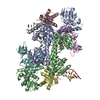
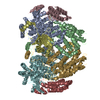
 Assembly
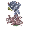

 Homo sapiens (human) / References: UniProt: Q9DER5
 Type: D-saccharide, beta linking / Mass: 221.208 Da / Num. of mol.: 8
 Sample preparation
 / Beamline: I03 / Wavelength: 0.97956 Å
 Processing