Movie
Movie Controller

[English] 日本語
 Yorodumi
- EMDB-12104: Cryo-EM structure of the dihydrolipoyl transacetylase cubic core ... -
+ Open data

- Basic information
| Entry | Database: EMDB / ID: EMD-12104 | |||||||||
|---|---|---|---|---|---|---|---|---|---|---|
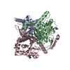
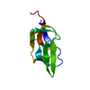
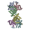
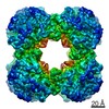
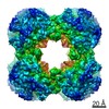
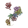
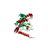
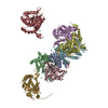
| Title | Cryo-EM structure of the dihydrolipoyl transacetylase cubic core of the E. coli pyruvate dehydrogenase complex including lipoyl domains | |||||||||
 Map data Map data | ||||||||||
 Sample Sample |
| |||||||||
| Function / homology |  Function and homology information Function and homology informationpyruvate catabolic process / dihydrolipoyllysine-residue acetyltransferase / dihydrolipoyllysine-residue acetyltransferase activity / acetyl-CoA biosynthetic process from pyruvate / lipoic acid binding / pyruvate dehydrogenase complex / pyruvate metabolic process / acetyltransferase activity / glycolytic process / cytoplasm Similarity search - Function | |||||||||
| Biological species |  | |||||||||
| Method | single particle reconstruction / cryo EM / Resolution: 3.16 Å | |||||||||
 Authors Authors | Skerlova J / Stenmark P | |||||||||
| Funding support |  Sweden, European Union, 2 items Sweden, European Union, 2 items
| |||||||||
 Citation Citation | Journal: Nat Commun / Year: 2021 Title: Structure of the native pyruvate dehydrogenase complex reveals the mechanism of substrate insertion. Authors: Jana Škerlová / Jens Berndtsson / Hendrik Nolte / Martin Ott / Pål Stenmark /  Abstract: The pyruvate dehydrogenase complex (PDHc) links glycolysis to the citric acid cycle by converting pyruvate into acetyl-coenzyme A. PDHc encompasses three enzymatically active subunits, namely ...The pyruvate dehydrogenase complex (PDHc) links glycolysis to the citric acid cycle by converting pyruvate into acetyl-coenzyme A. PDHc encompasses three enzymatically active subunits, namely pyruvate dehydrogenase, dihydrolipoyl transacetylase, and dihydrolipoyl dehydrogenase. Dihydrolipoyl transacetylase is a multidomain protein comprising a varying number of lipoyl domains, a peripheral subunit-binding domain, and a catalytic domain. It forms the structural core of the complex, provides binding sites for the other enzymes, and shuffles reaction intermediates between the active sites through covalently bound lipoyl domains. The molecular mechanism by which this shuttling occurs has remained elusive. Here, we report a cryo-EM reconstruction of the native E. coli dihydrolipoyl transacetylase core in a resting state. This structure provides molecular details of the assembly of the core and reveals how the lipoyl domains interact with the core at the active site. | |||||||||
| History |
|
- Structure visualization
| Movie |
Movie viewer |
|---|---|
| Structure viewer | EM map: SurfViewMolmilJmol/JSmol |
| Supplemental images |

- Downloads & links
-EMDB archive
| Map data | emd_12104.map.gz | 97.3 MB | EMDB map data format | |
|---|---|---|---|---|
| Header (meta data) | emd-12104-v30.xmlemd-12104.xml | 24.1 KB 24.1 KB | Display Display | EMDB header |
| FSC (resolution estimation) | emd_12104_fsc.xml | 10.5 KB | Display | FSC data file |
| Images | 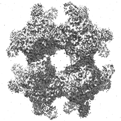 emd_12104.png emd_12104.png | 90.7 KB | ||
| Masks | emd_12104_msk_1.map | 103 MB | Mask map | |
| Others | emd_12104_half_map_1.map.gzemd_12104_half_map_2.map.gz | 95.5 MB 95.5 MB | ||
| Archive directory |  http://ftp.pdbj.org/pub/emdb/structures/EMD-12104ftp://ftp.pdbj.org/pub/emdb/structures/EMD-12104 http://ftp.pdbj.org/pub/emdb/structures/EMD-12104ftp://ftp.pdbj.org/pub/emdb/structures/EMD-12104 | HTTPS FTP |
-Validation report
| Summary document | emd_12104_validation.pdf.gz | 570.1 KB | Display | EMDB validaton report |
|---|---|---|---|---|
| Full document | emd_12104_full_validation.pdf.gz | 569.7 KB | Display | |
| Data in XML | emd_12104_validation.xml.gz | 18.2 KB | Display | |
| Data in CIF | emd_12104_validation.cif.gz | 23.3 KB | Display | |
| Arichive directory | https://ftp.pdbj.org/pub/emdb/validation_reports/EMD-12104ftp://ftp.pdbj.org/pub/emdb/validation_reports/EMD-12104 | HTTPS FTP |
-Related structure data
| Related structure data | 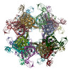 7b9kMC M: atomic model generated by this map C: citing same article ( |
|---|---|
| Similar structure data |


-Links
| EMDB pages | EMDB (EBI/PDBe) / EMDataResource |
|---|---|
| Related items in Molecule of the Month |


-Map
| File | Download / File: emd_12104.map.gz / Format: CCP4 / Size: 103 MB / Type: IMAGE STORED AS FLOATING POINT NUMBER (4 BYTES) | ||||||||||||||||||||||||||||||||||||||||||||||||||||||||||||
|---|---|---|---|---|---|---|---|---|---|---|---|---|---|---|---|---|---|---|---|---|---|---|---|---|---|---|---|---|---|---|---|---|---|---|---|---|---|---|---|---|---|---|---|---|---|---|---|---|---|---|---|---|---|---|---|---|---|---|---|---|---|
| Voxel size | X=Y=Z: 1.04 Å | ||||||||||||||||||||||||||||||||||||||||||||||||||||||||||||
| Density |
| ||||||||||||||||||||||||||||||||||||||||||||||||||||||||||||
| Symmetry | Space group: 1 | ||||||||||||||||||||||||||||||||||||||||||||||||||||||||||||
| Details | EMDB XML:
CCP4 map header:
| ||||||||||||||||||||||||||||||||||||||||||||||||||||||||||||
-Supplemental data
-Mask #1
| File | emd_12104_msk_1.map | ||||||||||||
|---|---|---|---|---|---|---|---|---|---|---|---|---|---|
| Projections & Slices |
| ||||||||||||
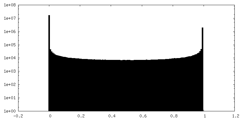
| Density Histograms |
 Z
Z Y
Y X
X


-Half map: #2
| File | emd_12104_half_map_1.map | ||||||||||||
|---|---|---|---|---|---|---|---|---|---|---|---|---|---|
| Projections & Slices |
| ||||||||||||
| Density Histograms |


-Half map: #1
| File | emd_12104_half_map_2.map | ||||||||||||
|---|---|---|---|---|---|---|---|---|---|---|---|---|---|
| Projections & Slices |
| ||||||||||||
| Density Histograms |


- Sample components
-Entire : dihydrolipoyl transacetylase cubic core of the pyruvate dehydroge...
| Entire | Name: dihydrolipoyl transacetylase cubic core of the pyruvate dehydrogenase complex |
|---|---|
| Components |
|
-Supramolecule #1: dihydrolipoyl transacetylase cubic core of the pyruvate dehydroge...
| Supramolecule | Name: dihydrolipoyl transacetylase cubic core of the pyruvate dehydrogenase complex type: complex / ID: 1 / Parent: 0 / Macromolecule list: all |
|---|---|
| Source (natural) | Organism: |
-Macromolecule #1: Dihydrolipoyllysine-residue acetyltransferase component of pyruva...
| Macromolecule | Name: Dihydrolipoyllysine-residue acetyltransferase component of pyruvate dehydrogenase complex type: protein_or_peptide / ID: 1 / Details: Lysine 248 covalently modified by dihydrolipoate / Number of copies: 24 / Enantiomer: LEVO / EC number: dihydrolipoyllysine-residue acetyltransferase |
|---|---|
| Source (natural) | Organism: |
| Molecular weight | Theoretical: 66.363203 KDa |
| Sequence | String: MAIEIKVPDI GADEVEITEI LVKVGDKVEA EQSLITVEGD KASMEVPSPQ AGIVKEIKVS VGDKTQTGAL IMIFDSADGA ADAAPAQAE EKKEAAPAAA PAAAAAKDVN VPDIGSDEVE VTEILVKVGD KVEAEQSLIT VEGDKASMEV PAPFAGTVKE I KVNVGDKV ...String: MAIEIKVPDI GADEVEITEI LVKVGDKVEA EQSLITVEGD KASMEVPSPQ AGIVKEIKVS VGDKTQTGAL IMIFDSADGA ADAAPAQAE EKKEAAPAAA PAAAAAKDVN VPDIGSDEVE VTEILVKVGD KVEAEQSLIT VEGDKASMEV PAPFAGTVKE I KVNVGDKV STGSLIMVFE VAGEAGAAAP AAKQEAAPAA APAPAAGVKE VNVPDIGGDE VEVTEVMVKV GDKVAAEQSL IT VEGD(LA2)AS MEVPAPFAGV VKELKVNVGD KVKTGSLIMI FEVEGAAPAA APAKQEAAAP APAAKAEAPA AAPAAKAEG KSEFAENDAY VHATPLIRRL AREFGVNLAK VKGTGRKGRI LREDVQAYVK EAIKRAEAAP AATGGGIPGM LPWPKVDFSK FGEIEEVEL GRIQKISGAN LSRNWVMIPH VTHFDKTDIT ELEAFRKQQN EEAAKRKLDV KITPVVFIMK AVAAALEQMP R FNSSLSED GQRLTLKKYI NIGVAVDTPN GLVVPVFKDV NKKGIIELSR ELMTISKKAR DGKLTAGEMQ GGCFTISSIG GL GTTHFAP IVNAPEVAIL GVSKSAMEPV WNGKEFVPRL MLPISLSFDH RVIDGADGAR FITIINNTLS DIRRLVM |
-Experimental details
-Structure determination
| Method | cryo EM |
|---|---|
 Processing Processing | single particle reconstruction |
| Aggregation state | particle |
-Sample preparation #1
| Preparation ID | 1 | |||||||||
|---|---|---|---|---|---|---|---|---|---|---|
| Concentration | 0.6 mg/mL | |||||||||
| Buffer | pH: 7.5 Component:
| |||||||||
| Grid | Model: Quantifoil R2/2 / Material: COPPER / Mesh: 300 / Support film - Film type ID: 1 / Support film - Material: CARBON / Support film - topology: HOLEY / Support film - Film thickness: 12.0 nm / Pretreatment - Type: GLOW DISCHARGE / Pretreatment - Atmosphere: AIR | |||||||||
| Vitrification | Cryogen name: ETHANE / Chamber humidity: 100 % / Chamber temperature: 277 K / Instrument: FEI VITROBOT MARK IV Details: 3 microliters of sample, blot force 0, 15 second wait time, 3 second blot time. |
-Sample preparation #2
| Preparation ID | 2 | |||||||||
|---|---|---|---|---|---|---|---|---|---|---|
| Concentration | 1.4 mg/mL | |||||||||
| Buffer | pH: 7.5 Component:
| |||||||||
| Grid | Model: Quantifoil R2/2 / Material: COPPER / Mesh: 300 / Support film - Film type ID: 1 / Support film - Material: CARBON / Support film - topology: HOLEY / Support film - Film thickness: 12.0 nm / Pretreatment - Type: GLOW DISCHARGE / Pretreatment - Atmosphere: AIR | |||||||||
| Vitrification | Cryogen name: ETHANE / Chamber humidity: 100 % / Chamber temperature: 277 K / Instrument: FEI VITROBOT MARK IV Details: 3 microliters of sample, blot force 0, 15 second wait time, 3 second blot time. |
-Sample preparation #3
| Preparation ID | 3 | |||||||||
|---|---|---|---|---|---|---|---|---|---|---|
| Concentration | 0.7 mg/mL | |||||||||
| Buffer | pH: 7.5 Component:
| |||||||||
| Grid | Model: Quantifoil R2/2 / Material: COPPER / Mesh: 300 / Support film - Film type ID: 1 / Support film - Material: CARBON / Support film - topology: HOLEY / Support film - Film thickness: 12.0 nm / Pretreatment - Type: GLOW DISCHARGE / Pretreatment - Atmosphere: AIR | |||||||||
| Vitrification | Cryogen name: ETHANE / Chamber humidity: 100 % / Chamber temperature: 277 K / Instrument: FEI VITROBOT MARK IV Details: 3 microliters of sample, blot force 0, 15 second wait time, 3 second blot time. |
- Electron microscopy #1
| Microscopy ID | 1 |
|---|---|
| Microscope | FEI TITAN KRIOS |
| Image recording | Image recording ID: 1 / Film or detector model: GATAN K2 QUANTUM (4k x 4k) / Detector mode: COUNTING / Number grids imaged: 1 / Number real images: 3708 / Average electron dose: 30.0 e/Å2 |
| Electron beam | Acceleration voltage: 300 kV / Electron source:  FIELD EMISSION GUN FIELD EMISSION GUN |
| Electron optics | Illumination mode: FLOOD BEAM / Imaging mode: BRIGHT FIELD / Cs: 2.7 mm |
| Experimental equipment |  Model: Titan Krios / Image courtesy: FEI Company |
-Electron microscopy #1~
| Microscopy ID | 1 |
|---|---|
| Microscope | FEI TITAN KRIOS |
| Image recording | Image recording ID: 2 / Film or detector model: GATAN K2 QUANTUM (4k x 4k) / Detector mode: COUNTING / Number grids imaged: 1 / Number real images: 3558 / Average electron dose: 28.11 e/Å2 |
| Electron beam | Acceleration voltage: 300 kV / Electron source: FIELD EMISSION GUN |
| Electron optics | Illumination mode: FLOOD BEAM / Imaging mode: BRIGHT FIELD / Cs: 2.7 mm |
| Experimental equipment | Model: Titan Krios / Image courtesy: FEI Company |
-Electron microscopy #1~~
| Microscopy ID | 1 |
|---|---|
| Microscope | FEI TITAN KRIOS |
| Image recording | Image recording ID: 3 / Film or detector model: GATAN K2 QUANTUM (4k x 4k) / Detector mode: COUNTING / Number grids imaged: 1 / Number real images: 12867 / Average electron dose: 28.11 e/Å2 |
| Electron beam | Acceleration voltage: 300 kV / Electron source: FIELD EMISSION GUN |
| Electron optics | Illumination mode: FLOOD BEAM / Imaging mode: BRIGHT FIELD / Cs: 2.7 mm |
| Experimental equipment | Model: Titan Krios / Image courtesy: FEI Company |