 Movie
Movie Controller

[English] 日本語
 Yorodumi
- PDB-2ihw: Crystal structure of a cubic core of the dihydrolipoamide acyltra... -
+ Open data

- Basic information
| Entry | Database: PDB / ID: 2ihw | ||||||
|---|---|---|---|---|---|---|---|
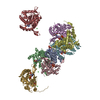
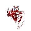
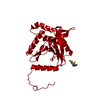
| Title | Crystal structure of a cubic core of the dihydrolipoamide acyltransferase (E2b) component in the branched-chain alpha-ketoacid dehydrogenase complex (BCKDC), apo form | ||||||
 Components Components | Lipoamide acyltransferase component of branched-chain alpha-keto acid dehydrogenase complex | ||||||
 Keywords Keywords | TRANSFERASE / cubic core / homo trimer / apo form | ||||||
| Function / homology |  Function and homology information Function and homology informationBranched-chain amino acid catabolism / BCKDH synthesizes BCAA-CoA from KIC, KMVA, KIV / RHOH GTPase cycle / dihydrolipoyllysine-residue (2-methylpropanoyl)transferase / dihydrolipoamide branched chain acyltransferase activity / Protein lipoylation / branched-chain alpha-keto acid decarboxylation to branched-chain acyl-CoA / branched-chain alpha-ketoacid dehydrogenase complex / Mitochondrial protein degradation / mitochondrial matrix ...Branched-chain amino acid catabolism / BCKDH synthesizes BCAA-CoA from KIC, KMVA, KIV / RHOH GTPase cycle / dihydrolipoyllysine-residue (2-methylpropanoyl)transferase / dihydrolipoamide branched chain acyltransferase activity / Protein lipoylation / branched-chain alpha-keto acid decarboxylation to branched-chain acyl-CoA / branched-chain alpha-ketoacid dehydrogenase complex / Mitochondrial protein degradation / mitochondrial matrix / mitochondrion / cytosol Similarity search - Function | ||||||
| Biological species |  | ||||||
| Method |  X-RAY DIFFRACTION / SYNCHROTRON / MOLECULAR REPLACEMENT / Resolution: 2.7 Å X-RAY DIFFRACTION / SYNCHROTRON / MOLECULAR REPLACEMENT / Resolution: 2.7 Å | ||||||
 Authors Authors | Kato, M. / Wynn, R.M. / Chuang, J.L. / Brautigam, C.A. / Custorio, M. / Chuang, D.T. | ||||||
 Citation Citation | Journal: Embo J. / Year: 2006 Title: A synchronized substrate-gating mechanism revealed by cubic-core structure of the bovine branched-chain alpha-ketoacid dehydrogenase complex. Authors: Kato, M. / Wynn, R.M. / Chuang, J.L. / Brautigam, C.A. / Custorio, M. / Chuang, D.T. | ||||||
| History |
|
- Structure visualization
| Structure viewer | Molecule: MolmilJmol/JSmol |
|---|
- Downloads & links
-Download
| PDBx/mmCIF format | 2ihw.cif.gz | 365.2 KB | Display | PDBx/mmCIF format |
|---|---|---|---|---|
| PDB format | pdb2ihw.ent.gz | 297.5 KB | Display | PDB format |
| PDBx/mmJSON format | 2ihw.json.gz | Tree view | PDBx/mmJSON format | |
| Others |  Other downloads Other downloads |
-Validation report
| Arichive directory | https://data.pdbj.org/pub/pdb/validation_reports/ih/2ihwftp://data.pdbj.org/pub/pdb/validation_reports/ih/2ihw | HTTPS FTP |
|---|
-Related structure data
| Related structure data |  2ii3C  2ii4C  2ii5C 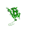 1eaaS C: citing same article ( S: Starting model for refinement |
|---|---|
| Similar structure data |


-Links
 PDBj
PDBj

- Assembly
| Deposited unit | 
| ||||||||||||||||||||||||||||||||||||||||||||||||||||||
|---|---|---|---|---|---|---|---|---|---|---|---|---|---|---|---|---|---|---|---|---|---|---|---|---|---|---|---|---|---|---|---|---|---|---|---|---|---|---|---|---|---|---|---|---|---|---|---|---|---|---|---|---|---|---|---|
| 1 | 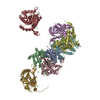
| ||||||||||||||||||||||||||||||||||||||||||||||||||||||
| Unit cell |
| ||||||||||||||||||||||||||||||||||||||||||||||||||||||
| Components on special symmetry positions |
| ||||||||||||||||||||||||||||||||||||||||||||||||||||||
| Noncrystallographic symmetry (NCS) | NCS domain:
NCS domain segments: Component-ID: 1 / Ens-ID: 1 / Beg auth comp-ID: GLY / Beg label comp-ID: GLY / End auth comp-ID: LYS / End label comp-ID: LYS / Refine code: 6 / Auth seq-ID: 188 - 421 / Label seq-ID: 29 - 262
| ||||||||||||||||||||||||||||||||||||||||||||||||||||||
| Details | A biological functional unit of this protein is a 24-meric cubic core, containing 8 homo trimers. The asymmetric unit in this crystal with the R3 space group contains 8 monomers (two trimers and two monomers from two another trimers). It is too complex to describe the symmetry operations to generate the cubic core from the asymmetric unit. Therefore, we highly recommend to download the coordinates of the biological functional unit (the 24-meric cubic core) from the PDB web site. |
-Components
| #1: Protein | Mass: 28726.576 Da / Num. of mol.: 8 / Fragment: core (catalytic) domain Source method: isolated from a genetically manipulated source Source: (gene. exp.)  References: UniProt: P11181, dihydrolipoyllysine-residue (2-methylpropanoyl)transferase #2: Chemical | ChemComp-ACT /   Mass: 59.044 Da / Num. of mol.: 16 / Source method: obtained synthetically / Formula: C2H3O2 Mass: 59.044 Da / Num. of mol.: 16 / Source method: obtained synthetically / Formula: C2H3O2#3: Chemical | ChemComp-CL /   Mass: 35.453 Da / Num. of mol.: 12 / Source method: obtained synthetically / Formula: Cl Mass: 35.453 Da / Num. of mol.: 12 / Source method: obtained synthetically / Formula: Cl#4: Water | ChemComp-HOH / |  Mass: 18.015 Da / Num. of mol.: 138 / Source method: isolated from a natural source / Formula: H2O Mass: 18.015 Da / Num. of mol.: 138 / Source method: isolated from a natural source / Formula: H2O |
|---|
-Experimental details
-Experiment
| Experiment | Method: X-RAY DIFFRACTION / Number of used crystals: 1 |
|---|
- Sample preparation
| Crystal | Density Matthews: 2.73 Å3/Da / Density % sol: 54.87 % |
|---|---|
| Crystal grow | Temperature: 292 K / Method: vapor diffusion, hanging drop / pH: 4.6 Details: 0.1 M Na-acetate (pH 4.6), 28% PEG 4000, 0.15 M NH4-acetate, VAPOR DIFFUSION, HANGING DROP, temperature 292K |
-Data collection
| Diffraction | Mean temperature: 100 K |
|---|---|
| Diffraction source | Source: SYNCHROTRON / Site: APS  / Beamline: 19-ID / Wavelength: 0.98 Å / Beamline: 19-ID / Wavelength: 0.98 Å |
| Detector | Type: ADSC QUANTUM 315 / Detector: CCD / Date: Dec 6, 2005 |
| Radiation | Monochromator: GRAPHITE / Protocol: SINGLE WAVELENGTH / Monochromatic (M) / Laue (L): M / Scattering type: x-ray |
| Radiation wavelength | Wavelength: 0.98 Å / Relative weight: 1 |
| Reflection | Resolution: 2.7→50 Å / Num. obs: 65296 / % possible obs: 97.8 % / Redundancy: 3 % / Rmerge(I) obs: 0.056 / Χ2: 1.121 / Net I/σ(I): 18.6 |
| Reflection shell | Resolution: 2.7→2.8 Å / Redundancy: 2.6 % / Rmerge(I) obs: 0.474 / Num. unique all: 6309 / Χ2: 1.234 / % possible all: 94.5 |
- Processing
| Software |
| ||||||||||||||||||||||||||||||||||||||||||||||||||||||||||||||||||||||||||||||||||||||||||||||||||||||||||||||||||||||||||||||||||||||||||||||||||||||||||||||||||||||||||||||||||||||||||||||||||||||||||||||||||||||||||||||||||||||||||
|---|---|---|---|---|---|---|---|---|---|---|---|---|---|---|---|---|---|---|---|---|---|---|---|---|---|---|---|---|---|---|---|---|---|---|---|---|---|---|---|---|---|---|---|---|---|---|---|---|---|---|---|---|---|---|---|---|---|---|---|---|---|---|---|---|---|---|---|---|---|---|---|---|---|---|---|---|---|---|---|---|---|---|---|---|---|---|---|---|---|---|---|---|---|---|---|---|---|---|---|---|---|---|---|---|---|---|---|---|---|---|---|---|---|---|---|---|---|---|---|---|---|---|---|---|---|---|---|---|---|---|---|---|---|---|---|---|---|---|---|---|---|---|---|---|---|---|---|---|---|---|---|---|---|---|---|---|---|---|---|---|---|---|---|---|---|---|---|---|---|---|---|---|---|---|---|---|---|---|---|---|---|---|---|---|---|---|---|---|---|---|---|---|---|---|---|---|---|---|---|---|---|---|---|---|---|---|---|---|---|---|---|---|---|---|---|---|---|---|---|---|---|---|---|---|---|---|---|---|---|---|---|---|---|---|---|
| Refinement | Method to determine structure: MOLECULAR REPLACEMENT Starting model: PDB Entry 1EAA Resolution: 2.7→50 Å / Cor.coef. Fo:Fc: 0.955 / Cor.coef. Fo:Fc free: 0.912 / SU B: 25.736 / SU ML: 0.253 / Isotropic thermal model: isotropic / Cross valid method: THROUGHOUT / σ(F): 0 / ESU R: 1.044 / ESU R Free: 0.345 / Stereochemistry target values: MAXIMUM LIKELIHOOD
| ||||||||||||||||||||||||||||||||||||||||||||||||||||||||||||||||||||||||||||||||||||||||||||||||||||||||||||||||||||||||||||||||||||||||||||||||||||||||||||||||||||||||||||||||||||||||||||||||||||||||||||||||||||||||||||||||||||||||||
| Solvent computation | Ion probe radii: 0.8 Å / Shrinkage radii: 0.8 Å / VDW probe radii: 1.4 Å / Solvent model: MASK | ||||||||||||||||||||||||||||||||||||||||||||||||||||||||||||||||||||||||||||||||||||||||||||||||||||||||||||||||||||||||||||||||||||||||||||||||||||||||||||||||||||||||||||||||||||||||||||||||||||||||||||||||||||||||||||||||||||||||||
| Displacement parameters | Biso mean: 54.357 Å2
| ||||||||||||||||||||||||||||||||||||||||||||||||||||||||||||||||||||||||||||||||||||||||||||||||||||||||||||||||||||||||||||||||||||||||||||||||||||||||||||||||||||||||||||||||||||||||||||||||||||||||||||||||||||||||||||||||||||||||||
| Refinement step | Cycle: LAST / Resolution: 2.7→50 Å
| ||||||||||||||||||||||||||||||||||||||||||||||||||||||||||||||||||||||||||||||||||||||||||||||||||||||||||||||||||||||||||||||||||||||||||||||||||||||||||||||||||||||||||||||||||||||||||||||||||||||||||||||||||||||||||||||||||||||||||
| Refine LS restraints |
| ||||||||||||||||||||||||||||||||||||||||||||||||||||||||||||||||||||||||||||||||||||||||||||||||||||||||||||||||||||||||||||||||||||||||||||||||||||||||||||||||||||||||||||||||||||||||||||||||||||||||||||||||||||||||||||||||||||||||||
| Refine LS restraints NCS | Ens-ID: 1 / Number: 1803 / Refine-ID: X-RAY DIFFRACTION
| ||||||||||||||||||||||||||||||||||||||||||||||||||||||||||||||||||||||||||||||||||||||||||||||||||||||||||||||||||||||||||||||||||||||||||||||||||||||||||||||||||||||||||||||||||||||||||||||||||||||||||||||||||||||||||||||||||||||||||
| LS refinement shell | Resolution: 2.7→2.77 Å / Total num. of bins used: 20
| ||||||||||||||||||||||||||||||||||||||||||||||||||||||||||||||||||||||||||||||||||||||||||||||||||||||||||||||||||||||||||||||||||||||||||||||||||||||||||||||||||||||||||||||||||||||||||||||||||||||||||||||||||||||||||||||||||||||||||
| Refinement TLS params. | Method: refined / Refine-ID: X-RAY DIFFRACTION
| ||||||||||||||||||||||||||||||||||||||||||||||||||||||||||||||||||||||||||||||||||||||||||||||||||||||||||||||||||||||||||||||||||||||||||||||||||||||||||||||||||||||||||||||||||||||||||||||||||||||||||||||||||||||||||||||||||||||||||
| Refinement TLS group | Refine-ID: X-RAY DIFFRACTION / Selection: ALL / Auth seq-ID: 188 - 421 / Label seq-ID: 29 - 262
|
