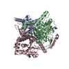
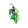
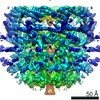
Journal: Nat Commun / Year: 2021 Title: Structure of the native pyruvate dehydrogenase complex reveals the mechanism of substrate insertion. Authors: Jana Škerlová / Jens Berndtsson / Hendrik Nolte / Martin Ott / Pål Stenmark / Abstract: The pyruvate dehydrogenase complex (PDHc) links glycolysis to the citric acid cycle by converting pyruvate into acetyl-coenzyme A. PDHc encompasses three enzymatically active subunits, namely ...The pyruvate dehydrogenase complex (PDHc) links glycolysis to the citric acid cycle by converting pyruvate into acetyl-coenzyme A. PDHc encompasses three enzymatically active subunits, namely pyruvate dehydrogenase, dihydrolipoyl transacetylase, and dihydrolipoyl dehydrogenase. Dihydrolipoyl transacetylase is a multidomain protein comprising a varying number of lipoyl domains, a peripheral subunit-binding domain, and a catalytic domain. It forms the structural core of the complex, provides binding sites for the other enzymes, and shuffles reaction intermediates between the active sites through covalently bound lipoyl domains. The molecular mechanism by which this shuttling occurs has remained elusive. Here, we report a cryo-EM reconstruction of the native E. coli dihydrolipoyl transacetylase core in a resting state. This structure provides molecular details of the assembly of the core and reveals how the lipoyl domains interact with the core at the active site.
History
Deposition
Dec 14, 2020
Deposition site: PDBE / Processing site: PDBE
Revision 1.0
Aug 11, 2021
Provider: repository / Type: Initial release
Revision 1.0
Aug 11, 2021
Data content type: EM metadata / Data content type: EM metadata / Provider: repository / Type: Initial release
Revision 1.0
Aug 11, 2021
Data content type: FSC / Data content type: FSC / Provider: repository / Type: Initial release
Revision 1.0
Aug 11, 2021
Data content type: Half map / Part number: 1 / Data content type: Half map / Provider: repository / Type: Initial release
Revision 1.0
Aug 11, 2021
Data content type: Half map / Part number: 2 / Data content type: Half map / Provider: repository / Type: Initial release
Revision 1.0
Aug 11, 2021
Data content type: Image / Data content type: Image / Provider: repository / Type: Initial release
Revision 1.0
Aug 11, 2021
Data content type: Mask / Data content type: Mask / Provider: repository / Type: Initial release
Revision 1.0
Aug 11, 2021
Data content type: Primary map / Data content type: Primary map / Provider: repository / Type: Initial release
Revision 1.0
Aug 11, 2021
Data content type: FSC / Data content type: FSC / Provider: repository / Type: Initial release
Revision 1.0
Aug 11, 2021
Data content type: Half map / Part number: 1 / Data content type: Half map / Provider: repository / Type: Initial release
Revision 1.0
Aug 11, 2021
Data content type: Half map / Part number: 2 / Data content type: Half map / Provider: repository / Type: Initial release
Revision 1.0
Aug 11, 2021
Data content type: Image / Data content type: Image / Provider: repository / Type: Initial release
Revision 1.0
Aug 11, 2021
Data content type: Mask / Data content type: Mask / Provider: repository / Type: Initial release
Revision 1.0
Aug 11, 2021
Data content type: Primary map / Data content type: Primary map / Provider: repository / Type: Initial release
Data content type: EM metadata / Data content type: EM metadata / EM metadata / EM metadata Group: Data processing / Experimental summary / Refinement description Data content type: EM metadata / EM metadata / EM metadata Category: em_3d_fitting_list / em_admin / pdbx_initial_refinement_model Data content type: EM metadata / EM metadata ...EM metadata / EM metadata / EM metadata / EM metadata / EM metadata Item: _em_3d_fitting_list.accession_code / _em_3d_fitting_list.initial_refinement_model_id ..._em_3d_fitting_list.accession_code / _em_3d_fitting_list.initial_refinement_model_id / _em_3d_fitting_list.source_name / _em_3d_fitting_list.type / _em_admin.last_update
In the structure databanks used in Yorodumi, some data are registered as the other names, "COVID-19 virus" and "2019-nCoV". Here are the details of the virus and the list of structure data.
Jan 31, 2019. EMDB accession codes are about to change! (news from PDBe EMDB page)
EMDB accession codes are about to change! (news from PDBe EMDB page)
The allocation of 4 digits for EMDB accession codes will soon come to an end. Whilst these codes will remain in use, new EMDB accession codes will include an additional digit and will expand incrementally as the available range of codes is exhausted. The current 4-digit format prefixed with “EMD-” (i.e. EMD-XXXX) will advance to a 5-digit format (i.e. EMD-XXXXX), and so on. It is currently estimated that the 4-digit codes will be depleted around Spring 2019, at which point the 5-digit format will come into force.
The EM Navigator/Yorodumi systems omit the EMD- prefix.
Related info.:Q: What is EMD? / ID/Accession-code notation in Yorodumi/EM Navigator
Yorodumi is a browser for structure data from EMDB, PDB, SASBDB, etc.
This page is also the successor to EM Navigator detail page, and also detail information page/front-end page for Omokage search.
The word "yorodu" (or yorozu) is an old Japanese word meaning "ten thousand". "mi" (miru) is to see.
Related info.:EMDB / PDB / SASBDB / Comparison of 3 databanks / Yorodumi Search / Aug 31, 2016. New EM Navigator & Yorodumi / Yorodumi Papers / Jmol/JSmol / Function and homology information / Changes in new EM Navigator and Yorodumi
 Movie
Movie Controller

 Yorodumi
 Open data
 Basic information
 Components
 Keywords
 Function and homology information
 Authors
 Sweden, European Union, 2items
  Citation
 Structure visualization
 UCSF Chimera
 Downloads & links
 Other downloads
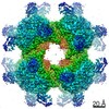

 PDBj
PDBj

 Assembly
 Sample preparation
 Electron microscopy imaging
 Processing