 Movie
Movie Controller


+ Open data

- Basic information
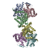
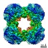
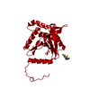
| Entry | Database: PDB / ID: 1e2o | ||||||
|---|---|---|---|---|---|---|---|
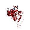
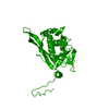
| Title | CATALYTIC DOMAIN FROM DIHYDROLIPOAMIDE SUCCINYLTRANSFERASE | ||||||
 Components Components | DIHYDROLIPOAMIDE SUCCINYLTRANSFERASE | ||||||
 Keywords Keywords | TRANSFERASE / ACYLTRANSFERASE / KETOGLUTARATE DEHYDROGENASE MULTIENZYME COMPLEX | ||||||
| Function / homology |  Function and homology information Function and homology information: / dihydrolipoyllysine-residue succinyltransferase / dihydrolipoyllysine-residue succinyltransferase activity / lipoic acid binding / oxoglutarate dehydrogenase complex / tricarboxylic acid cycle / cytosol / cytoplasm Similarity search - Function | ||||||
| Biological species |  | ||||||
| Method |  X-RAY DIFFRACTION / SYNCHROTRON / MOLECULAR REPLACEMENT / Resolution: 3 Å X-RAY DIFFRACTION / SYNCHROTRON / MOLECULAR REPLACEMENT / Resolution: 3 Å | ||||||
 Authors Authors | Knapp, J.E. / Mitchell, D.T. / Yazdi, M.A. / Ernst, S.R. / Reed, L.J. / Hackert, M.L. | ||||||
 Citation Citation | Journal: J.Mol.Biol. / Year: 1998 Title: Crystal structure of the truncated cubic core component of the Escherichia coli 2-oxoglutarate dehydrogenase multienzyme complex. Authors: Knapp, J.E. / Mitchell, D.T. / Yazdi, M.A. / Ernst, S.R. / Reed, L.J. / Hackert, M.L. #1: Journal: Science / Year: 1992Title: Atomic Structure of the Cubic Core of the Pyruvate Dehydrogenase Multienzyme Complex Authors: Mattevi, A. / Obmolova, G. / Schulze, E. / Kalk, K.H. / Westphal, A.H. / De Kok, A. / Hol, W.G. #2: Journal: Eur.J.Biochem. / Year: 1984Title: Nucleotide Sequence of the Sucb Gene Encoding the Dihydrolipoamide Succinyltransferase of Escherichia Coli K12 and Homology with the Corresponding Acetyltransferase Authors: Spencer, M.E. / Darlison, M.G. / Stephens, P.E. / Duckenfield, I.K. / Guest, J.R. #3: Journal: Proc.Natl.Acad.Sci.USA / Year: 1971Title: Crystallization and Preliminary Structural Analysis of Dihydrolipoyl Transsuccinylase, the Core of the 2-Oxoglutarate Dehydrogenase Complex Authors: Derosier, D.J. / Oliver, R.M. / Reed, L.J. | ||||||
| History |
|
- Structure visualization
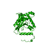
| Structure viewer | Molecule: MolmilJmol/JSmol |
|---|
- Downloads & links
-Download
| PDBx/mmCIF format | 1e2o.cif.gz | 52.8 KB | Display | PDBx/mmCIF format |
|---|---|---|---|---|
| PDB format | pdb1e2o.ent.gz | 39.8 KB | Display | PDB format |
| PDBx/mmJSON format | 1e2o.json.gz | Tree view | PDBx/mmJSON format | |
| Others |  Other downloads Other downloads |
-Validation report
| Arichive directory | https://data.pdbj.org/pub/pdb/validation_reports/e2/1e2oftp://data.pdbj.org/pub/pdb/validation_reports/e2/1e2o | HTTPS FTP |
|---|
-Related structure data
| Related structure data |  1eaaS S: Starting model for refinement |
|---|---|
| Similar structure data |


-Links
 PDBj
PDBj

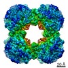
- Assembly
| Deposited unit | 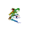
| ||||||||
|---|---|---|---|---|---|---|---|---|---|
| 1 | x 24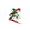
| ||||||||
| Unit cell |
| ||||||||
| Components on special symmetry positions |
|
-Components
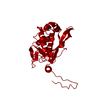
| #1: Protein | Mass: 26107.420 Da / Num. of mol.: 1 / Fragment: CATALYTIC DOMAIN, RESIDUES 172 - 404 Source method: isolated from a genetically manipulated source Source: (gene. exp.) References: UniProt: P07016, UniProt: P0AFG6*PLUS, dihydrolipoyllysine-residue succinyltransferase |
|---|---|
| #2: Chemical | ChemComp-SO4 /   Mass: 96.063 Da / Num. of mol.: 1 / Source method: obtained synthetically / Formula: SO4 Mass: 96.063 Da / Num. of mol.: 1 / Source method: obtained synthetically / Formula: SO4 |
| #3: Water | ChemComp-HOH /  Mass: 18.015 Da / Num. of mol.: 16 / Source method: isolated from a natural source / Formula: H2O Mass: 18.015 Da / Num. of mol.: 16 / Source method: isolated from a natural source / Formula: H2O |
-Experimental details
-Experiment
| Experiment | Method: X-RAY DIFFRACTION / Number of used crystals: 1 |
|---|
- Sample preparation
| Crystal | Density Matthews: 4.4 Å3/Da / Density % sol: 72 % | ||||||||||||||||||||||||||||||||||||||||
|---|---|---|---|---|---|---|---|---|---|---|---|---|---|---|---|---|---|---|---|---|---|---|---|---|---|---|---|---|---|---|---|---|---|---|---|---|---|---|---|---|---|
| Crystal grow | pH: 7 Details: PROTEIN WAS CRYSTALLIZED FROM 1.2M AMMONIUM SULFATE, 1% ETHANOL, 50 MM POTASSIUM PHOSPHATE, PH 7.0 | ||||||||||||||||||||||||||||||||||||||||
| Crystal grow | *PLUS pH: 7.3 / Method: vapor diffusion, hanging drop | ||||||||||||||||||||||||||||||||||||||||
| Components of the solutions | *PLUS
|
-Data collection
| Diffraction | Mean temperature: 298 K |
|---|---|
| Diffraction source | Source: SYNCHROTRON / Site: SSRL  / Beamline: BL7-1 / Wavelength: 1.08 / Beamline: BL7-1 / Wavelength: 1.08 |
| Detector | Type: MARRESEARCH / Detector: IMAGE PLATE / Date: Mar 1, 1994 / Details: MIRRORS |
| Radiation | Monochromator: SI(111) / Monochromatic (M) / Laue (L): M / Scattering type: x-ray |
| Radiation wavelength | Wavelength: 1.08 Å / Relative weight: 1 |
| Reflection | Resolution: 3→20 Å / Num. obs: 9906 / % possible obs: 98.5 % / Observed criterion σ(I): 3 / Redundancy: 4.1 % / Rsym value: 0.046 / Net I/σ(I): 13 |
| Reflection shell | Resolution: 3→3.16 Å / Redundancy: 4.5 % / Mean I/σ(I) obs: 4.9 / Rsym value: 0.145 / % possible all: 100 |
| Reflection | *PLUS Rmerge(I) obs: 0.046 |
| Reflection shell | *PLUS % possible obs: 100 % / Rmerge(I) obs: 0.145 |
- Processing
| Software |
| ||||||||||||||||||||||||||||||||||||||||||||||||||||||||||||
|---|---|---|---|---|---|---|---|---|---|---|---|---|---|---|---|---|---|---|---|---|---|---|---|---|---|---|---|---|---|---|---|---|---|---|---|---|---|---|---|---|---|---|---|---|---|---|---|---|---|---|---|---|---|---|---|---|---|---|---|---|---|
| Refinement | Method to determine structure: MOLECULAR REPLACEMENT Starting model: PDB ENTRY 1EAA Resolution: 3→20 Å / Rfactor Rfree error: 0.008 / Data cutoff high absF: 10000000 / Data cutoff low absF: 0.001 / Isotropic thermal model: GROUP / Cross valid method: THROUGHOUT / σ(F): 0 Details: BULK SOLVENT MODEL AND RESOLUTION-DEPENDENT WEIGHTING SCHEME USED.
| ||||||||||||||||||||||||||||||||||||||||||||||||||||||||||||
| Displacement parameters | Biso mean: 33.6 Å2
| ||||||||||||||||||||||||||||||||||||||||||||||||||||||||||||
| Refine analyze |
| ||||||||||||||||||||||||||||||||||||||||||||||||||||||||||||
| Refinement step | Cycle: LAST / Resolution: 3→20 Å
| ||||||||||||||||||||||||||||||||||||||||||||||||||||||||||||
| Refine LS restraints |
| ||||||||||||||||||||||||||||||||||||||||||||||||||||||||||||
| LS refinement shell | Resolution: 3→3.19 Å / Rfactor Rfree error: 0.028 / Total num. of bins used: 6
| ||||||||||||||||||||||||||||||||||||||||||||||||||||||||||||
| Xplor file |
| ||||||||||||||||||||||||||||||||||||||||||||||||||||||||||||
| Software | *PLUS Name: X-PLOR / Version: 3.851 / Classification: refinement | ||||||||||||||||||||||||||||||||||||||||||||||||||||||||||||
| Refine LS restraints | *PLUS
|
