 Movie
Movie Controller

[English] 日本語
 Yorodumi
- PDB-6h05: Cryo-electron microscopic structure of the dihydrolipoamide succi... -
+ Open data

- Basic information
| Entry | Database: PDB / ID: 6h05 | |||||||||||||||||||||
|---|---|---|---|---|---|---|---|---|---|---|---|---|---|---|---|---|---|---|---|---|---|---|
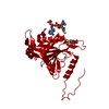
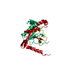
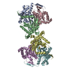
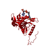
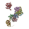
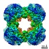
| Title | Cryo-electron microscopic structure of the dihydrolipoamide succinyltransferase (E2) component of the human alpha-ketoglutarate (2-oxoglutarate) dehydrogenase complex [residues 218-453] | |||||||||||||||||||||
 Components Components | Dihydrolipoyllysine-residue succinyltransferase component of 2-oxoglutarate dehydrogenase complex, mitochondrial | |||||||||||||||||||||
 Keywords Keywords | TRANSFERASE / alpha-ketoglutarate dehydrogenase complex / 2-oxoglutarate dehydrogenase complex / dihydrolipoamide succinyltransferase / E2 component | |||||||||||||||||||||
| Function / homology |  Function and homology information Function and homology informationOGDH complex synthesizes succinyl-CoA from 2-OG / OADH complex synthesizes glutaryl-CoA from 2-OA / 2-oxoglutarate decarboxylation to succinyl-CoA / oxoadipate dehydrogenase complex / Glycine degradation / : / succinyl-CoA metabolic process / dihydrolipoyllysine-residue succinyltransferase / dihydrolipoyllysine-residue succinyltransferase activity / oxoglutarate dehydrogenase complex ...OGDH complex synthesizes succinyl-CoA from 2-OG / OADH complex synthesizes glutaryl-CoA from 2-OA / 2-oxoglutarate decarboxylation to succinyl-CoA / oxoadipate dehydrogenase complex / Glycine degradation / : / succinyl-CoA metabolic process / dihydrolipoyllysine-residue succinyltransferase / dihydrolipoyllysine-residue succinyltransferase activity / oxoglutarate dehydrogenase complex / Protein lipoylation / 2-oxoglutarate metabolic process / acyltransferase activity / tricarboxylic acid cycle / generation of precursor metabolites and energy / mitochondrial matrix / mitochondrion / nucleoplasm / membrane / nucleus / cytosol Similarity search - Function | |||||||||||||||||||||
| Biological species |  Homo sapiens (human) Homo sapiens (human) | |||||||||||||||||||||
| Method | ELECTRON MICROSCOPY / single particle reconstruction / cryo EM / Resolution: 2.9 Å | |||||||||||||||||||||
 Authors Authors | Nagy, B. / Zambo, Z. / Hubert, A. / Polak, M. / Nemeria, N.S. / Novacek, J. / Jordan, F. / Adam-Vizi, V. / Ambrus, A. | |||||||||||||||||||||
| Funding support |  Hungary, Hungary,  United States, 6items United States, 6items
| |||||||||||||||||||||
 Citation Citation | Journal: Biochim Biophys Acta Gen Subj / Year: 2021 Title: Structure of the dihydrolipoamide succinyltransferase (E2) component of the human alpha-ketoglutarate dehydrogenase complex (hKGDHc) revealed by cryo-EM and cross-linking mass spectrometry: ...Title: Structure of the dihydrolipoamide succinyltransferase (E2) component of the human alpha-ketoglutarate dehydrogenase complex (hKGDHc) revealed by cryo-EM and cross-linking mass spectrometry: Implications for the overall hKGDHc structure. Authors: Balint Nagy / Martin Polak / Oliver Ozohanics / Zsofia Zambo / Eszter Szabo / Agnes Hubert / Frank Jordan / Jiří Novaček / Vera Adam-Vizi / Attila Ambrus /  Abstract: BACKGROUND: The human mitochondrial alpha-ketoglutarate dehydrogenase complex (hKGDHc) converts KG to succinyl-CoA and NADH. Malfunction of and reactive oxygen species generation by the hKGDHc as ...BACKGROUND: The human mitochondrial alpha-ketoglutarate dehydrogenase complex (hKGDHc) converts KG to succinyl-CoA and NADH. Malfunction of and reactive oxygen species generation by the hKGDHc as well as its E1-E2 subcomplex are implicated in neurodegenerative disorders, ischemia-reperfusion injury, E3-deficiency and cancers. METHODS: We performed cryo-EM, cross-linking mass spectrometry (CL-MS) and molecular modeling analyses to determine the structure of the E2 component of the hKGDHc (hE2k); hE2k transfers a succinyl ...METHODS: We performed cryo-EM, cross-linking mass spectrometry (CL-MS) and molecular modeling analyses to determine the structure of the E2 component of the hKGDHc (hE2k); hE2k transfers a succinyl group to CoA and forms the structural core of hKGDHc. We also assessed the overall structure of the hKGDHc by negative-stain EM and modeling. RESULTS: We report the 2.9 Å resolution cryo-EM structure of the hE2k component. The cryo-EM map comprises density for hE2k residues 151-386 - the entire (inner) core catalytic domain plus a few ...RESULTS: We report the 2.9 Å resolution cryo-EM structure of the hE2k component. The cryo-EM map comprises density for hE2k residues 151-386 - the entire (inner) core catalytic domain plus a few additional residues -, while residues 1-150 are not observed due to the inherent flexibility of the N-terminal region. The structure of the latter segment was also determined by CL-MS and homology modeling. Negative-stain EM on in vitro assembled hKGDHc and previous data were used to build a putative overall structural model of the hKGDHc. CONCLUSIONS: The E2 core of the hKGDHc is composed of 24 hE2k chains organized in octahedral (8 × 3 type) assembly. Each lipoyl domain is oriented towards the core domain of an adjacent chain in ...CONCLUSIONS: The E2 core of the hKGDHc is composed of 24 hE2k chains organized in octahedral (8 × 3 type) assembly. Each lipoyl domain is oriented towards the core domain of an adjacent chain in the hE2k homotrimer. hE1k and hE3 are most likely tethered at the edges and faces, respectively, of the cubic hE2k assembly. GENERAL SIGNIFICANCE: The revealed structural information will support the future pharmacologically targeting of the hKGDHc. | |||||||||||||||||||||
| History |
|
- Structure visualization
| Movie |
Movie viewer |
|---|---|
| Structure viewer | Molecule: MolmilJmol/JSmol |
- Downloads & links
-Download
| PDBx/mmCIF format | 6h05.cif.gz | 58.8 KB | Display | PDBx/mmCIF format |
|---|---|---|---|---|
| PDB format | pdb6h05.ent.gz | 39.2 KB | Display | PDB format |
| PDBx/mmJSON format | 6h05.json.gz | Tree view | PDBx/mmJSON format | |
| Others |  Other downloads Other downloads |
-Validation report
| Arichive directory | https://data.pdbj.org/pub/pdb/validation_reports/h0/6h05ftp://data.pdbj.org/pub/pdb/validation_reports/h0/6h05 | HTTPS FTP |
|---|
-Related structure data
| Related structure data | 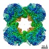 0108MC M: map data used to model this data C: citing same article ( |
|---|---|
| Similar structure data |


-Links
 PDBj
PDBj

- Assembly
| Deposited unit | 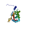
|
|---|---|
| 1 | x 24
|
-Components
| #1: Protein | Mass: 45543.004 Da / Num. of mol.: 1 Source method: isolated from a genetically manipulated source Details: N-terminal Twin-Strep (affinity) tag with proteolytic cleavage site and linkers: MASWSHPQFEKGGGSGGGSGGSAWSHPQFEKLEVLFQGPG Density was found for 236 residues [218-453] Source: (gene. exp.) Homo sapiens (human) / Gene: DLST, DLTS / Plasmid: pET52b+ / Production host:  References: UniProt: P36957, dihydrolipoyllysine-residue succinyltransferase |
|---|
-Experimental details
-Experiment
| Experiment | Method: ELECTRON MICROSCOPY |
|---|---|
| EM experiment | Aggregation state: PARTICLE / 3D reconstruction method: single particle reconstruction |
- Sample preparation
| Component | Name: Affinity purified human dihydrolipoamide succinyltransferase Type: COMPLEX Details: Protein in 100 mM Tris-HCl, 150 mM NaCl, 1 mM EDTA, pH 8.0. Entity ID: all / Source: RECOMBINANT | ||||||||||||||||||||
|---|---|---|---|---|---|---|---|---|---|---|---|---|---|---|---|---|---|---|---|---|---|
| Molecular weight | Experimental value: NO | ||||||||||||||||||||
| Source (natural) | Organism: Homo sapiens (human) | ||||||||||||||||||||
| Source (recombinant) | Organism: | ||||||||||||||||||||
| Buffer solution | pH: 8 | ||||||||||||||||||||
| Buffer component |
| ||||||||||||||||||||
| Specimen | Conc.: 1 mg/ml / Embedding applied: NO / Shadowing applied: NO / Staining applied: NO / Vitrification applied: YES / Details: This sample was highly purified and monodisperse. | ||||||||||||||||||||
| Specimen support | Grid material: COPPER / Grid mesh size: 200 divisions/in. / Grid type: Quantifoil R2/1 | ||||||||||||||||||||
| Vitrification | Instrument: FEI VITROBOT MARK IV / Cryogen name: ETHANE / Humidity: 100 % / Chamber temperature: 298 K |
- Electron microscopy imaging
| Experimental equipment |  Model: Titan Krios / Image courtesy: FEI Company |
|---|---|
| Microscopy | Model: FEI TITAN KRIOS |
| Electron gun | Electron source:  FIELD EMISSION GUN / Accelerating voltage: 300 kV / Illumination mode: OTHER FIELD EMISSION GUN / Accelerating voltage: 300 kV / Illumination mode: OTHER |
| Electron lens | Mode: BRIGHT FIELD / Nominal defocus max: 3000 nm / Nominal defocus min: 1000 nm |
| Image recording | Average exposure time: 1 sec. / Electron dose: 48.8 e/Å2 / Detector mode: INTEGRATING / Film or detector model: FEI FALCON II (4k x 4k) / Num. of grids imaged: 1 |
- Processing
| EM software |
| ||||||||||||||||||
|---|---|---|---|---|---|---|---|---|---|---|---|---|---|---|---|---|---|---|---|
| CTF correction | Type: PHASE FLIPPING AND AMPLITUDE CORRECTION | ||||||||||||||||||
| Particle selection | Num. of particles selected: 1533026 Details: Manual picking for the templates was performed by the EMAN2 E2boxer program. Automated selection was then applied by CryoSPARC. | ||||||||||||||||||
| Symmetry | Point symmetry: O (octahedral) | ||||||||||||||||||
| 3D reconstruction | Resolution: 2.9 Å / Resolution method: FSC 0.143 CUT-OFF / Num. of particles: 740101 / Algorithm: FOURIER SPACE / Num. of class averages: 1 / Symmetry type: POINT | ||||||||||||||||||
| Atomic model building | Protocol: RIGID BODY FIT Details: COOT was used for structure manipulations, while MOLPROBITY was applied for validation. |
