Entry Database : PDB / ID : 6pbrTitle Catalytic domain of E.coli dihydrolipoamide succinyltransferase in I4 space group Dihydrolipoyllysine-residue succinyltransferase component of 2-oxoglutarate dehydrogenase complex Keywords / / / / / Function / homology Function Domain/homology Component
/ / / / / / / / / / / / / / / / / / / / / / / / / / / / / / / / / / / Biological species Escherichia coli (E. coli)Method / / / Resolution : 3 Å Authors Andi, B. / Soares, A.S. / Shi, W. / Fuchs, M.R. / McSweeney, S. / Liu, Q. Funding support Organization Grant number Country National Institutes of Health/National Institute of General Medical Sciences (NIH/NIGMS) P41GM111244 Department of Energy (DOE, United States) KP1605010
Journal : Acta Crystallogr.,Sect.F / Year : 2019Title : Structure of the dihydrolipoamide succinyltransferase catalytic domain from Escherichia coli in a novel crystal form: a tale of a common protein crystallization contaminant.Authors : Andi, B. / Soares, A.S. / Shi, W. / Fuchs, M.R. / McSweeney, S. / Liu, Q. History Deposition Jun 14, 2019 Deposition site / Processing site Revision 1.0 Jun 26, 2019 Provider / Type Revision 1.1 Sep 11, 2019 Group / Database references / Category / citation_authorItem _citation.country / _citation.journal_abbrev ... _citation.country / _citation.journal_abbrev / _citation.journal_id_ASTM / _citation.journal_id_CSD / _citation.journal_id_ISSN / _citation.journal_volume / _citation.page_first / _citation.page_last / _citation.pdbx_database_id_DOI / _citation.pdbx_database_id_PubMed / _citation.title / _citation.year Revision 1.2 Sep 18, 2019 Group / Structure summary / Category Revision 1.3 Dec 4, 2019 Group / Category / Item Revision 1.4 Oct 11, 2023 Group / Database references / Refinement descriptionCategory chem_comp_atom / chem_comp_bond ... chem_comp_atom / chem_comp_bond / database_2 / pdbx_initial_refinement_model / struct_ncs_dom_lim Item _database_2.pdbx_DOI / _database_2.pdbx_database_accession ... _database_2.pdbx_DOI / _database_2.pdbx_database_accession / _struct_ncs_dom_lim.beg_auth_comp_id / _struct_ncs_dom_lim.beg_label_asym_id / _struct_ncs_dom_lim.beg_label_comp_id / _struct_ncs_dom_lim.beg_label_seq_id / _struct_ncs_dom_lim.end_auth_comp_id / _struct_ncs_dom_lim.end_label_asym_id / _struct_ncs_dom_lim.end_label_comp_id / _struct_ncs_dom_lim.end_label_seq_id
Show all Show less
 Movie
Movie Controller

 Yorodumi
 Open data
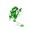
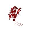
 Basic information
 Components
 Keywords
 Function and homology information
 X-RAY DIFFRACTION /
  Authors
 United States, 2items
  Citation
 Structure visualization
 Downloads & links
 Other downloads
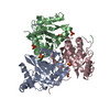


 PDBj
PDBj

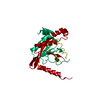
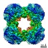
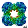
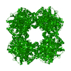
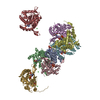
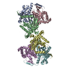
 Assembly

 Mass: 22.990 Da / Num. of mol.: 6 / Source method: obtained synthetically / Formula: Na
 Mass: 18.015 Da / Num. of mol.: 6 / Source method: isolated from a natural source / Formula: H2O
 Sample preparation
 Processing