 Movie
Movie Controller

[English] 日本語
 Yorodumi
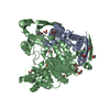
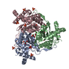
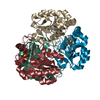
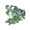
- PDB-1c4t: CATALYTIC DOMAIN FROM TRIMERIC DIHYDROLIPOAMIDE SUCCINYLTRANSFERASE -
+ Open data

- Basic information
| Entry | Database: PDB / ID: 1c4t | ||||||
|---|---|---|---|---|---|---|---|
| Title | CATALYTIC DOMAIN FROM TRIMERIC DIHYDROLIPOAMIDE SUCCINYLTRANSFERASE | ||||||
 Components Components | PROTEIN (DIHYDROLIPOAMIDE SUCCINYLTRANSFERASE) | ||||||
 Keywords Keywords | TRANSFERASE / ACYLTRANSFERASE / KETOGLUTARATE DEHYDROGENASE MULTIENZYME COMPLEX | ||||||
| Function / homology |  Function and homology information Function and homology information: / dihydrolipoyllysine-residue succinyltransferase / dihydrolipoyllysine-residue succinyltransferase activity / lipoic acid binding / oxoglutarate dehydrogenase complex / tricarboxylic acid cycle / cytosol / cytoplasm Similarity search - Function | ||||||
| Biological species |  | ||||||
| Method |  X-RAY DIFFRACTION / MOLECULAR REPLACEMENT / Resolution: 3 Å X-RAY DIFFRACTION / MOLECULAR REPLACEMENT / Resolution: 3 Å | ||||||
 Authors Authors | Knapp, J.E. / Carroll, D. / Lawson, J.E. / Ernst, S.R. / Reed, L.J. / Hackert, M.L. | ||||||
 Citation Citation | Journal: Protein Sci. / Year: 2000 Title: Expression, purification, and structural analysis of the trimeric form of the catalytic domain of the Escherichia coli dihydrolipoamide succinyltransferase. Authors: Knapp, J.E. / Carroll, D. / Lawson, J.E. / Ernst, S.R. / Reed, L.J. / Hackert, M.L. #1: Journal: J.Mol.Biol. / Year: 1998Title: Crystal Structure of the Truncated Cubic Core Component of the Escherichia Coli 2-Oxoglutarate Dehydrogenase Multienzyme Complex Authors: Knapp, J.E. / Mitchell, D.T. / Yazdi, M.A. / Ernst, S.R. / Reed, L.J. / Hackert, M.L. #2: Journal: Eur.J.Biochem. / Year: 1984Title: Nucleotide Sequence of the Sucb Gene Encoding the Dihydrolipoamide Succinyltransferase of Escherichia Coli K12 and Homology with the Corresponding Acetyltransferase Authors: Spencer, M.E. / Darlison, M.G. / Stephens, P.E. / Duckenfield, I.K. / Guest, J.R. | ||||||
| History |
|
- Structure visualization
| Structure viewer | Molecule: MolmilJmol/JSmol |
|---|
- Downloads & links
-Download
| PDBx/mmCIF format | 1c4t.cif.gz | 126.8 KB | Display | PDBx/mmCIF format |
|---|---|---|---|---|
| PDB format | pdb1c4t.ent.gz | 99.5 KB | Display | PDB format |
| PDBx/mmJSON format | 1c4t.json.gz | Tree view | PDBx/mmJSON format | |
| Others |  Other downloads Other downloads |
-Validation report
| Arichive directory | https://data.pdbj.org/pub/pdb/validation_reports/c4/1c4tftp://data.pdbj.org/pub/pdb/validation_reports/c4/1c4t | HTTPS FTP |
|---|
-Related structure data
| Related structure data | 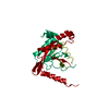 1e2oS S: Starting model for refinement |
|---|---|
| Similar structure data |


-Links
 PDBj
PDBj

- Assembly
| Deposited unit | 
| ||||||||||||
|---|---|---|---|---|---|---|---|---|---|---|---|---|---|
| 1 |
| ||||||||||||
| Unit cell |
| ||||||||||||
| Noncrystallographic symmetry (NCS) | NCS oper:
|
-Components
| #1: Protein | Mass: 26107.420 Da / Num. of mol.: 3 / Fragment: CATALYTIC DOMAIN, RESIDUES 172 - 404 Source method: isolated from a genetically manipulated source Source: (gene. exp.) References: UniProt: P07016, UniProt: P0AFG6*PLUS, dihydrolipoyllysine-residue succinyltransferase #2: Chemical | ChemComp-SO4 /   Mass: 96.063 Da / Num. of mol.: 6 / Source method: obtained synthetically / Formula: SO4 Mass: 96.063 Da / Num. of mol.: 6 / Source method: obtained synthetically / Formula: SO4#3: Water | ChemComp-HOH / |  Mass: 18.015 Da / Num. of mol.: 28 / Source method: isolated from a natural source / Formula: H2O Mass: 18.015 Da / Num. of mol.: 28 / Source method: isolated from a natural source / Formula: H2O |
|---|
-Experimental details
-Experiment
| Experiment | Method: X-RAY DIFFRACTION / Number of used crystals: 1 |
|---|
- Sample preparation
| Crystal | Density Matthews: 2.9 Å3/Da / Density % sol: 57.6 % | |||||||||||||||||||||||||
|---|---|---|---|---|---|---|---|---|---|---|---|---|---|---|---|---|---|---|---|---|---|---|---|---|---|---|
| Crystal grow | pH: 7.5 / Details: pH 7.5 | |||||||||||||||||||||||||
| Crystal grow | *PLUS Method: vapor diffusion, hanging drop | |||||||||||||||||||||||||
| Components of the solutions | *PLUS
|
-Data collection
| Diffraction | Mean temperature: 298 K |
|---|---|
| Diffraction source | Source: ROTATING ANODE / Type: RIGAKU RU200 / Wavelength: 1.5418 |
| Detector | Type: RIGAKU RAXIS IV / Detector: IMAGE PLATE / Date: Aug 1, 1996 / Details: MSC MIRRORS |
| Radiation | Monochromator: NI FILTER / Protocol: SINGLE WAVELENGTH / Monochromatic (M) / Laue (L): M / Scattering type: x-ray |
| Radiation wavelength | Wavelength: 1.5418 Å / Relative weight: 1 |
| Reflection | Resolution: 3→10 Å / Num. obs: 18669 / % possible obs: 95.9 % / Redundancy: 2.9 % / Biso Wilson estimate: 57.3 Å2 / Rsym value: 10.3 / Net I/σ(I): 13.8 |
| Reflection shell | Resolution: 3→3.1 Å / Redundancy: 1.8 % / Mean I/σ(I) obs: 2.6 / Rsym value: 30.9 / % possible all: 93.5 |
| Reflection | *PLUS Num. obs: 18677 / % possible obs: 95.8 % / Rmerge(I) obs: 0.103 |
| Reflection shell | *PLUS % possible obs: 93.5 % / Num. unique obs: 1798 / Rmerge(I) obs: 0.309 |
- Processing
| Software |
| ||||||||||||||||||||||||||||||||||||||||||||||||||||||||||||||||||||||||||||||||
|---|---|---|---|---|---|---|---|---|---|---|---|---|---|---|---|---|---|---|---|---|---|---|---|---|---|---|---|---|---|---|---|---|---|---|---|---|---|---|---|---|---|---|---|---|---|---|---|---|---|---|---|---|---|---|---|---|---|---|---|---|---|---|---|---|---|---|---|---|---|---|---|---|---|---|---|---|---|---|---|---|---|
| Refinement | Method to determine structure: MOLECULAR REPLACEMENT Starting model: PDB ENTRY 1E2O Resolution: 3→10 Å / Rfactor Rfree error: 0.007 / Data cutoff high absF: 10000000 / Data cutoff low absF: 0.001 / Isotropic thermal model: RESTRAINED / Cross valid method: THROUGHOUT / σ(F): 0 / Details: BULK SOLVENT MODEL USED
| ||||||||||||||||||||||||||||||||||||||||||||||||||||||||||||||||||||||||||||||||
| Displacement parameters | Biso mean: 49.6 Å2
| ||||||||||||||||||||||||||||||||||||||||||||||||||||||||||||||||||||||||||||||||
| Refine analyze |
| ||||||||||||||||||||||||||||||||||||||||||||||||||||||||||||||||||||||||||||||||
| Refinement step | Cycle: LAST / Resolution: 3→10 Å
| ||||||||||||||||||||||||||||||||||||||||||||||||||||||||||||||||||||||||||||||||
| Refine LS restraints |
| ||||||||||||||||||||||||||||||||||||||||||||||||||||||||||||||||||||||||||||||||
| LS refinement shell | Resolution: 3→3.18 Å / Rfactor Rfree error: 0.024 / Total num. of bins used: 6
| ||||||||||||||||||||||||||||||||||||||||||||||||||||||||||||||||||||||||||||||||
| Xplor file |
| ||||||||||||||||||||||||||||||||||||||||||||||||||||||||||||||||||||||||||||||||
| Software | *PLUS Name: X-PLOR / Version: 3.851 / Classification: refinement | ||||||||||||||||||||||||||||||||||||||||||||||||||||||||||||||||||||||||||||||||
| Refine LS restraints | *PLUS
|
