 Movie
Movie Controller

[English] 日本語
 Yorodumi
- PDB-4ofs: Crystal structure of a truncated catalytic core of the 2-oxoacid ... -
+ Open data

- Basic information
| Entry | Database: PDB / ID: 4ofs | ||||||
|---|---|---|---|---|---|---|---|
| Title | Crystal structure of a truncated catalytic core of the 2-oxoacid dehydrogenase multienzyme complex from Thermoplasma acidophilum | ||||||
 Components Components | Probable lipoamide acyltransferase | ||||||
 Keywords Keywords | OXIDOREDUCTASE / ALPHA BETA FOLD/ACYL-TRANSFERASE/TRANSFERASE / 2-Oxoacid dehydrogenase | ||||||
| Function / homology |  Function and homology information Function and homology informationdihydrolipoyllysine-residue (2-methylpropanoyl)transferase / dihydrolipoamide branched chain acyltransferase activity / lipoic acid binding / acetyltransferase activity / identical protein binding / cytoplasm Similarity search - Function | ||||||
| Biological species |   Thermoplasma acidophilum (acidophilic) Thermoplasma acidophilum (acidophilic) | ||||||
| Method |  X-RAY DIFFRACTION / SYNCHROTRON / MOLECULAR REPLACEMENT / Resolution: 4.1 Å X-RAY DIFFRACTION / SYNCHROTRON / MOLECULAR REPLACEMENT / Resolution: 4.1 Å | ||||||
 Authors Authors | Marrot, N.L. / Marshall, J.J.T. / Svergun, D.I. / Crennell, S.J. / Hough, D.W. / van den Elsen, J.M.H. / Danson, M.J. | ||||||
 Citation Citation | Journal: Biochem.J. / Year: 2014 Title: Why are the 2-oxoacid dehydrogenase complexes so large? Generation of an active trimeric complex. Authors: Marrott, N.L. / Marshall, J.J. / Svergun, D.I. / Crennell, S.J. / Hough, D.W. / van den Elsen, J.M. / Danson, M.J. #1: Journal: Febs J. / Year: 2012Title: The catalytic core of an archaeal 2-oxoacid dehydrogenase multienzyme complex is a 42-mer protein assembly. Authors: Marrott, N.L. / Marshall, J.J. / Svergun, D.I. / Crennell, S.J. / Hough, D.W. / Danson, M.J. / van den Elsen, J.M. | ||||||
| History |
|
- Structure visualization
| Structure viewer | Molecule: MolmilJmol/JSmol |
|---|
- Downloads & links
-Download
| PDBx/mmCIF format | 4ofs.cif.gz | 516.1 KB | Display | PDBx/mmCIF format |
|---|---|---|---|---|
| PDB format | pdb4ofs.ent.gz | 435.5 KB | Display | PDB format |
| PDBx/mmJSON format | 4ofs.json.gz | Tree view | PDBx/mmJSON format | |
| Others |  Other downloads Other downloads |
-Validation report
| Arichive directory | https://data.pdbj.org/pub/pdb/validation_reports/of/4ofsftp://data.pdbj.org/pub/pdb/validation_reports/of/4ofs | HTTPS FTP |
|---|
-Related structure data
| Related structure data | 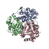 3rqcS S: Starting model for refinement |
|---|---|
| Similar structure data |


-Links
 PDBj
PDBj

- Assembly
| Deposited unit | 
| ||||||||||||||||||||||||||||
|---|---|---|---|---|---|---|---|---|---|---|---|---|---|---|---|---|---|---|---|---|---|---|---|---|---|---|---|---|---|
| 1 | 
| ||||||||||||||||||||||||||||
| 2 | 
| ||||||||||||||||||||||||||||
| Unit cell |
| ||||||||||||||||||||||||||||
| Noncrystallographic symmetry (NCS) | NCS oper:
| ||||||||||||||||||||||||||||
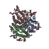
| Details | This is the truncated version of the enzyme that naturally forms a 42-mer multienzyme complex. In this structure a crystallographic dimer of trimer is observed which only assembles into an active trimer in solution |
-Components
| #1: Protein | Mass: 24857.930 Da / Num. of mol.: 6 Source method: isolated from a genetically manipulated source Source: (gene. exp.) Thermoplasma acidophilum (acidophilic) / Strain: DSM 1728 / Gene: Ta1436 / Plasmid: PET24A / Production host:  References: UniProt: Q9HIA5, 3-methyl-2-oxobutanoate dehydrogenase (2-methylpropanoyl-transferring) |
|---|
-Experimental details
-Experiment
| Experiment | Method: X-RAY DIFFRACTION / Number of used crystals: 1 |
|---|
- Sample preparation
| Crystal | Density Matthews: 2.65 Å3/Da / Density % sol: 53.61 % |
|---|---|
| Crystal grow | Temperature: 291 K / Method: vapor diffusion, hanging drop / pH: 6.5 Details: 8% (w/v) PEG4K; 3% (w/v) PGA; 0.1M Na Cacodylate pH 6.5, VAPOR DIFFUSION, HANGING DROP, temperature 291K |
-Data collection
| Diffraction | Mean temperature: 100 K |
|---|---|
| Diffraction source | Source: SYNCHROTRON / Site: Diamond  / Beamline: I04 / Wavelength: 0.97949 Å / Beamline: I04 / Wavelength: 0.97949 Å |
| Detector | Type: ADSC QUANTUM 315 / Detector: CCD / Date: Oct 8, 2011 |
| Radiation | Protocol: SINGLE WAVELENGTH / Monochromatic (M) / Laue (L): M / Scattering type: x-ray |
| Radiation wavelength | Wavelength: 0.97949 Å / Relative weight: 1 |
| Reflection | Resolution: 4.1→50 Å / Num. all: 12902 / Num. obs: 12274 / % possible obs: 99.2 % / Observed criterion σ(F): 0 / Observed criterion σ(I): 0 / Redundancy: 5.3 % / Biso Wilson estimate: 0 Å2 / Rmerge(I) obs: 0.122 / Net I/σ(I): 13.06 |
| Reflection shell | Resolution: 4.1→4.17 Å / Redundancy: 5.1 % / Rmerge(I) obs: 0.551 / Mean I/σ(I) obs: 2.48 / % possible all: 97.4 |
- Processing
| Software |
| |||||||||||||||||||||||||||||||||||||||||||||||||||||||||||||||||||||||||||||||||||||||||||||||||||||||||||||||||||||||||||||||||||||||||||||||||||||||||||||||||||||||||||||||
|---|---|---|---|---|---|---|---|---|---|---|---|---|---|---|---|---|---|---|---|---|---|---|---|---|---|---|---|---|---|---|---|---|---|---|---|---|---|---|---|---|---|---|---|---|---|---|---|---|---|---|---|---|---|---|---|---|---|---|---|---|---|---|---|---|---|---|---|---|---|---|---|---|---|---|---|---|---|---|---|---|---|---|---|---|---|---|---|---|---|---|---|---|---|---|---|---|---|---|---|---|---|---|---|---|---|---|---|---|---|---|---|---|---|---|---|---|---|---|---|---|---|---|---|---|---|---|---|---|---|---|---|---|---|---|---|---|---|---|---|---|---|---|---|---|---|---|---|---|---|---|---|---|---|---|---|---|---|---|---|---|---|---|---|---|---|---|---|---|---|---|---|---|---|---|---|---|
| Refinement | Method to determine structure: MOLECULAR REPLACEMENT Starting model: PDB entry 3RQC Resolution: 4.1→48.87 Å / Cor.coef. Fo:Fc: 0.949 / Cor.coef. Fo:Fc free: 0.915 / SU B: 164.294 / SU ML: 0.894 / Cross valid method: THROUGHOUT / ESU R Free: 1.086 / Stereochemistry target values: MAXIMUM LIKELIHOOD / Details: HYDROGENS HAVE BEEN USED IF PRESENT IN THE INPUT
| |||||||||||||||||||||||||||||||||||||||||||||||||||||||||||||||||||||||||||||||||||||||||||||||||||||||||||||||||||||||||||||||||||||||||||||||||||||||||||||||||||||||||||||||
| Solvent computation | Ion probe radii: 0.8 Å / Shrinkage radii: 0.8 Å / VDW probe radii: 1.2 Å / Solvent model: MASK | |||||||||||||||||||||||||||||||||||||||||||||||||||||||||||||||||||||||||||||||||||||||||||||||||||||||||||||||||||||||||||||||||||||||||||||||||||||||||||||||||||||||||||||||
| Displacement parameters | Biso mean: 74.61 Å2
| |||||||||||||||||||||||||||||||||||||||||||||||||||||||||||||||||||||||||||||||||||||||||||||||||||||||||||||||||||||||||||||||||||||||||||||||||||||||||||||||||||||||||||||||
| Refinement step | Cycle: LAST / Resolution: 4.1→48.87 Å
| |||||||||||||||||||||||||||||||||||||||||||||||||||||||||||||||||||||||||||||||||||||||||||||||||||||||||||||||||||||||||||||||||||||||||||||||||||||||||||||||||||||||||||||||
| Refine LS restraints |
| |||||||||||||||||||||||||||||||||||||||||||||||||||||||||||||||||||||||||||||||||||||||||||||||||||||||||||||||||||||||||||||||||||||||||||||||||||||||||||||||||||||||||||||||
| LS refinement shell | Resolution: 4.104→4.21 Å / Total num. of bins used: 20
| |||||||||||||||||||||||||||||||||||||||||||||||||||||||||||||||||||||||||||||||||||||||||||||||||||||||||||||||||||||||||||||||||||||||||||||||||||||||||||||||||||||||||||||||
| Refinement TLS params. | Method: refined / Refine-ID: X-RAY DIFFRACTION
| |||||||||||||||||||||||||||||||||||||||||||||||||||||||||||||||||||||||||||||||||||||||||||||||||||||||||||||||||||||||||||||||||||||||||||||||||||||||||||||||||||||||||||||||
| Refinement TLS group |
|
