Loading
PDBj
MenuPDBj@FacebookPDBj@TwitterPDBj@YouTubewwPDB FoundationwwPDB
RCSB PDBPDBeBMRBAdv. SearchSearch help
Search by PDB author
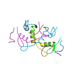
4R2Y
DownloadVisualize
BU of 4r2y by Molmil
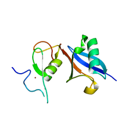
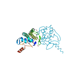
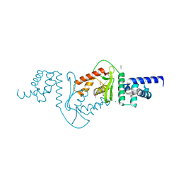
Crystal structure of APC11 RING domain
Descriptor: Anaphase-promoting complex subunit 11, ZINC ION
Authors:Brown, N.G, Watson, E.R, Weissmann, F, Jarvis, M.A, Vanderlinden, R, Grace, C.R.R, Frye, J.J, Dube, P, Qiao, R, Petzold, G, Cho, S.E, Alsharif, O, Bao, J, Zheng, J, Nourse, A, Kurinov, I, Peters, J.M, Stark, H, Schulman, B.A.
Deposit date:2014-08-13
Release date:2014-10-29
Last modified:2024-02-28
Method:X-RAY DIFFRACTION (1.755 Å)
Cite:Mechanism of Polyubiquitination by Human Anaphase-Promoting Complex: RING Repurposing for Ubiquitin Chain Assembly.
Mol.Cell, 56, 2014
5KHR
DownloadVisualize
BU of 5khr by Molmil
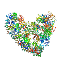
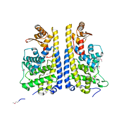
Model of human Anaphase-promoting complex/Cyclosome complex (APC15 deletion mutant) in complex with the E2 UBE2C/UBCH10 poised for ubiquitin ligation to substrate (APC/C-CDC20-substrate-UBE2C)
Descriptor: Anaphase-promoting complex subunit 1, Anaphase-promoting complex subunit 10, Anaphase-promoting complex subunit 11, ...
Authors:VanderLinden, R, Yamaguchi, M, Dube, P, Haselbach, D, Stark, H, Schulman, B.A.
Deposit date:2016-06-15
Release date:2016-08-24
Last modified:2024-03-06
Method:ELECTRON MICROSCOPY (6.1 Å)
Cite:Cryo-EM of Mitotic Checkpoint Complex-Bound APC/C Reveals Reciprocal and Conformational Regulation of Ubiquitin Ligation.
Mol.Cell, 63, 2016
5L9U
DownloadVisualize
BU of 5l9u by Molmil
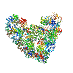
Model of human Anaphase-promoting complex/Cyclosome (APC/C-CDH1) with a cross linked Ubiquitin variant-substrate-UBE2C (UBCH10) complex representing key features of multiubiquitination
Descriptor: Anaphase-promoting complex subunit 1, Anaphase-promoting complex subunit 10, Anaphase-promoting complex subunit 11, ...
Authors:Brown, N.G, VanderLinden, R, Dube, P, Haselbach, D, Peters, J.M, Stark, H, Schulman, B.A.
Deposit date:2016-06-11
Release date:2016-09-14
Last modified:2024-05-08
Method:ELECTRON MICROSCOPY (6.4 Å)
Cite:Dual RING E3 Architectures Regulate Multiubiquitination and Ubiquitin Chain Elongation by APC/C.
Cell, 165, 2016
5KHU
DownloadVisualize
BU of 5khu by Molmil
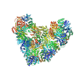
Model of human Anaphase-promoting complex/Cyclosome (APC15 deletion mutant), in complex with the Mitotic checkpoint complex (APC/C-CDC20-MCC) based on cryo EM data at 4.8 Angstrom resolution
Descriptor: Anaphase-promoting complex subunit 1, Anaphase-promoting complex subunit 10, Anaphase-promoting complex subunit 11, ...
Authors:Yamaguchi, M, VanderLinden, R, Dube, P, Stark, H, Schulman, B.
Deposit date:2016-06-15
Release date:2016-09-07
Last modified:2024-03-06
Method:ELECTRON MICROSCOPY (4.8 Å)
Cite:Cryo-EM of Mitotic Checkpoint Complex-Bound APC/C Reveals Reciprocal and Conformational Regulation of Ubiquitin Ligation.
Mol.Cell, 63, 2016
5L9T
DownloadVisualize
BU of 5l9t by Molmil
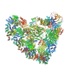
Model of human Anaphase-promoting complex/Cyclosome (APC/C-CDH1) with E2 UBE2S poised for polyubiquitination where UBE2S, APC2, and APC11 are modeled into low resolution density
Descriptor: Anaphase-promoting complex subunit 1, Anaphase-promoting complex subunit 10, Anaphase-promoting complex subunit 11, ...
Authors:Brown, N.G, VanderLinden, R, Dube, P, Haselbach, D, Peters, J.M, Stark, H, Schulman, B.A.
Deposit date:2016-06-11
Release date:2016-10-26
Last modified:2024-05-08
Method:ELECTRON MICROSCOPY (6.4 Å)
Cite:Dual RING E3 Architectures Regulate Multiubiquitination and Ubiquitin Chain Elongation by APC/C.
Cell, 165, 2016
6XFN
DownloadVisualize
BU of 6xfn by Molmil
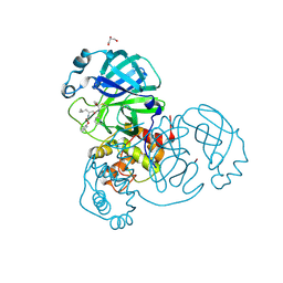
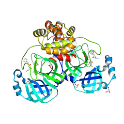
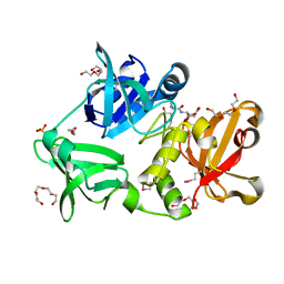
Crystal structure of the SARS-CoV-2 (COVID-19) main protease in complex with UAW243
Descriptor: 3C-like proteinase, GLYCEROL, UAW243
Authors:Sacco, M, Ma, C, Wang, J, Chen, Y.
Deposit date:2020-06-15
Release date:2020-06-24
Last modified:2023-11-15
Method:X-RAY DIFFRACTION (1.7 Å)
Cite:Structure and inhibition of the SARS-CoV-2 main protease reveal strategy for developing dual inhibitors against M pro and cathepsin L.
Sci Adv, 6, 2020
6XBI
DownloadVisualize
BU of 6xbi by Molmil
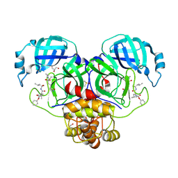
Crystal structure of the SARS-CoV-2 (COVID-19) main protease in complex with inhibitor UAW248
Descriptor: 3C-like proteinase, DIMETHYL SULFOXIDE, GLYCEROL, ...
Authors:Sacco, M, Ma, C, Wang, J, Chen, Y.
Deposit date:2020-06-06
Release date:2020-06-17
Last modified:2023-11-15
Method:X-RAY DIFFRACTION (1.7 Å)
Cite:Structure and inhibition of the SARS-CoV-2 main protease reveal strategy for developing dual inhibitors against M pro and cathepsin L.
Sci Adv, 6, 2020
6XA4
DownloadVisualize
BU of 6xa4 by Molmil
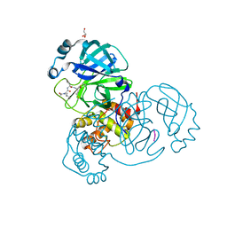
Crystal structure of the SARS-CoV-2 (COVID-19) main protease in complex with UAW241
Descriptor: 3C-like proteinase, GLYCEROL, inhibitor UAW241
Authors:Sacco, M, Ma, C, Wang, J, Chen, Y.
Deposit date:2020-06-03
Release date:2020-06-17
Last modified:2023-11-15
Method:X-RAY DIFFRACTION (1.65 Å)
Cite:Structure and inhibition of the SARS-CoV-2 main protease reveal strategy for developing dual inhibitors against M pro and cathepsin L.
Sci Adv, 6, 2020
6XBH
DownloadVisualize
BU of 6xbh by Molmil
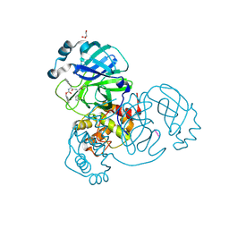
Crystal structure of the SARS-CoV-2 (COVID-19) main protease in complex with inhibitor UAW247
Descriptor: 3C-like proteinase, GLYCEROL, SODIUM ION, ...
Authors:Sacco, M, Ma, C, Wang, J, Chen, Y.
Deposit date:2020-06-06
Release date:2020-06-17
Last modified:2023-11-15
Method:X-RAY DIFFRACTION (1.6 Å)
Cite:Structure and inhibition of the SARS-CoV-2 main protease reveal strategy for developing dual inhibitors against M pro and cathepsin L.
Sci Adv, 6, 2020
6XBG
DownloadVisualize
BU of 6xbg by Molmil
Crystal structure of the SARS-CoV-2 (COVID-19) main protease in complex with inhibitor UAW246
Descriptor: 3C-like proteinase, GLYCEROL, SODIUM ION, ...
Authors:Sacco, M, Ma, C, Wang, J, Chen, Y.
Deposit date:2020-06-05
Release date:2020-06-17
Last modified:2023-11-15
Method:X-RAY DIFFRACTION (1.45 Å)
Cite:Structure and inhibition of the SARS-CoV-2 main protease reveal strategy for developing dual inhibitors against M pro and cathepsin L.
Sci Adv, 6, 2020
2MT5
DownloadVisualize
BU of 2mt5 by Molmil
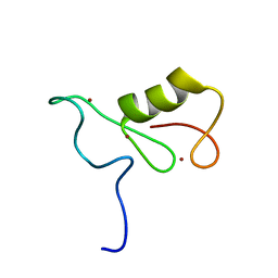
Isolated Ring domain
Descriptor: Anaphase-promoting complex subunit 11, ZINC ION
Authors:Brown, N.G, Watson, E.R, Weissman, F, Royappa, G, Schulman, B, Jarvis, M, Vanderlinden, R, Frye, J.J, Qiao, R, Petzold, G, Peters, J, Stark, H.
Deposit date:2014-08-13
Release date:2014-10-29
Last modified:2024-05-15
Method:SOLUTION NMR
Cite:Mechanism of Polyubiquitination by Human Anaphase-Promoting Complex: RING Repurposing for Ubiquitin Chain Assembly.
Mol.Cell, 56, 2014
5JG6
DownloadVisualize
BU of 5jg6 by Molmil
APC11-Ubv shows role of noncovalent RING-Ubiquitin interactions in processive multiubiquitination and Ubiquitin chain elongation by APC/C
Descriptor: Anaphase-promoting complex subunit 11, Polyubiquitin-B, ZINC ION
Authors:Brown, N.G, Zhang, W, Yu, S, Miller, D.J, Sidhu, S.S, Schulman, B.A.
Deposit date:2016-04-19
Release date:2016-06-15
Last modified:2023-09-27
Method:X-RAY DIFFRACTION (2.0013 Å)
Cite:Dual RING E3 Architectures Regulate Multiubiquitination and Ubiquitin Chain Elongation by APC/C.
Cell, 165, 2016
2A6A
DownloadVisualize
BU of 2a6a by Molmil
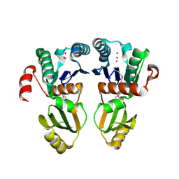
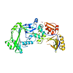
Crystal structure of Glycoprotein endopeptidase (tm0874) from THERMOTOGA MARITIMA at 2.50 A resolution
Descriptor: UNKNOWN LIGAND, hypothetical protein TM0874
Authors:Joint Center for Structural Genomics (JCSG)
Deposit date:2005-07-02
Release date:2005-07-19
Last modified:2023-01-25
Method:X-RAY DIFFRACTION (2.5 Å)
Cite:Structure of an essential bacterial protein YeaZ (TM0874) from Thermotoga maritima at 2.5 A resolution.
Acta Crystallogr.,Sect.F, 66, 2010
3KK7
DownloadVisualize
BU of 3kk7 by Molmil
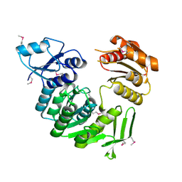
Crystal structure of Putative cell invasion protein with MAC/Perforin domain (NP_812351.1) from BACTERIODES THETAIOTAOMICRON VPI-5482 at 2.46 A resolution
Descriptor: (4S)-2-METHYL-2,4-PENTANEDIOL, 1,2-ETHANEDIOL, CHLORIDE ION, ...
Authors:Joint Center for Structural Genomics (JCSG)
Deposit date:2009-11-04
Release date:2009-11-24
Last modified:2023-02-01
Method:X-RAY DIFFRACTION (2.46 Å)
Cite:Structure of a membrane-attack complex/perforin (MACPF) family protein from the human gut symbiont Bacteroides thetaiotaomicron.
Acta Crystallogr.,Sect.F, 66, 2010
3L5O
DownloadVisualize
BU of 3l5o by Molmil
Crystal structure of protein with unknown function from DUF364 family (ZP_00559375.1) from Desulfitobacterium hafniense DCB-2 at 2.01 A resolution
Descriptor: 1,2-ETHANEDIOL, CHLORIDE ION, IMIDAZOLE, ...
Authors:Joint Center for Structural Genomics, Joint Center for Structural Genomics (JCSG)
Deposit date:2009-12-22
Release date:2010-02-02
Last modified:2023-02-01
Method:X-RAY DIFFRACTION (2.01 Å)
Cite:Structure of the first representative of Pfam family PF04016 (DUF364) reveals enolase and Rossmann-like folds that combine to form a unique active site with a possible role in heavy-metal chelation.
Acta Crystallogr.,Sect.F, 66, 2010
1J6U
DownloadVisualize
BU of 1j6u by Molmil
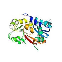
Crystal structure of UDP-N-acetylmuramate-alanine ligase MurC (TM0231) from Thermotoga maritima at 2.3 A resolution
Descriptor: UDP-N-acetylmuramate-alanine ligase MurC
Authors:Joint Center for Structural Genomics (JCSG)
Deposit date:2002-08-29
Release date:2002-11-06
Last modified:2023-01-25
Method:X-RAY DIFFRACTION (2.3 Å)
Cite:Crystal structure of an Udp-n-acetylmuramate-alanine ligase MurC (TM0231) from Thermotoga maritima at 2.3 A resolution.
Proteins, 55, 2004
3H0N
DownloadVisualize
BU of 3h0n by Molmil
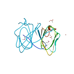
Crystal structure of a duf1470 family protein (jann_2411) from jannaschia sp. ccs1 at 1.45 A resolution
Descriptor: ACETATE ION, GLYCEROL, NICKEL (II) ION, ...
Authors:Joint Center for Structural Genomics (JCSG)
Deposit date:2009-04-09
Release date:2009-04-21
Last modified:2023-02-01
Method:X-RAY DIFFRACTION (1.45 Å)
Cite:The structure of Jann_2411 (DUF1470) from Jannaschia sp. at 1.45  resolution reveals a new fold (the ABATE domain) and suggests its possible role as a transcription regulator.
Acta Crystallogr.,Sect.F, 66, 2010
3DEE
DownloadVisualize
BU of 3dee by Molmil
CRYSTAL STRUCTURE OF A PUTATIVE REGULATORY PROTEIN INVOLVED IN TRANSCRIPTION (NGO1945) FROM NEISSERIA GONORRHOEAE FA 1090 AT 2.25 A RESOLUTION
Descriptor: CHLORIDE ION, IMIDAZOLE, Putative regulatory protein
Authors:Joint Center for Structural Genomics (JCSG)
Deposit date:2008-06-09
Release date:2008-08-05
Last modified:2023-02-01
Method:X-RAY DIFFRACTION (2.1 Å)
Cite:The structure of the first representative of Pfam family PF09836 reveals a two-domain organization and suggests involvement in transcriptional regulation.
Acta Crystallogr.,Sect.F, 66, 2010
3EQX
DownloadVisualize
BU of 3eqx by Molmil
CRYSTAL STRUCTURE OF A FIC FAMILY PROTEIN (SO_4266) FROM SHEWANELLA ONEIDENSIS AT 1.6 A RESOLUTION
Descriptor: FIC DOMAIN CONTAINING TRANSCRIPTIONAL REGULATOR, TRIETHYLENE GLYCOL
Authors:Joint Center for Structural Genomics (JCSG)
Deposit date:2008-10-01
Release date:2008-10-14
Last modified:2023-02-01
Method:X-RAY DIFFRACTION (1.6 Å)
Cite:Crystal structure of the Fic (Filamentation induced by cAMP) family protein SO4266 (gi|24375750) from Shewanella oneidensis MR-1 at 1.6 A resolution.
Proteins, 75, 2009
3H41
DownloadVisualize
BU of 3h41 by Molmil
CRYSTAL STRUCTURE OF A NLPC/P60 FAMILY PROTEIN (BCE_2878) FROM BACILLUS CEREUS ATCC 10987 AT 1.79 A RESOLUTION
Descriptor: ALANINE, D-GLUTAMIC ACID, NLP/P60 family protein, ...
Authors:Joint Center for Structural Genomics (JCSG)
Deposit date:2009-04-17
Release date:2009-05-05
Last modified:2023-02-01
Method:X-RAY DIFFRACTION (1.79 Å)
Cite:Structure of the gamma-D-glutamyl-L-diamino acid endopeptidase YkfC from Bacillus cereus in complex with L-Ala-[gamma]-D-Glu: insights into substrate recognition by NlpC/P60 cysteine peptidases
Acta Crystallogr.,Sect.F, 66, 2010
3H50
DownloadVisualize
BU of 3h50 by Molmil
CRYSTAL STRUCTURE OF A TETRACENOMYCIN POLYKETIDE SYNTHESIS PROTEIN (TCMJ) FROM XANTHOMONAS CAMPESTRIS PV. CAMPESTRIS AT 1.60 A RESOLUTION
Descriptor: ACETATE ION, Tetracenomycin polyketide synthesis protein, ZINC ION
Authors:Joint Center for Structural Genomics (JCSG)
Deposit date:2009-04-21
Release date:2009-05-05
Last modified:2023-02-01
Method:X-RAY DIFFRACTION (1.6 Å)
Cite:Conformational changes associated with the binding of zinc acetate at the putative active site of XcTcmJ, a cupin from Xanthomonas campestris pv. campestris.
Acta Crystallogr.,Sect.F, 66, 2010
3HSA
DownloadVisualize
BU of 3hsa by Molmil
Crystal structure of pleckstrin homology domain (YP_926556.1) from SHEWANELLA AMAZONENSIS SB2B at 1.99 A resolution
Descriptor: DI(HYDROXYETHYL)ETHER, GLYCEROL, pleckstrin homology domain
Authors:Joint Center for Structural Genomics (JCSG)
Deposit date:2009-06-10
Release date:2009-06-23
Last modified:2023-02-01
Method:X-RAY DIFFRACTION (1.99 Å)
Cite:Bacterial pleckstrin homology domains: a prokaryotic origin for the PH domain.
J.Mol.Biol., 396, 2010
3HBZ
DownloadVisualize
BU of 3hbz by Molmil
Crystal structure of a putative glycoside hydrolase (bt_2081) from bacteroides thetaiotaomicron vpi-5482 at 2.05 A resolution
Descriptor: ACETATE ION, CACODYLATE ION, CALCIUM ION, ...
Authors:Joint Center for Structural Genomics (JCSG)
Deposit date:2009-05-05
Release date:2009-05-26
Last modified:2023-02-01
Method:X-RAY DIFFRACTION (2.05 Å)
Cite:Structure of Bacteroides thetaiotaomicron BT2081 at 2.05 A resolution: the first structural representative of a new protein family that may play a role in carbohydrate metabolism.
Acta Crystallogr.,Sect.F, 66, 2010
3K5J
DownloadVisualize
BU of 3k5j by Molmil
Crystal structure of Putative SUFU (suppressor of fused protein) homolog (YP_208451.1) from Neisseria gonorrhoeae FA 1090 at 1.40 A resolution
Descriptor: GLYCEROL, SULFATE ION, Suppressor of fused family protein
Authors:Joint Center for Structural Genomics (JCSG)
Deposit date:2009-10-07
Release date:2010-01-26
Last modified:2019-07-17
Method:X-RAY DIFFRACTION (1.4 Å)
Cite:The crystal structure of a bacterial Sufu-like protein defines a novel group of bacterial proteins that are similar to the N-terminal domain of human Sufu.
Protein Sci., 19, 2010
3BYQ
DownloadVisualize
BU of 3byq by Molmil
Crystal structure of a duf1185 family protein (bb2672) from bordetella bronchiseptica rb50 at 1.70 A resolution
Descriptor: 1,2-ETHANEDIOL, CHLORIDE ION, TETRAETHYLENE GLYCOL, ...
Authors:Joint Center for Structural Genomics (JCSG)
Deposit date:2008-01-16
Release date:2008-01-29
Last modified:2023-02-01
Method:X-RAY DIFFRACTION (1.7 Å)
Cite:Structures of the first representatives of Pfam family PF06684 (DUF1185) reveal a novel variant of the Bacillus chorismate mutase fold and suggest a role in amino-acid metabolism.
Acta Crystallogr.,Sect.F, 66, 2010


1234>

225158

PDB entries from 2024-09-18

PDB statisticsPDBj update infoContact PDBjnumon