Loading
PDBj
メニューPDBj@FacebookPDBj@TwitterPDBj@YouTubewwPDB FoundationwwPDB
RCSB PDBPDBeBMRBAdv. SearchSearch help

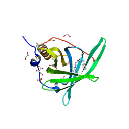
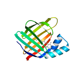
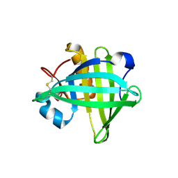
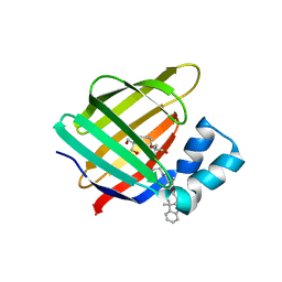
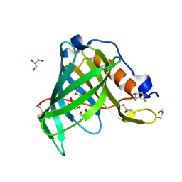
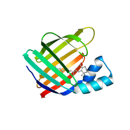
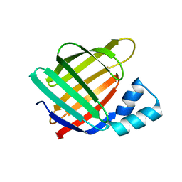
6MOQ
DownloadVisualize
BU of 6moq by Molmil
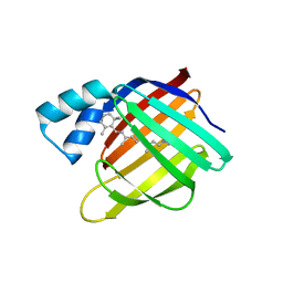
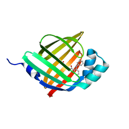
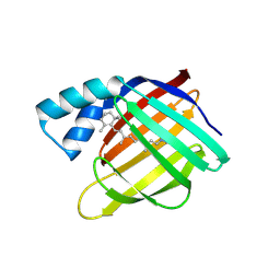
Crystal Structure of the All-trans Retinal-Bound R111K:Y134F:T54V:R132Q:P39Y:R59Y:L121W Mutant of Human Cellular Retinoic Acid Binding Protein II in the Dark at 2.2 Angstrom Resolution
分子名称: Cellular retinoic acid-binding protein 2, RETINAL
著者Ghanbarpour, A, Geiger, J.
登録日2018-10-04
公開日2019-01-09
最終更新日2023-10-11
実験手法X-RAY DIFFRACTION (2.198 Å)
主引用文献Mimicking Microbial Rhodopsin Isomerization in a Single Crystal.
J. Am. Chem. Soc., 141, 2019
6MOW
DownloadVisualize
BU of 6mow by Molmil
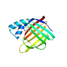
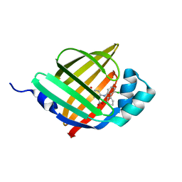
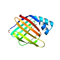
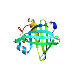
Crystal Structure of the apo R111K:Y134F:T54V:R132Q:P39Y:R59Y:L121Q mutant of Human Cellular Retinoic Acid Binding Protein II at 2.3 Angstrom Resolution
分子名称: Cellular retinoic acid-binding protein 2
著者Ghanbarpour, A, Geiger, J.
登録日2018-10-04
公開日2019-10-09
最終更新日2023-10-11
実験手法X-RAY DIFFRACTION (2.354 Å)
主引用文献Crystal Structure of the apo R111K:Y134F:T54V:R132Q:P39Y:R59Y:L121Q mutant of Human Cellular Retinoic Acid Binding Protein II at 2.3 Angstrom Resolution
To Be Published
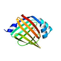
6MOV
DownloadVisualize
BU of 6mov by Molmil
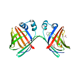
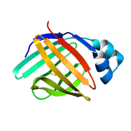
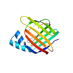
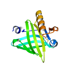
Crystal Structure of the All-trans Retinal bound R111K:Y134F:T54V:R132Q:P39Y:R59Y:L121Q Human Cellular Retinoic Acid Binding Protein II in the Dark at 1.75 Angstrom Resolution
分子名称: Cellular retinoic acid-binding protein 2, RETINAL
著者Ghanbarpour, A, Geiger, J.
登録日2018-10-04
公開日2019-01-09
最終更新日2023-10-11
実験手法X-RAY DIFFRACTION (1.752 Å)
主引用文献Mimicking Microbial Rhodopsin Isomerization in a Single Crystal.
J. Am. Chem. Soc., 141, 2019
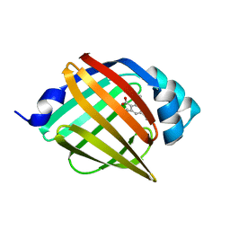
6MOX
DownloadVisualize
BU of 6mox by Molmil
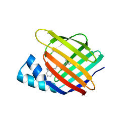
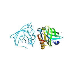
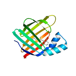
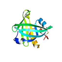
Crystal Structure of the All-trans Retinal-Bound R111K:Y134F:T54V:R132Q:P39Q:R59Y:L121E Human Cellular Retinoic Acid Binding Protein II Mutant in the Dark at 2.18 Angstrom Resolution
分子名称: Cellular retinoic acid-binding protein 2, RETINAL
著者Ghanbarpour, A, Geiger, J.
登録日2018-10-04
公開日2019-01-09
最終更新日2023-10-11
実験手法X-RAY DIFFRACTION (2.18 Å)
主引用文献Mimicking Microbial Rhodopsin Isomerization in a Single Crystal.
J. Am. Chem. Soc., 141, 2019
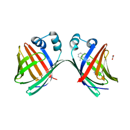
6MLB
DownloadVisualize
BU of 6mlb by Molmil
Crystal structure of the holo retinal-bound domain-swapped dimer Q108K:K40L:T51F mutant of human cellular retinol binding protein II
分子名称: ACETATE ION, GLYCEROL, RETINAL, ...
著者Ghanbarpour, A, Geiger, J.
登録日2018-09-27
公開日2019-10-16
最終更新日2023-10-11
実験手法X-RAY DIFFRACTION (2.15 Å)
主引用文献Engineering the hCRBPII Domain-Swapped Dimer into a New Class of Protein Switches.
J.Am.Chem.Soc., 141, 2019
6MR0
DownloadVisualize
BU of 6mr0 by Molmil
Crystal Structure of the All-trans Retinal Bound R111K:Y134F:T54V:R132Q:P39Q:R59Y:L121E Human Cellular Retinoic Acid Binding Protein II Mutant After 5 Minutes UV irradiation at 2.6 Angstrom Resolution
分子名称: Cellular retinoic acid-binding protein 2, RETINAL
著者Ghanbarpour, A, Geiger, J.
登録日2018-10-11
公開日2019-01-09
最終更新日2023-10-11
実験手法X-RAY DIFFRACTION (2.649 Å)
主引用文献Mimicking Microbial Rhodopsin Isomerization in a Single Crystal.
J. Am. Chem. Soc., 141, 2019
4PSQ
DownloadVisualize
BU of 4psq by Molmil
Crystal Structure of Retinol-Binding Protein 4 (RBP4) in complex with a non-retinoid ligand
分子名称: (1-benzyl-1H-imidazol-4-yl)[4-(2-chlorophenyl)piperazin-1-yl]methanone, 1,2-ETHANEDIOL, PHOSPHATE ION, ...
著者Wang, Z, Johnstone, S, Walker, N.
登録日2014-03-07
公開日2014-07-02
実験手法X-RAY DIFFRACTION (2.4 Å)
主引用文献Structure-assisted discovery of the first non-retinoid ligands for Retinol-Binding Protein 4.
Bioorg.Med.Chem.Lett., 24, 2014
6MKV
DownloadVisualize
BU of 6mkv by Molmil
Crystal structure of Retinal-bound holo Q108K:K40L:T51W domain-swapped dimer of human cellular retinol binding protein 2
分子名称: ACETATE ION, GLYCEROL, RETINAL, ...
著者Ghanbarpour, A, Geiger, J.
登録日2018-09-26
公開日2019-10-16
最終更新日2023-10-11
実験手法X-RAY DIFFRACTION (2.107 Å)
主引用文献Engineering the hCRBPII Domain-Swapped Dimer into a New Class of Protein Switches.
J.Am.Chem.Soc., 141, 2019
4NNS
DownloadVisualize
BU of 4nns by Molmil
Crystal structure of FABP4 in complex with novel inhibitor
分子名称: 2,4,6-tri(propan-2-yl)benzenesulfonic acid, Fatty acid-binding protein, adipocyte
著者Liu, Q.F, Chen, T.T, Xu, Y.C.
登録日2013-11-19
公開日2014-11-19
最終更新日2024-03-20
実験手法X-RAY DIFFRACTION (1.53 Å)
主引用文献The complex structure of FABP4 with novel inhibitors
To be Published
4NNT
DownloadVisualize
BU of 4nnt by Molmil
Crystal structure of FABP4 in complex with novel inhibitor
分子名称: 2,4,6-tri(propan-2-yl)benzoic acid, Fatty acid-binding protein, adipocyte
著者Liu, Q.F, Chen, T.T, Xu, Y.C.
登録日2013-11-19
公開日2014-11-19
最終更新日2024-03-20
実験手法X-RAY DIFFRACTION (1.53 Å)
主引用文献The complex structure of FABP4 with novel inhibitors
To be Published
6LJW
DownloadVisualize
BU of 6ljw by Molmil
Crystal structure of human FABP4 in complex with a novel inhibitor
分子名称: 1,2-ETHANEDIOL, 2-phenylazanylbenzoic acid, Fatty acid-binding protein, ...
著者Su, H.X, Zhang, X.L, Li, M.J, Xu, Y.C.
登録日2019-12-17
公開日2020-04-15
最終更新日2023-11-22
実験手法X-RAY DIFFRACTION (1.4 Å)
主引用文献Exploration of Fragment Binding Poses Leading to Efficient Discovery of Highly Potent and Orally Effective Inhibitors of FABP4 for Anti-inflammation.
J.Med.Chem., 63, 2020
6LJT
DownloadVisualize
BU of 6ljt by Molmil
Crystal structure of human FABP4 in complex with a novel inhibitor
分子名称: 1,2-ETHANEDIOL, 2-[(3-chloranyl-2-phenyl-phenyl)amino]benzoic acid, Fatty acid-binding protein, ...
著者Su, H.X, Zhang, X.L, Li, M.J, Xu, Y.C.
登録日2019-12-17
公開日2020-04-15
最終更新日2023-11-22
実験手法X-RAY DIFFRACTION (1.45 Å)
主引用文献Exploration of Fragment Binding Poses Leading to Efficient Discovery of Highly Potent and Orally Effective Inhibitors of FABP4 for Anti-inflammation.
J.Med.Chem., 63, 2020
6L7K
DownloadVisualize
BU of 6l7k by Molmil
solution structure of hIFABP V60C/Y70C variant.
分子名称: Fatty acid-binding protein, intestinal
著者Fan, J, Yang, D.
登録日2019-11-01
公開日2020-11-04
最終更新日2023-06-14
実験手法SOLUTION NMR
主引用文献Ligand Entry into Fatty Acid Binding Protein via Local Unfolding Instead of Gap Widening.
Biophys.J., 118, 2020
4LZU
DownloadVisualize
BU of 4lzu by Molmil
Bovine beta-lactoglobulin crystallized in the presence of 2 mM zinc chloride
分子名称: Beta-lactoglobulin, ZINC ION
著者Oelker, M, Zocher, G, Stehle, T.
登録日2013-08-01
公開日2014-08-06
最終更新日2023-09-20
実験手法X-RAY DIFFRACTION (2.4 Å)
主引用文献Non-Classical Pathways of Protein Crystallization in the Presence of Multivalent Metal Ions
To be Published
6MQI
DownloadVisualize
BU of 6mqi by Molmil
Crystal Structure of the All-trans Retinal bound R111K:Y134F:T54V:R132Q:P39Y:R59Y:L121Q mutant of Human Cellular Retinoic Acid Binding Protein II Irradiated with 400 nm Laser for 5 minutes at 1.87 Angstrom Resolution
分子名称: Cellular retinoic acid-binding protein 2, RETINAL
著者Ghanbarpour, A, Geiger, J.
登録日2018-10-09
公開日2019-01-09
最終更新日2023-10-11
実験手法X-RAY DIFFRACTION (1.87 Å)
主引用文献Mimicking Microbial Rhodopsin Isomerization in a Single Crystal.
J. Am. Chem. Soc., 141, 2019
6MQZ
DownloadVisualize
BU of 6mqz by Molmil
Crystal Structure of the 13-cis Product of the All-trans Retinal-Bound R111K:Y134F:T54V:R132Q:P39Y:R59Y:L121E Mutant of Human Cellular Retinoic Acid Binding Protein II Irradiated with 400 nm Laser (5 minutes) at 2.07 Angstrom
分子名称: Cellular retinoic acid-binding protein 2, GLYCEROL, RETINAL
著者Ghanbarpour, A, Geiger, J.
登録日2018-10-11
公開日2019-01-09
最終更新日2023-10-11
実験手法X-RAY DIFFRACTION (2.07 Å)
主引用文献Mimicking Microbial Rhodopsin Isomerization in a Single Crystal.
J. Am. Chem. Soc., 141, 2019
6MQX
DownloadVisualize
BU of 6mqx by Molmil
Crystal Structure of All-trans Retinal-Bound R111K:Y134F:T54V:R132Q:P39Y:R59Y:L121E Human Cellular Retinoic Acid Binding Protein II Irradiated with 400 nm Laser (30 seconds) and Subsequently Dark Adapted (25 minutes) at 2.0 Angstrom Resolution
分子名称: Cellular retinoic acid-binding protein 2, GLYCEROL, RETINAL
著者Ghanbarpour, A, Geiger, J.
登録日2018-10-11
公開日2019-01-09
最終更新日2023-10-11
実験手法X-RAY DIFFRACTION (2.01 Å)
主引用文献Mimicking Microbial Rhodopsin Isomerization in a Single Crystal.
J. Am. Chem. Soc., 141, 2019
6MOR
DownloadVisualize
BU of 6mor by Molmil
Crystal Structure of the All-Trans Retinal-Bound R111K:Y134F:T54V:R132Q:P39Y:R59Y:L121Y Mutant of Human Cellular Retinoic Acid Binding Protein II in the Dark at 1.79 Angstrom Resolution
分子名称: Cellular retinoic acid-binding protein 2, RETINAL
著者Ghanbarpour, A, Geiger, J.
登録日2018-10-04
公開日2019-01-09
最終更新日2023-10-11
実験手法X-RAY DIFFRACTION (1.79 Å)
主引用文献Mimicking Microbial Rhodopsin Isomerization in a Single Crystal.
J. Am. Chem. Soc., 141, 2019
6MQW
DownloadVisualize
BU of 6mqw by Molmil
Crystal Structure of All-trans Retinal-Bound R111K:Y134F:T54V:R132Q:P39Y:R59Y:L121E Human Cellular Retinoic Acid Binding Protein II Irradiated with 400 nm laser (30 seconds) and subsequently dark adapted (10 minutes) at 2.1 Angstrom Resolution
分子名称: Cellular retinoic acid-binding protein 2, RETINAL
著者Ghanbarpour, A, Geiger, J.
登録日2018-10-11
公開日2019-01-09
最終更新日2023-10-11
実験手法X-RAY DIFFRACTION (2.09 Å)
主引用文献Mimicking Microbial Rhodopsin Isomerization in a Single Crystal.
J. Am. Chem. Soc., 141, 2019
4OS3
DownloadVisualize
BU of 4os3 by Molmil
Three-dimensional structure of the C65A-W112F double mutant of Human lipocalin-type Prostaglandin D Synthase apo-form
分子名称: Prostaglandin-H2 D-isomerase
著者Perduca, M, Bovi, M, Bertinelli, M, Bertini, E, Destefanis, L, Carrizo, M.E, Capaldi, S, Monaco, H.L.
登録日2014-02-12
公開日2014-08-06
最終更新日2014-12-17
実験手法X-RAY DIFFRACTION (1.4 Å)
主引用文献High-resolution structures of mutants of residues that affect access to the ligand-binding cavity of human lipocalin-type prostaglandin D synthase.
Acta Crystallogr.,Sect.D, 70, 2014
4ODD
DownloadVisualize
BU of 4odd by Molmil
Crystal structure of a dog lipocalin allergen
分子名称: LIPOCALIN ALLERGEN
著者Niemi, M.H, Rouvinen, J.
登録日2014-01-10
公開日2014-06-04
実験手法X-RAY DIFFRACTION (2.6 Å)
主引用文献Structural aspects of dog allergies: The crystal structure of a dog dander allergen Can f 4.
Mol.Immunol., 61, 2014
4ORW
DownloadVisualize
BU of 4orw by Molmil
Three-dimensional structure of the C65A-K59A double mutant of Human lipocalin-type Prostaglandin D Synthase apo-form
分子名称: Prostaglandin-H2 D-isomerase
著者Perduca, M, Bovi, M, Bertinelli, M, Bertini, E, Destefanis, L, Carrizo, M.E, Capaldi, S, Monaco, H.L.
登録日2014-02-12
公開日2014-08-06
最終更新日2023-09-20
実験手法X-RAY DIFFRACTION (1.664 Å)
主引用文献High-resolution structures of mutants of residues that affect access to the ligand-binding cavity of human lipocalin-type prostaglandin D synthase.
Acta Crystallogr.,Sect.D, 70, 2014
4OS8
DownloadVisualize
BU of 4os8 by Molmil
Three-dimensional structure of the C65A-W54F-W112F triple mutant of Human lipocalin-type Prostaglandin D Synthase apo-form
分子名称: Prostaglandin-H2 D-isomerase
著者Perduca, M, Bovi, M, Bertinelli, M, Bertini, E, Destefanis, L, Carrizo, M.E, Capaldi, S, Monaco, H.L.
登録日2014-02-12
公開日2014-08-06
最終更新日2020-02-26
実験手法X-RAY DIFFRACTION (1.69 Å)
主引用文献High-resolution structures of mutants of residues that affect access to the ligand-binding cavity of human lipocalin-type prostaglandin D synthase.
Acta Crystallogr.,Sect.D, 70, 2014
4QGW
DownloadVisualize
BU of 4qgw by Molmil
Crystal sturcture of the R132K:R111L:L121D mutant of Cellular Retinoic Acid Binding ProteinII complexed with a synthetic ligand (Merocyanine) at 1.77 angstrom resolution
分子名称: (2E,4E,6E)-3-methyl-6-(1,3,3-trimethyl-1,3-dihydro-2H-indol-2-ylidene)hexa-2,4-dienal, 2-[3-(2-HYDROXY-1,1-DIHYDROXYMETHYL-ETHYLAMINO)-PROPYLAMINO]-2-HYDROXYMETHYL-PROPANE-1,3-DIOL, Cellular retinoic acid-binding protein 2
著者Nosrati, M, Yapici, I, Geiger, J.H.
登録日2014-05-26
公開日2015-01-28
最終更新日2015-02-11
実験手法X-RAY DIFFRACTION (1.77 Å)
主引用文献"Turn-on" protein fluorescence: in situ formation of cyanine dyes.
J.Am.Chem.Soc., 137, 2015
1JYD
DownloadVisualize
BU of 1jyd by Molmil
Crystal Structure of Recombinant Human Serum Retinol-Binding Protein at 1.7 A Resolution
分子名称: GLYCEROL, PLASMA RETINOL-BINDING PROTEIN
著者Greene, L.H, Chrysina, E.D, Irons, L.I, Papageorgiou, A.C, Acharya, K.R, Brew, K.
登録日2001-09-12
公開日2003-07-01
最終更新日2023-08-16
実験手法X-RAY DIFFRACTION (1.7 Å)
主引用文献Role of conserved residues in structure and stability: Tryptophans of human serum retinol-binding protein, a model for the lipocalin superfamily
Protein Sci., 10, 2001

221371

件を2024-06-19に公開中

PDB statisticsPDBj update infoContact PDBjnumon