 Movie
Movie Controller

[English] 日本語
 Yorodumi
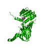
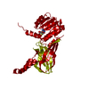
- PDB-4hc4: Human HMT1 hnRNP methyltransferase-like protein 6 (S. cerevisiae) -
+ Open data

- Basic information
| Entry | Database: PDB / ID: 4hc4 | ||||||
|---|---|---|---|---|---|---|---|
| Title | Human HMT1 hnRNP methyltransferase-like protein 6 (S. cerevisiae) | ||||||
 Components Components | Protein arginine N-methyltransferase 6 | ||||||
 Keywords Keywords |  TRANSFERASE / HRMT1L6 / METHYLTRANSFERASE / S-ADENOSYL-L-HOMOCYSTEINE / STRUCTURAL GENOMICS / STRUCTURAL GENOMICS CONSORTIUM / SGC TRANSFERASE / HRMT1L6 / METHYLTRANSFERASE / S-ADENOSYL-L-HOMOCYSTEINE / STRUCTURAL GENOMICS / STRUCTURAL GENOMICS CONSORTIUM / SGC | ||||||
| Function / homology |  Function and homology information Function and homology informationhistone H2AR3 methyltransferase activity / peptidyl-arginine methylation, to asymmetrical-dimethyl arginine / protein-arginine omega-N monomethyltransferase activity / histone H4R3 methyltransferase activity / histone H3R2 methyltransferase activity / type I protein arginine methyltransferase / protein-arginine omega-N asymmetric methyltransferase activity / regulation of megakaryocyte differentiation / histone arginine N-methyltransferase activity / protein-arginine N-methyltransferase activity ...histone H2AR3 methyltransferase activity / peptidyl-arginine methylation, to asymmetrical-dimethyl arginine / protein-arginine omega-N monomethyltransferase activity / histone H4R3 methyltransferase activity / histone H3R2 methyltransferase activity / type I protein arginine methyltransferase / protein-arginine omega-N asymmetric methyltransferase activity / regulation of megakaryocyte differentiation / histone arginine N-methyltransferase activity / protein-arginine N-methyltransferase activity / regulation of mitochondrion organization / histone H3 methyltransferase activity / histone methyltransferase activity / negative regulation of ubiquitin-dependent protein catabolic process / regulation of signal transduction by p53 class mediator / base-excision repair / protein modification process / RUNX1 regulates genes involved in megakaryocyte differentiation and platelet function / RMTs methylate histone arginines / cellular senescence / histone binding / negative regulation of DNA-templated transcription / chromatin binding / nucleolus / negative regulation of transcription by RNA polymerase II / nucleoplasm / nucleusSimilarity search - Function | ||||||
| Biological species |  Homo sapiens (human) Homo sapiens (human) | ||||||
| Method | X-RAY DIFFRACTION / SYNCHROTRON / MOLECULAR REPLACEMENT / Resolution: 1.97 Å | ||||||
 Authors Authors | Dong, A. / Zeng, H. / He, H. / El Bakkouri, M. / Bountra, C. / Arrowsmith, C.H. / Edwards, A.M. / Brown, P.J. / Wu, H. / Structural Genomics Consortium (SGC) | ||||||
 Citation Citation | Journal: Biochem. J. / Year: 2016 Title: Structural basis of arginine asymmetrical dimethylation by PRMT6. Authors: Wu, H. / Zheng, W. / Eram, M.S. / Vhuiyan, M. / Dong, A. / Zeng, H. / He, H. / Brown, P. / Frankel, A. / Vedadi, M. / Luo, M. / Min, J. | ||||||
| History |
|
- Structure visualization
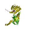
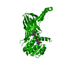
| Structure viewer | Molecule: MolmilJmol/JSmol |
|---|
- Downloads & links
-Download
| PDBx/mmCIF format | 4hc4.cif.gz | 88.6 KB | Display | PDBx/mmCIF format |
|---|---|---|---|---|
| PDB format | pdb4hc4.ent.gz | 64.6 KB | Display | PDB format |
| PDBx/mmJSON format | 4hc4.json.gz | Tree view | PDBx/mmJSON format | |
| Others |  Other downloads Other downloads |
-Validation report
| Arichive directory | https://data.pdbj.org/pub/pdb/validation_reports/hc/4hc4ftp://data.pdbj.org/pub/pdb/validation_reports/hc/4hc4 | HTTPS FTP |
|---|
-Related structure data
| Related structure data |  4qqkC  5hzmC 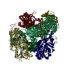 1g6qS S: Starting model for refinement C: citing same article ( |
|---|---|
| Similar structure data |


-Links
 PDBj
PDBj- Assembly
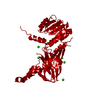
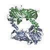
| Deposited unit | 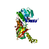
| ||||||||
|---|---|---|---|---|---|---|---|---|---|
| 1 | 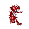
| ||||||||
| Unit cell |
| ||||||||
| Details | AUTHORS STATE THAT THE BIOLOGICAL UNIT IS UNKNOWN. |
-Components
| #1: Protein | Mass: 42074.559 Da / Num. of mol.: 1 / Mutation: A194V Source method: isolated from a genetically manipulated source Details: pFBOH-MHL / Source: (gene. exp.) Homo sapiens (human) / Gene: PRMT6, HRMT1L6 / Production host:  unidentified baculovirus unidentified baculovirusReferences: UniProt: Q96LA8, Transferases; Transferring one-carbon groups; Methyltransferases, EC: 2.1.1.125 |
|---|---|
| #2: Chemical | ChemComp-SAH / S-Adenosyl-L-homocysteine  Type: L-peptide linking / Mass: 384.411 Da / Num. of mol.: 1 / Source method: obtained synthetically / Formula: C14H20N6O5S Type: L-peptide linking / Mass: 384.411 Da / Num. of mol.: 1 / Source method: obtained synthetically / Formula: C14H20N6O5S |
| #3: Chemical | ChemComp-EDO / Ethylene glycol  Mass: 62.068 Da / Num. of mol.: 1 / Source method: obtained synthetically / Formula: C2H6O2 Mass: 62.068 Da / Num. of mol.: 1 / Source method: obtained synthetically / Formula: C2H6O2 |
| #4: Chemical | ChemComp-GOL / Glycerol  Mass: 92.094 Da / Num. of mol.: 1 / Source method: obtained synthetically / Formula: C3H8O3 Mass: 92.094 Da / Num. of mol.: 1 / Source method: obtained synthetically / Formula: C3H8O3 |
| #5: Water | ChemComp-HOH / Water Mass: 18.015 Da / Num. of mol.: 229 / Source method: isolated from a natural source / Formula: H2O Mass: 18.015 Da / Num. of mol.: 229 / Source method: isolated from a natural source / Formula: H2O |
-Experimental details
-Experiment
| Experiment | Method: X-RAY DIFFRACTION / Number of used crystals: 1 |
|---|
- Sample preparation
| Crystal | Density Matthews: 2.86 Å3/Da / Density % sol: 56.95 % |
|---|---|
| Crystal grow | Temperature: 293 K / Method: vapor diffusion, hanging drop / pH: 7 Details: Purified HRMT1L6 was complexed with SAH at 1:5 molar ratio of protein:SAH and crystallized by mixing 1 ul of the protein solution with 1 ul of the reservoir solution containing 15% PEG3350, ...Details: Purified HRMT1L6 was complexed with SAH at 1:5 molar ratio of protein:SAH and crystallized by mixing 1 ul of the protein solution with 1 ul of the reservoir solution containing 15% PEG3350, 0.1 M succinate acid, pH 7.0, vapor diffusion, hanging drop, temperature 293K |
-Data collection
| Diffraction | Mean temperature: 100 K |
|---|---|
| Diffraction source | Source: SYNCHROTRON / Site: APS  / Beamline: 19-ID / Wavelength: 0.97934 Å / Beamline: 19-ID / Wavelength: 0.97934 Å |
| Detector | Type: ADSC QUANTUM 315r / Detector: CCD / Date: Aug 16, 2012 |
| Radiation | Protocol: SINGLE WAVELENGTH / Monochromatic (M) / Laue (L): M / Scattering type: x-ray |
| Radiation wavelength | Wavelength: 0.97934 Å / Relative weight: 1 |
| Reflection | Resolution: 1.97→50 Å / Num. all: 33354 / Num. obs: 33354 / % possible obs: 99.9 % / Observed criterion σ(F): 0 / Observed criterion σ(I): 0 / Redundancy: 10 % / Rsym value: 0.061 / Net I/σ(I): 42.1 |
| Reflection shell | Resolution: 1.97→2 Å / Redundancy: 10 % / Mean I/σ(I) obs: 2.8 / Rsym value: 0.746 / % possible all: 100 |
- Processing
| Software |
| ||||||||||||||||||||||||||||||||||||||||||||||||||||||||||||||||||||||||||||||||||||||||||||||||||||||||||||||||||||||||||||||||||||||||||||||||||||||||||||||||||||||||||
|---|---|---|---|---|---|---|---|---|---|---|---|---|---|---|---|---|---|---|---|---|---|---|---|---|---|---|---|---|---|---|---|---|---|---|---|---|---|---|---|---|---|---|---|---|---|---|---|---|---|---|---|---|---|---|---|---|---|---|---|---|---|---|---|---|---|---|---|---|---|---|---|---|---|---|---|---|---|---|---|---|---|---|---|---|---|---|---|---|---|---|---|---|---|---|---|---|---|---|---|---|---|---|---|---|---|---|---|---|---|---|---|---|---|---|---|---|---|---|---|---|---|---|---|---|---|---|---|---|---|---|---|---|---|---|---|---|---|---|---|---|---|---|---|---|---|---|---|---|---|---|---|---|---|---|---|---|---|---|---|---|---|---|---|---|---|---|---|---|---|---|---|
| Refinement | Method to determine structure: MOLECULAR REPLACEMENT Starting model: PDB entry 1G6Q Resolution: 1.97→33.25 Å / Cor.coef. Fo:Fc: 0.963 / Cor.coef. Fo:Fc free: 0.953 / Occupancy max: 1 / Occupancy min: 0.3 / SU B: 3.378 / SU ML: 0.096 / SU R Cruickshank DPI: 0.1408 / Cross valid method: THROUGHOUT / ESU R: 0.141 / ESU R Free: 0.132 / Stereochemistry target values: MAXIMUM LIKELIHOOD / Details: HYDROGENS HAVE BEEN USED IF PRESENT IN THE INPUT
| ||||||||||||||||||||||||||||||||||||||||||||||||||||||||||||||||||||||||||||||||||||||||||||||||||||||||||||||||||||||||||||||||||||||||||||||||||||||||||||||||||||||||||
| Solvent computation | Ion probe radii: 0.8 Å / Shrinkage radii: 0.8 Å / VDW probe radii: 1.2 Å / Solvent model: MASK | ||||||||||||||||||||||||||||||||||||||||||||||||||||||||||||||||||||||||||||||||||||||||||||||||||||||||||||||||||||||||||||||||||||||||||||||||||||||||||||||||||||||||||
| Displacement parameters | Biso mean: 38.407 Å2
| ||||||||||||||||||||||||||||||||||||||||||||||||||||||||||||||||||||||||||||||||||||||||||||||||||||||||||||||||||||||||||||||||||||||||||||||||||||||||||||||||||||||||||
| Refinement step | Cycle: LAST / Resolution: 1.97→33.25 Å
| ||||||||||||||||||||||||||||||||||||||||||||||||||||||||||||||||||||||||||||||||||||||||||||||||||||||||||||||||||||||||||||||||||||||||||||||||||||||||||||||||||||||||||
| Refine LS restraints |
| ||||||||||||||||||||||||||||||||||||||||||||||||||||||||||||||||||||||||||||||||||||||||||||||||||||||||||||||||||||||||||||||||||||||||||||||||||||||||||||||||||||||||||
| LS refinement shell | Resolution: 1.97→2.021 Å / Total num. of bins used: 20
|
