 Movie
Movie Controller

[English] 日本語
 Yorodumi
- PDB-4qqk: Human HMT1 hnRNP methyltransferase-like protein 6 (S. cerevisiae)... -
+ Open data

- Basic information
| Entry | Database: PDB / ID: 4qqk | ||||||
|---|---|---|---|---|---|---|---|
| Title | Human HMT1 hnRNP methyltransferase-like protein 6 (S. cerevisiae) with GMS | ||||||
 Components Components | Protein arginine N-methyltransferase 6 | ||||||
 Keywords Keywords |  TRANSFERASE / HRMT1L6 / Protein arginine N-methyltransferase 6 / Structural Genomics / Structural Genomics Consortium / SGC TRANSFERASE / HRMT1L6 / Protein arginine N-methyltransferase 6 / Structural Genomics / Structural Genomics Consortium / SGC | ||||||
| Function / homology |  Function and homology information Function and homology informationhistone H2AR3 methyltransferase activity / peptidyl-arginine methylation, to asymmetrical-dimethyl arginine / protein-arginine omega-N monomethyltransferase activity / histone H4R3 methyltransferase activity / histone H3R2 methyltransferase activity / type I protein arginine methyltransferase / protein-arginine omega-N asymmetric methyltransferase activity / regulation of megakaryocyte differentiation / histone arginine N-methyltransferase activity / protein-arginine N-methyltransferase activity ...histone H2AR3 methyltransferase activity / peptidyl-arginine methylation, to asymmetrical-dimethyl arginine / protein-arginine omega-N monomethyltransferase activity / histone H4R3 methyltransferase activity / histone H3R2 methyltransferase activity / type I protein arginine methyltransferase / protein-arginine omega-N asymmetric methyltransferase activity / regulation of megakaryocyte differentiation / histone arginine N-methyltransferase activity / protein-arginine N-methyltransferase activity / regulation of mitochondrion organization / histone H3 methyltransferase activity / histone methyltransferase activity / negative regulation of ubiquitin-dependent protein catabolic process / regulation of signal transduction by p53 class mediator / base-excision repair / protein modification process / RUNX1 regulates genes involved in megakaryocyte differentiation and platelet function / RMTs methylate histone arginines / cellular senescence / histone binding / negative regulation of DNA-templated transcription / chromatin binding / nucleolus / negative regulation of transcription by RNA polymerase II / nucleoplasm / nucleusSimilarity search - Function | ||||||
| Biological species |  Homo sapiens (human) Homo sapiens (human) | ||||||
| Method | X-RAY DIFFRACTION / SYNCHROTRON / MOLECULAR REPLACEMENT / Resolution: 1.88 Å | ||||||
 Authors Authors | Dong, A. / Zeng, H. / He, H. / Wernimont, A. / Bountra, C. / Arrowsmith, C.H. / Edwards, A.M. / Brown, P.J. / Min, J. / Luo, M. ...Dong, A. / Zeng, H. / He, H. / Wernimont, A. / Bountra, C. / Arrowsmith, C.H. / Edwards, A.M. / Brown, P.J. / Min, J. / Luo, M. / Wu, H. / Structural Genomics Consortium (SGC) | ||||||
 Citation Citation | Journal: Biochem. J. / Year: 2016 Title: Structural basis of arginine asymmetrical dimethylation by PRMT6. Authors: Wu, H. / Zheng, W. / Eram, M.S. / Vhuiyan, M. / Dong, A. / Zeng, H. / He, H. / Brown, P. / Frankel, A. / Vedadi, M. / Luo, M. / Min, J. | ||||||
| History |
|
- Structure visualization
| Structure viewer | Molecule: MolmilJmol/JSmol |
|---|
- Downloads & links
-Download
| PDBx/mmCIF format | 4qqk.cif.gz | 86.1 KB | Display | PDBx/mmCIF format |
|---|---|---|---|---|
| PDB format | pdb4qqk.ent.gz | 61.9 KB | Display | PDB format |
| PDBx/mmJSON format | 4qqk.json.gz | Tree view | PDBx/mmJSON format | |
| Others |  Other downloads Other downloads |
-Validation report
| Arichive directory | https://data.pdbj.org/pub/pdb/validation_reports/qq/4qqkftp://data.pdbj.org/pub/pdb/validation_reports/qq/4qqk | HTTPS FTP |
|---|
-Related structure data
| Related structure data |  4hc4SC  5hzmC S: Starting model for refinement C: citing same article ( |
|---|---|
| Similar structure data |


-Links
 PDBj
PDBj- Assembly
| Deposited unit | 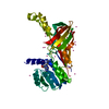
| ||||||||
|---|---|---|---|---|---|---|---|---|---|
| 1 | 
| ||||||||
| Unit cell |
|
-Components
| #1: Protein | Mass: 42074.559 Da / Num. of mol.: 1 Source method: isolated from a genetically manipulated source Source: (gene. exp.) Homo sapiens (human) / Gene: PRMT6, HRMT1L6 / Plasmid: pFBOH-MHL / Production host:   Spodoptera frugiperda (fall armyworm) / Strain (production host): SF9 Spodoptera frugiperda (fall armyworm) / Strain (production host): SF9References: UniProt: Q96LA8, Transferases; Transferring one-carbon groups; Methyltransferases, EC: 2.1.1.125 | ||
|---|---|---|---|
| #2: Chemical | ChemComp-37H / (  Mass: 437.454 Da / Num. of mol.: 1 / Source method: obtained synthetically / Formula: C17H27N9O5 Mass: 437.454 Da / Num. of mol.: 1 / Source method: obtained synthetically / Formula: C17H27N9O5 | ||
| #3: Chemical | ChemComp-GOL / Glycerol  Mass: 92.094 Da / Num. of mol.: 1 / Source method: obtained synthetically / Formula: C3H8O3 Mass: 92.094 Da / Num. of mol.: 1 / Source method: obtained synthetically / Formula: C3H8O3 | ||
| #4: Chemical | ChemComp-UNX /   Num. of mol.: 21 / Source method: obtained synthetically Num. of mol.: 21 / Source method: obtained synthetically#5: Water | ChemComp-HOH / | Water Mass: 18.015 Da / Num. of mol.: 179 / Source method: isolated from a natural source / Formula: H2O Mass: 18.015 Da / Num. of mol.: 179 / Source method: isolated from a natural source / Formula: H2O |
-Experimental details
-Experiment
| Experiment | Method: X-RAY DIFFRACTION / Number of used crystals: 1 |
|---|
- Sample preparation
| Crystal | Density Matthews: 2.92 Å3/Da / Density % sol: 57.85 % |
|---|---|
| Crystal grow | Temperature: 291 K / Method: vapor diffusion, hanging drop Details: 20% PEG 3350, 0.2 M KSCN, vapor diffusion hanging drop, temperature 291K |
-Data collection
| Diffraction | Mean temperature: 100 K | |||||||||||||||||||||||||||||||||||||||||||||||||||||||||||||||||||||||||||||||||||||||||||||||||||||||||||||||||||||||||||||||||||||||||||||||||||
|---|---|---|---|---|---|---|---|---|---|---|---|---|---|---|---|---|---|---|---|---|---|---|---|---|---|---|---|---|---|---|---|---|---|---|---|---|---|---|---|---|---|---|---|---|---|---|---|---|---|---|---|---|---|---|---|---|---|---|---|---|---|---|---|---|---|---|---|---|---|---|---|---|---|---|---|---|---|---|---|---|---|---|---|---|---|---|---|---|---|---|---|---|---|---|---|---|---|---|---|---|---|---|---|---|---|---|---|---|---|---|---|---|---|---|---|---|---|---|---|---|---|---|---|---|---|---|---|---|---|---|---|---|---|---|---|---|---|---|---|---|---|---|---|---|---|---|---|---|
| Diffraction source | Source: SYNCHROTRON / Site: APS  / Beamline: 19-ID / Wavelength: 0.97912 Å / Beamline: 19-ID / Wavelength: 0.97912 Å | |||||||||||||||||||||||||||||||||||||||||||||||||||||||||||||||||||||||||||||||||||||||||||||||||||||||||||||||||||||||||||||||||||||||||||||||||||
| Detector | Type: ADSC QUANTUM 315r / Detector: CCD / Date: Nov 23, 2012 | |||||||||||||||||||||||||||||||||||||||||||||||||||||||||||||||||||||||||||||||||||||||||||||||||||||||||||||||||||||||||||||||||||||||||||||||||||
| Radiation | Protocol: SINGLE WAVELENGTH / Monochromatic (M) / Laue (L): M / Scattering type: x-ray | |||||||||||||||||||||||||||||||||||||||||||||||||||||||||||||||||||||||||||||||||||||||||||||||||||||||||||||||||||||||||||||||||||||||||||||||||||
| Radiation wavelength | Wavelength: 0.97912 Å / Relative weight: 1 | |||||||||||||||||||||||||||||||||||||||||||||||||||||||||||||||||||||||||||||||||||||||||||||||||||||||||||||||||||||||||||||||||||||||||||||||||||
| Reflection | Resolution: 1.88→71.52 Å / Num. obs: 39024 / % possible obs: 100 % / Redundancy: 38.2 % / Biso Wilson estimate: 26.2 Å2 / Rmerge(I) obs: 0.06 / Χ2: 1.091 / Net I/σ(I): 10.7 | |||||||||||||||||||||||||||||||||||||||||||||||||||||||||||||||||||||||||||||||||||||||||||||||||||||||||||||||||||||||||||||||||||||||||||||||||||
| Reflection shell |
|
- Processing
| Software |
| |||||||||||||||||||||||||||||||||||||||||||||||||||||||||||||||||||||||||||
|---|---|---|---|---|---|---|---|---|---|---|---|---|---|---|---|---|---|---|---|---|---|---|---|---|---|---|---|---|---|---|---|---|---|---|---|---|---|---|---|---|---|---|---|---|---|---|---|---|---|---|---|---|---|---|---|---|---|---|---|---|---|---|---|---|---|---|---|---|---|---|---|---|---|---|---|---|
| Refinement | Method to determine structure: MOLECULAR REPLACEMENT Starting model: 4HC4 Resolution: 1.88→71.52 Å / Cor.coef. Fo:Fc: 0.963 / Cor.coef. Fo:Fc free: 0.951 / SU B: 2.507 / SU ML: 0.074 / Cross valid method: THROUGHOUT / σ(F): 0 / ESU R: 0.109 / ESU R Free: 0.108 / Stereochemistry target values: MAXIMUM LIKELIHOOD Details: HYDROGENS HAVE BEEN ADDED IN THE RIDING POSITIONS U VALUES : REFINED INDIVIDUALLY
| |||||||||||||||||||||||||||||||||||||||||||||||||||||||||||||||||||||||||||
| Solvent computation | Ion probe radii: 0.8 Å / Shrinkage radii: 0.8 Å / VDW probe radii: 1.2 Å / Solvent model: MASK | |||||||||||||||||||||||||||||||||||||||||||||||||||||||||||||||||||||||||||
| Displacement parameters | Biso max: 73.7 Å2 / Biso mean: 32.467 Å2 / Biso min: 18.49 Å2
| |||||||||||||||||||||||||||||||||||||||||||||||||||||||||||||||||||||||||||
| Refinement step | Cycle: LAST / Resolution: 1.88→71.52 Å
| |||||||||||||||||||||||||||||||||||||||||||||||||||||||||||||||||||||||||||
| Refine LS restraints |
| |||||||||||||||||||||||||||||||||||||||||||||||||||||||||||||||||||||||||||
| LS refinement shell | Resolution: 1.882→1.931 Å / Total num. of bins used: 20
|
