 Movie
Movie Controller


+ Open data

- Basic information
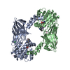
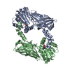
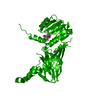
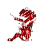
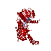
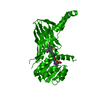
| Entry | Database: PDB / ID: 6sq4 | ||||||
|---|---|---|---|---|---|---|---|
| Title | Crystal structure of mouse PRMT6 in complex with inhibitor U2 | ||||||
 Components Components | Protein arginine N-methyltransferase 6 | ||||||
 Keywords Keywords |  TRANSFERASE / SAM binding domain / arginine methylation TRANSFERASE / SAM binding domain / arginine methylation | ||||||
| Function / homology |  Function and homology information Function and homology informationhistone H2AR3 methyltransferase activity / peptidyl-arginine methylation, to asymmetrical-dimethyl arginine / protein-arginine omega-N monomethyltransferase activity / histone H4R3 methyltransferase activity / histone H3R2 methyltransferase activity / RUNX1 regulates genes involved in megakaryocyte differentiation and platelet function / RMTs methylate histone arginines / type I protein arginine methyltransferase / protein-arginine omega-N asymmetric methyltransferase activity / histone arginine N-methyltransferase activity ...histone H2AR3 methyltransferase activity / peptidyl-arginine methylation, to asymmetrical-dimethyl arginine / protein-arginine omega-N monomethyltransferase activity / histone H4R3 methyltransferase activity / histone H3R2 methyltransferase activity / RUNX1 regulates genes involved in megakaryocyte differentiation and platelet function / RMTs methylate histone arginines / type I protein arginine methyltransferase / protein-arginine omega-N asymmetric methyltransferase activity / histone arginine N-methyltransferase activity / protein-arginine N-methyltransferase activity / regulation of mitochondrion organization / histone H3 methyltransferase activity / histone methyltransferase activity / negative regulation of ubiquitin-dependent protein catabolic process / regulation of signal transduction by p53 class mediator / protein modification process / cellular senescence / histone binding / DNA repair / negative regulation of DNA-templated transcription / chromatin binding / nucleolus / negative regulation of transcription by RNA polymerase II / nucleoplasm / nucleusSimilarity search - Function | ||||||
| Biological species |  Mus musculus (house mouse) Mus musculus (house mouse) | ||||||
| Method | X-RAY DIFFRACTION / SAD / Resolution: 1.7 Å | ||||||
 Authors Authors | Bonnefond, L. / Cavarelli, J. | ||||||
 Citation Citation | Journal: To be published Title: Crystal structure of mouse PRMT6 in complex with inhibitors Authors: Bonnefond, L. / Cavarelli, J. | ||||||
| History |
|
- Structure visualization
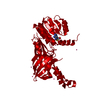
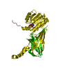
| Structure viewer | Molecule: MolmilJmol/JSmol |
|---|
- Downloads & links
-Download
| PDBx/mmCIF format | 6sq4.cif.gz | 388.3 KB | Display | PDBx/mmCIF format |
|---|---|---|---|---|
| PDB format | pdb6sq4.ent.gz | 322.9 KB | Display | PDB format |
| PDBx/mmJSON format | 6sq4.json.gz | Tree view | PDBx/mmJSON format | |
| Others |  Other downloads Other downloads |
-Validation report
| Arichive directory | https://data.pdbj.org/pub/pdb/validation_reports/sq/6sq4ftp://data.pdbj.org/pub/pdb/validation_reports/sq/6sq4 | HTTPS FTP |
|---|
-Related structure data


-Links
 PDBj
PDBj
- Assembly
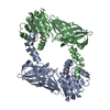
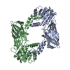
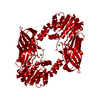
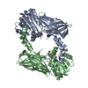
| Deposited unit | 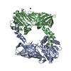
| ||||||||
|---|---|---|---|---|---|---|---|---|---|
| 1 |
| ||||||||
| Unit cell |
|
-Components
| #1: Protein | Mass: 42821.301 Da / Num. of mol.: 2 / Fragment: mouse PRMT6 / Mutation: F315L Source method: isolated from a genetically manipulated source Source: (gene. exp.) Mus musculus (house mouse) / Gene: Prmt6, Hrmt1l6 / Plasmid: pET15b / Production host:  Escherichia coli BL21(DE3) (bacteria) Escherichia coli BL21(DE3) (bacteria)References: UniProt: Q6NZB1, type I protein arginine methyltransferase #2: Chemical | Iodide  Mass: 126.904 Da / Num. of mol.: 2 / Source method: obtained synthetically / Formula: I Mass: 126.904 Da / Num. of mol.: 2 / Source method: obtained synthetically / Formula: I#3: Chemical |   Mass: 337.358 Da / Num. of mol.: 2 / Source method: obtained synthetically / Formula: C13H21N8O3 / Feature type: SUBJECT OF INVESTIGATION Mass: 337.358 Da / Num. of mol.: 2 / Source method: obtained synthetically / Formula: C13H21N8O3 / Feature type: SUBJECT OF INVESTIGATION#4: Water | ChemComp-HOH / | Water Mass: 18.015 Da / Num. of mol.: 444 / Source method: isolated from a natural source / Formula: H2O Mass: 18.015 Da / Num. of mol.: 444 / Source method: isolated from a natural source / Formula: H2OHas ligand of interest | Y | |
|---|
-Experimental details
-Experiment
| Experiment | Method: X-RAY DIFFRACTION / Number of used crystals: 1 |
|---|
- Sample preparation
| Crystal | Density Matthews: 2.34 Å3/Da / Density % sol: 47.53 % / Mosaicity: 0.2 ° |
|---|---|
| Crystal grow | Temperature: 298 K / Method: vapor diffusion / pH: 7.5 / Details: PEG 3,350 20%, NaI 200 mM, Tris-HCl pH 7.5 100 mM |
-Data collection
| Diffraction | Mean temperature: 100 K / Serial crystal experiment: N |
|---|---|
| Diffraction source | Source: ROTATING ANODE / Type: RIGAKU FR-X / Wavelength: 1.54178 Å |
| Detector | Type: DECTRIS PILATUS 300K / Detector: PIXEL / Date: Apr 6, 2017 |
| Radiation | Protocol: SINGLE WAVELENGTH / Monochromatic (M) / Laue (L): M / Scattering type: x-ray |
| Radiation wavelength | Wavelength: 1.54178 Å / Relative weight: 1 |
| Reflection | Resolution: 1.7→45.17 Å / Num. obs: 74687 / % possible obs: 99.8 % / Redundancy: 4.8 % / Biso Wilson estimate: 20.39 Å2 / CC1/2: 0.999 / Rmerge(I) obs: 0.053 / Rpim(I) all: 0.023 / Rrim(I) all: 0.058 / Net I/σ(I): 16.5 / Num. measured all: 361459 / Scaling rejects: 160 |
| Reflection shell | Resolution: 1.7→1.73 Å / Redundancy: 3 % / Rmerge(I) obs: 0.934 / Mean I/σ(I) obs: 1.2 / Num. unique obs: 3909 / CC1/2: 0.454 / Rpim(I) all: 0.621 / Rrim(I) all: 1.125 / % possible all: 98.7 |
- Processing
| Software |
| |||||||||||||||||||||||||||||||||||||||||||||||||||||||||||||||||||||||||||
|---|---|---|---|---|---|---|---|---|---|---|---|---|---|---|---|---|---|---|---|---|---|---|---|---|---|---|---|---|---|---|---|---|---|---|---|---|---|---|---|---|---|---|---|---|---|---|---|---|---|---|---|---|---|---|---|---|---|---|---|---|---|---|---|---|---|---|---|---|---|---|---|---|---|---|---|---|
| Refinement | Method to determine structure: SAD / Resolution: 1.7→35.058 Å / Cross valid method: THROUGHOUT / σ(F): 1.37
| |||||||||||||||||||||||||||||||||||||||||||||||||||||||||||||||||||||||||||
| Displacement parameters | Biso max: 95.61 Å2 / Biso mean: 30.2832 Å2 / Biso min: 11.89 Å2 | |||||||||||||||||||||||||||||||||||||||||||||||||||||||||||||||||||||||||||
| Refinement step | Cycle: final / Resolution: 1.7→35.058 Å
| |||||||||||||||||||||||||||||||||||||||||||||||||||||||||||||||||||||||||||
| Refinement TLS params. | Method: refined / Refine-ID: X-RAY DIFFRACTION
| |||||||||||||||||||||||||||||||||||||||||||||||||||||||||||||||||||||||||||
| Refinement TLS group |
|
