 Movie
Movie Controller

[English] 日本語
 Yorodumi
- PDB-5e8r: Human HMT1 hnRNP methyltransferase-like protein 6 (S. cerevisiae) -
+ Open data

- Basic information
| Entry | Database: PDB / ID: 5e8r | ||||||
|---|---|---|---|---|---|---|---|
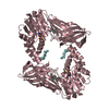
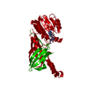
| Title | Human HMT1 hnRNP methyltransferase-like protein 6 (S. cerevisiae) | ||||||
 Components Components | Protein arginine N-methyltransferase 6 | ||||||
 Keywords Keywords |  TRANSFERASE / HRMT1L6 / MS-023 / Structural Genomics / Structural Genomics Consortium / SGC TRANSFERASE / HRMT1L6 / MS-023 / Structural Genomics / Structural Genomics Consortium / SGC | ||||||
| Function / homology |  Function and homology information Function and homology informationhistone H2AR3 methyltransferase activity / peptidyl-arginine methylation, to asymmetrical-dimethyl arginine / protein-arginine omega-N monomethyltransferase activity / histone H4R3 methyltransferase activity / histone H3R2 methyltransferase activity / type I protein arginine methyltransferase / protein-arginine omega-N asymmetric methyltransferase activity / regulation of megakaryocyte differentiation / histone arginine N-methyltransferase activity / protein-arginine N-methyltransferase activity ...histone H2AR3 methyltransferase activity / peptidyl-arginine methylation, to asymmetrical-dimethyl arginine / protein-arginine omega-N monomethyltransferase activity / histone H4R3 methyltransferase activity / histone H3R2 methyltransferase activity / type I protein arginine methyltransferase / protein-arginine omega-N asymmetric methyltransferase activity / regulation of megakaryocyte differentiation / histone arginine N-methyltransferase activity / protein-arginine N-methyltransferase activity / regulation of mitochondrion organization / histone H3 methyltransferase activity / histone methyltransferase activity / negative regulation of ubiquitin-dependent protein catabolic process / regulation of signal transduction by p53 class mediator / base-excision repair / protein modification process / RUNX1 regulates genes involved in megakaryocyte differentiation and platelet function / RMTs methylate histone arginines / cellular senescence / histone binding / negative regulation of DNA-templated transcription / chromatin binding / nucleolus / negative regulation of transcription by RNA polymerase II / nucleoplasm / nucleusSimilarity search - Function | ||||||
| Biological species |  Homo sapiens (human) Homo sapiens (human) | ||||||
| Method | X-RAY DIFFRACTION / SYNCHROTRON / Resolution: 2.55 Å | ||||||
 Authors Authors | DONG, A. / ZENG, H. / LIU, J. / TEMPEL, W. / Seitova, A. / Hutchinson, A. / Bountra, C. / Arrowsmith, C.H. / Edwards, A.M. / JIN, J. ...DONG, A. / ZENG, H. / LIU, J. / TEMPEL, W. / Seitova, A. / Hutchinson, A. / Bountra, C. / Arrowsmith, C.H. / Edwards, A.M. / JIN, J. / BROWN, P.J. / WU, H. / Structural Genomics Consortium (SGC) | ||||||
 Citation Citation | Journal: Acs Chem.Biol. / Year: 2016 Title: A Potent, Selective, and Cell-Active Inhibitor of Human Type I Protein Arginine Methyltransferases. Authors: Eram, M.S. / Shen, Y. / Szewczyk, M.M. / Wu, H. / Senisterra, G. / Li, F. / Butler, K.V. / Kaniskan, H.U. / Speed, B.A. / Dela Sena, C. / Dong, A. / Zeng, H. / Schapira, M. / Brown, P.J. / ...Authors: Eram, M.S. / Shen, Y. / Szewczyk, M.M. / Wu, H. / Senisterra, G. / Li, F. / Butler, K.V. / Kaniskan, H.U. / Speed, B.A. / Dela Sena, C. / Dong, A. / Zeng, H. / Schapira, M. / Brown, P.J. / Arrowsmith, C.H. / Barsyte-Lovejoy, D. / Liu, J. / Vedadi, M. / Jin, J. | ||||||
| History |
|
- Structure visualization
| Structure viewer | Molecule: MolmilJmol/JSmol |
|---|
- Downloads & links
-Download
| PDBx/mmCIF format | 5e8r.cif.gz | 146.7 KB | Display | PDBx/mmCIF format |
|---|---|---|---|---|
| PDB format | pdb5e8r.ent.gz | 111.5 KB | Display | PDB format |
| PDBx/mmJSON format | 5e8r.json.gz | Tree view | PDBx/mmJSON format | |
| Others |  Other downloads Other downloads |
-Validation report
| Arichive directory | https://data.pdbj.org/pub/pdb/validation_reports/e8/5e8rftp://data.pdbj.org/pub/pdb/validation_reports/e8/5e8r | HTTPS FTP |
|---|
-Related structure data
| Similar structure data |
|---|
-Links
 PDBj
PDBj- Assembly
| Deposited unit | 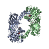
| ||||||||
|---|---|---|---|---|---|---|---|---|---|
| 1 |
| ||||||||
| Unit cell |
| ||||||||
| Details | As per the authors the biological assembly is unknown |
-Components
-Protein , 1 types, 2 molecules AB
| #1: Protein | Mass: 42074.559 Da / Num. of mol.: 2 Source method: isolated from a genetically manipulated source Source: (gene. exp.) Homo sapiens (human) / Gene: PRMT6, HRMT1L6 / Plasmid: pFBOH-MHL / Production host:   Spodoptera frugiperda (fall armyworm) / Strain (production host): Sf9 Spodoptera frugiperda (fall armyworm) / Strain (production host): Sf9References: UniProt: Q96LA8, Transferases; Transferring one-carbon groups; Methyltransferases, EC: 2.1.1.125 |
|---|
-Non-polymers , 5 types, 83 molecules 


| #2: Chemical | S-Adenosyl-L-homocysteine Type: L-peptide linking / Mass: 384.411 Da / Num. of mol.: 2 / Source method: obtained synthetically / Formula: C14H20N6O5S Type: L-peptide linking / Mass: 384.411 Da / Num. of mol.: 2 / Source method: obtained synthetically / Formula: C14H20N6O5S#3: Chemical |  Mass: 287.400 Da / Num. of mol.: 2 / Source method: obtained synthetically / Formula: C17H25N3O Mass: 287.400 Da / Num. of mol.: 2 / Source method: obtained synthetically / Formula: C17H25N3O#4: Chemical | ChemComp-CL / Chloride Mass: 35.453 Da / Num. of mol.: 5 / Source method: obtained synthetically / Formula: Cl Mass: 35.453 Da / Num. of mol.: 5 / Source method: obtained synthetically / Formula: Cl#5: Chemical | ChemComp-UNX /  Num. of mol.: 9 / Source method: obtained synthetically Num. of mol.: 9 / Source method: obtained synthetically#6: Water | ChemComp-HOH / | WaterMass: 18.015 Da / Num. of mol.: 65 / Source method: isolated from a natural source / Formula: H2O |
|---|
-Experimental details
-Experiment
| Experiment | Method: X-RAY DIFFRACTION / Number of used crystals: 1 |
|---|
- Sample preparation
| Crystal | Density Matthews: 2.68 Å3/Da / Density % sol: 54.15 % |
|---|---|
| Crystal grow | Temperature: 295 K / Method: vapor diffusion, sitting drop / pH: 5.6 Details: 10% PEG 3350, 0.2M MgCl2, 0.1M Sodium Cacadylate, pH6.5 |
-Data collection
| Diffraction | Mean temperature: 100 K | |||||||||||||||||||||||||||||||||||||||||||||||||||||||||||||||||||||||||||||||||||||||||||||||||||||||||||||||||||||||||||||||||||||||||||||||||||||||||||||||||||||||||||||||||||||||||||||
|---|---|---|---|---|---|---|---|---|---|---|---|---|---|---|---|---|---|---|---|---|---|---|---|---|---|---|---|---|---|---|---|---|---|---|---|---|---|---|---|---|---|---|---|---|---|---|---|---|---|---|---|---|---|---|---|---|---|---|---|---|---|---|---|---|---|---|---|---|---|---|---|---|---|---|---|---|---|---|---|---|---|---|---|---|---|---|---|---|---|---|---|---|---|---|---|---|---|---|---|---|---|---|---|---|---|---|---|---|---|---|---|---|---|---|---|---|---|---|---|---|---|---|---|---|---|---|---|---|---|---|---|---|---|---|---|---|---|---|---|---|---|---|---|---|---|---|---|---|---|---|---|---|---|---|---|---|---|---|---|---|---|---|---|---|---|---|---|---|---|---|---|---|---|---|---|---|---|---|---|---|---|---|---|---|---|---|---|---|---|---|
| Diffraction source | Source: SYNCHROTRON / Site: APS  / Beamline: 24-ID-E / Wavelength: 0.97921 Å / Beamline: 24-ID-E / Wavelength: 0.97921 Å | |||||||||||||||||||||||||||||||||||||||||||||||||||||||||||||||||||||||||||||||||||||||||||||||||||||||||||||||||||||||||||||||||||||||||||||||||||||||||||||||||||||||||||||||||||||||||||||
| Detector | Type: ADSC QUANTUM 315 / Detector: CCD / Date: Jul 10, 2015 | |||||||||||||||||||||||||||||||||||||||||||||||||||||||||||||||||||||||||||||||||||||||||||||||||||||||||||||||||||||||||||||||||||||||||||||||||||||||||||||||||||||||||||||||||||||||||||||
| Radiation | Protocol: SINGLE WAVELENGTH / Monochromatic (M) / Laue (L): M / Scattering type: x-ray | |||||||||||||||||||||||||||||||||||||||||||||||||||||||||||||||||||||||||||||||||||||||||||||||||||||||||||||||||||||||||||||||||||||||||||||||||||||||||||||||||||||||||||||||||||||||||||||
| Radiation wavelength | Wavelength: 0.97921 Å / Relative weight: 1 | |||||||||||||||||||||||||||||||||||||||||||||||||||||||||||||||||||||||||||||||||||||||||||||||||||||||||||||||||||||||||||||||||||||||||||||||||||||||||||||||||||||||||||||||||||||||||||||
| Reflection | Resolution: 2.55→50 Å / Num. obs: 28929 / % possible obs: 99.3 % / Redundancy: 3.2 % / Rmerge(I) obs: 0.171 / Rpim(I) all: 0.116 / Rrim(I) all: 0.198 / Χ2: 1.693 / Net I/av σ(I): 8.89 / Net I/σ(I): 6.5 / Num. measured all: 91551 | |||||||||||||||||||||||||||||||||||||||||||||||||||||||||||||||||||||||||||||||||||||||||||||||||||||||||||||||||||||||||||||||||||||||||||||||||||||||||||||||||||||||||||||||||||||||||||||
| Reflection shell | Diffraction-ID: 1 / Rejects: 0
|
- Processing
| Software |
| |||||||||||||||||||||||||||||||||||||||||||||||||||||||||||||||||||||||||||
|---|---|---|---|---|---|---|---|---|---|---|---|---|---|---|---|---|---|---|---|---|---|---|---|---|---|---|---|---|---|---|---|---|---|---|---|---|---|---|---|---|---|---|---|---|---|---|---|---|---|---|---|---|---|---|---|---|---|---|---|---|---|---|---|---|---|---|---|---|---|---|---|---|---|---|---|---|
| Refinement | Resolution: 2.55→50 Å / Cor.coef. Fo:Fc: 0.912 / Cor.coef. Fo:Fc free: 0.875 / WRfactor Rfree: 0.2218 / WRfactor Rwork: 0.1778 / FOM work R set: 0.7859 / SU B: 11.493 / SU ML: 0.247 / SU R Cruickshank DPI: 0.4657 / SU Rfree: 0.2787 / Cross valid method: THROUGHOUT / σ(F): 0 / ESU R: 0.499 / ESU R Free: 0.287 / Stereochemistry target values: MAXIMUM LIKELIHOOD Details: HYDROGENS HAVE BEEN ADDED IN THE RIDING POSITIONS U VALUES : REFINED INDIVIDUALLY
| |||||||||||||||||||||||||||||||||||||||||||||||||||||||||||||||||||||||||||
| Solvent computation | Ion probe radii: 0.8 Å / Shrinkage radii: 0.8 Å / VDW probe radii: 1.2 Å / Solvent model: MASK | |||||||||||||||||||||||||||||||||||||||||||||||||||||||||||||||||||||||||||
| Displacement parameters | Biso max: 67.11 Å2 / Biso mean: 25.338 Å2 / Biso min: 9.69 Å2
| |||||||||||||||||||||||||||||||||||||||||||||||||||||||||||||||||||||||||||
| Refinement step | Cycle: final / Resolution: 2.55→50 Å
| |||||||||||||||||||||||||||||||||||||||||||||||||||||||||||||||||||||||||||
| Refine LS restraints |
| |||||||||||||||||||||||||||||||||||||||||||||||||||||||||||||||||||||||||||
| LS refinement shell | Resolution: 2.551→2.617 Å / Total num. of bins used: 20
|
