 Movie
Movie Controller


+ Open data

- Basic information
| Entry | Database: PDB / ID: 1g6q | ||||||
|---|---|---|---|---|---|---|---|
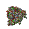
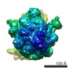
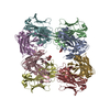
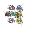
| Title | CRYSTAL STRUCTURE OF YEAST ARGININE METHYLTRANSFERASE, HMT1 | ||||||
 Components Components | HNRNP ARGININE N-METHYLTRANSFERASE | ||||||
 Keywords Keywords |  TRANSFERASE / SAM-binding domain / beta-barrel / mixed alpha-beta / hexamer / dimer TRANSFERASE / SAM-binding domain / beta-barrel / mixed alpha-beta / hexamer / dimer | ||||||
| Function / homology |  Function and homology information Function and homology informationnegative regulation of termination of DNA-templated transcription / protein-arginine omega-N monomethyltransferase activity / peptidyl-arginine methylation / protein-arginine omega-N asymmetric methyltransferase activity / folic acid biosynthetic process / RMTs methylate histone arginines / Estrogen-dependent gene expression / mRNA export from nucleus / Transferases; Transferring one-carbon groups; Methyltransferases / positive regulation of transcription elongation by RNA polymerase II ...negative regulation of termination of DNA-templated transcription / protein-arginine omega-N monomethyltransferase activity / peptidyl-arginine methylation / protein-arginine omega-N asymmetric methyltransferase activity / folic acid biosynthetic process / RMTs methylate histone arginines / Estrogen-dependent gene expression / mRNA export from nucleus / Transferases; Transferring one-carbon groups; Methyltransferases / positive regulation of transcription elongation by RNA polymerase II / identical protein binding / nucleusSimilarity search - Function | ||||||
| Biological species |  Saccharomyces cerevisiae (brewer's yeast) Saccharomyces cerevisiae (brewer's yeast) | ||||||
| Method | X-RAY DIFFRACTION / SYNCHROTRON / MAD / Resolution: 2.9 Å | ||||||
 Authors Authors | Weiss, V.H. / McBride, A.E. / Soriano, M.A. / Filman, D.J. / Silver, P.A. / Hogle, J.M. | ||||||
 Citation Citation | Journal: Nat.Struct.Biol. / Year: 2000 Title: The structure and oligomerization of the yeast arginine methyltransferase, Hmt1. Authors: Weiss, V.H. / McBride, A.E. / Soriano, M.A. / Filman, D.J. / Silver, P.A. / Hogle, J.M. #1: Journal: J.Biol.Chem. / Year: 2000Title: Analysis of the yeast arginine methyltransferase Hmt1p/Rmt1p and its in vivo function. Cofactor binding and substrate interactions. Authors: McBride, A.E. / Weiss, V.H. / Kim, H.K. / Hogle, J.M. / Silver, P.A. #2: Journal: rna / Year: 1999Title: Arginine methylation and binding of Hrp1p to the efficiency element for mRNA 3'-end formation. Authors: valentini, s.r. / weiss, v.h. / silver, p.a. #3: Journal: Mol.Cell.Biol. / Year: 1996Title: A novel methyltransferase (Hmt1p) modifies poly(A)+RNA binding proteins. Authors: henry, m.f. / silver, p.a. #4: Journal: Genes Dev. / Year: 1998Title: Arginine methylation facilitates the nuclear export of hnRNP proteins. Authors: shen, e.c. / Henry, M.F. / Weiss, V.H. / Valentini, S.R. / Silver, P.A. / Lee, M.S. | ||||||
| History |
|
- Structure visualization
| Structure viewer | Molecule: MolmilJmol/JSmol |
|---|
- Downloads & links
-Download
| PDBx/mmCIF format | 1g6q.cif.gz | 348.1 KB | Display | PDBx/mmCIF format |
|---|---|---|---|---|
| PDB format | pdb1g6q.ent.gz | 290.4 KB | Display | PDB format |
| PDBx/mmJSON format | 1g6q.json.gz | Tree view | PDBx/mmJSON format | |
| Others |  Other downloads Other downloads |
-Validation report
| Arichive directory | https://data.pdbj.org/pub/pdb/validation_reports/g6/1g6qftp://data.pdbj.org/pub/pdb/validation_reports/g6/1g6q | HTTPS FTP |
|---|
-Related structure data
| Similar structure data |
|---|


-Links
 PDBj
PDBj


- Assembly
| Deposited unit | 
| ||||||||
|---|---|---|---|---|---|---|---|---|---|
| 1 |
| ||||||||
| 2 | 
| ||||||||
| 3 | 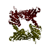
| ||||||||
| 4 | 
| ||||||||
| Unit cell |
| ||||||||
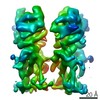
| Details | hexamer has very approximate 32 symmetry. Monomers exhibit varying degrees of order. |
-Components
| #1: Protein | Mass: 37692.613 Da / Num. of mol.: 6 / Fragment: MISSING 20 AMINOACYL RESIDUES FROM N-TERMINUS / Mutation: Q21D, H22Y, N25D Source method: isolated from a genetically manipulated source Source: (gene. exp.) Saccharomyces cerevisiae (brewer's yeast)Gene: HMT1 Plasmid details: MODIFIED PET15B VECTOR CONTAINING INSERT PRECISSION PROTEASE SITE AFTER HIS TAG (PPS2193) Plasmid: PPS2193 / Production host:  Escherichia coli (E. coli) / Strain (production host): BL21 (DE3) C41 Escherichia coli (E. coli) / Strain (production host): BL21 (DE3) C41References: UniProt: P38074, Transferases; Transferring one-carbon groups; Methyltransferases |
|---|
-Experimental details
-Experiment
| Experiment | Method: X-RAY DIFFRACTION / Number of used crystals: 1 |
|---|
- Sample preparation
| Crystal | Density Matthews: 2.38 Å3/Da / Density % sol: 48.32 % | ||||||||||||||||||||||||||||||||||||||||||||||||
|---|---|---|---|---|---|---|---|---|---|---|---|---|---|---|---|---|---|---|---|---|---|---|---|---|---|---|---|---|---|---|---|---|---|---|---|---|---|---|---|---|---|---|---|---|---|---|---|---|---|
| Crystal grow | Temperature: 295 K / Method: vapor diffusion, hanging drop / pH: 7.5 Details: 2 microliters of Hmt1 (8mg/ml) in 50 mM Tris, pH 7.0, 50 mM NaCl, 1 mM EDTA, 1 mM DTT + 2 microliters of reservoir:50 mM sodium hepes, pH 7.5, 14% v/v PEG 400, 100 mM CaCl2 + microseeds in 1 ...Details: 2 microliters of Hmt1 (8mg/ml) in 50 mM Tris, pH 7.0, 50 mM NaCl, 1 mM EDTA, 1 mM DTT + 2 microliters of reservoir:50 mM sodium hepes, pH 7.5, 14% v/v PEG 400, 100 mM CaCl2 + microseeds in 1 microliter of 20 mM sodium citrate, pH 5.6, 15% w/v PEG 4000, 100 mM ammonium acetate, VAPOR DIFFUSION, HANGING DROP, temperature 22K | ||||||||||||||||||||||||||||||||||||||||||||||||
| Crystal grow | *PLUS | ||||||||||||||||||||||||||||||||||||||||||||||||
| Components of the solutions | *PLUS
|
-Data collection
| Diffraction | Mean temperature: 113 K | |||||||||||||||
|---|---|---|---|---|---|---|---|---|---|---|---|---|---|---|---|---|
| Diffraction source | Source: SYNCHROTRON / Site: NSLS  / Beamline: X12C / Wavelength: 0.9784,1.0100,0.9500,0.9788 / Beamline: X12C / Wavelength: 0.9784,1.0100,0.9500,0.9788 | |||||||||||||||
| Detector | Type: BRANDEIS / Detector: CCD / Date: Mar 2, 2000 / Details: monochromator | |||||||||||||||
| Radiation | Monochromator: pair of parallel silicon crystals / Protocol: MAD / Monochromatic (M) / Laue (L): M / Scattering type: x-ray | |||||||||||||||
| Radiation wavelength |
| |||||||||||||||
| Reflection | Resolution: 2.9→28 Å / Num. all: 91268 / Num. obs: 90403 / % possible obs: 99.1 % / Observed criterion σ(F): 0 / Observed criterion σ(I): 0 / Redundancy: 3.1 % / Biso Wilson estimate: 56.6 Å2 / Rmerge(I) obs: 0.062 / Net I/σ(I): 17.4 | |||||||||||||||
| Reflection shell | Resolution: 2.89→2.99 Å / Redundancy: 1.8 % / Rmerge(I) obs: 0.235 / Mean I/σ(I) obs: 5 / Num. unique all: 8739 / Rsym value: 0.235 / % possible all: 95.9 | |||||||||||||||
| Reflection | *PLUS Num. measured all: 277416 | |||||||||||||||
| Reflection shell | *PLUS % possible obs: 95.9 % |
- Processing
| Software |
| |||||||||||||||||||||||||
|---|---|---|---|---|---|---|---|---|---|---|---|---|---|---|---|---|---|---|---|---|---|---|---|---|---|---|
| Refinement | Method to determine structure: MAD / Resolution: 2.9→28 Å / Isotropic thermal model: isotropic / Cross valid method: THROUGHOUT / σ(F): 0 / σ(I): 0 / Stereochemistry target values: param19x Details: 65 overlapping resolution-dependent bin scales, plus 1 or 2 grouped B values per aminoacyl residue. With bin-scales present, the refined B's do not correspond to rms displacements. NCS ...Details: 65 overlapping resolution-dependent bin scales, plus 1 or 2 grouped B values per aminoacyl residue. With bin-scales present, the refined B's do not correspond to rms displacements. NCS restraints were applied separately to the SAM-binding domains and the beta-barrel domains, with structurally variable segments of the polypeptide excluded.
| |||||||||||||||||||||||||
| Refinement step | Cycle: LAST / Resolution: 2.9→28 Å
| |||||||||||||||||||||||||
| Refine LS restraints |
| |||||||||||||||||||||||||
| LS refinement shell | Resolution: 2.9→2.97 Å /
| |||||||||||||||||||||||||
| Software | *PLUS Name: X-PLOR / Version: 3.851 / Classification: refinement | |||||||||||||||||||||||||
| Refine LS restraints | *PLUS
|
