2WUR
 
 | |
3ADF
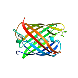
 | | Crystal structure of a monomeric green fluorescent protein, Azami-Green (mAG) | | 分子名称: | Monomeric Azami Green | | 著者 | Ebisawa, T, Yamamura, A, Kameda, Y, Hayakawa, K, Nagata, K, Tanokura, M. | | 登録日 | 2010-01-20 | | 公開日 | 2010-05-19 | | 最終更新日 | 2023-11-15 | | 実験手法 | X-RAY DIFFRACTION (2.2 Å) | | 主引用文献 | The structure of mAG, a monomeric mutant of the green fluorescent protein Azami-Green, reveals the structural basis of its stable green emission
Acta Crystallogr.,Sect.F, 66, 2010
|
|
3I19
 | |
3OGO
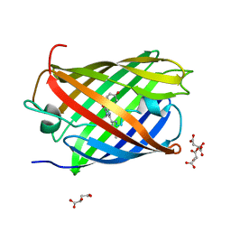
 | | Structure of the GFP:GFP-nanobody complex at 2.8 A resolution in spacegroup P21212 | | 分子名称: | GFP-nanobody, Green fluorescent protein, ISOPROPYL ALCOHOL | | 著者 | Kubala, M.H, Kovtun, O, Alexandrov, K, Collins, B.M. | | 登録日 | 2010-08-17 | | 公開日 | 2010-08-25 | | 最終更新日 | 2024-02-21 | | 実験手法 | X-RAY DIFFRACTION (2.8 Å) | | 主引用文献 | Structural and thermodynamic analysis of the GFP:GFP-nanobody complex.
Protein Sci., 19, 2010
|
|
3O77
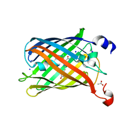
 | | The structure of Ca2+ Sensor (Case-16) | | 分子名称: | CALCIUM ION, CHLORIDE ION, Myosin light chain kinase, ... | | 著者 | Leder, L, Stark, W, Freuler, F, Marsh, M, Meyerhofer, M, Stettler, T, Mayr, L.M, Britanova, O.V, Strukova, L.A, Chudakov, D.M. | | 登録日 | 2010-07-30 | | 公開日 | 2010-09-29 | | 最終更新日 | 2017-06-21 | | 実験手法 | X-RAY DIFFRACTION (2.35 Å) | | 主引用文献 | The structure of Ca2+ sensor Case16 reveals the mechanism of reaction to low Ca2+ concentrations
Sensors (Basel), 10, 2010
|
|
3O78
 | | The structure of Ca2+ Sensor (Case-12) | | 分子名称: | CALCIUM ION, Myosin light chain kinase, smooth muscle,Green fluorescent protein,Green fluorescent protein,Calmodulin-1 | | 著者 | Leder, L, Stark, W, Freuler, F, Marsh, M, Meyerhofer, M, Stettler, T, Mayr, L.M, Britanova, O.V, Strukova, L.A, Chudakov, D.M. | | 登録日 | 2010-07-30 | | 公開日 | 2010-09-29 | | 最終更新日 | 2023-12-06 | | 実験手法 | X-RAY DIFFRACTION (2.6 Å) | | 主引用文献 | The structure of Ca2+ sensor Case16 reveals the mechanism of reaction to low Ca2+ concentrations
Sensors (Basel), 10, 2010
|
|
3AI5
 | | Crystal structure of yeast enhanced green fluorescent protein-ubiquitin fusion protein | | 分子名称: | 1,2-ETHANEDIOL, yeast enhanced green fluorescent protein,Ubiquitin | | 著者 | Suzuki, N, Wakatsuki, S, Kawasaki, M. | | 登録日 | 2010-05-10 | | 公開日 | 2010-09-29 | | 最終更新日 | 2023-11-15 | | 実験手法 | X-RAY DIFFRACTION (1.4 Å) | | 主引用文献 | Crystallization of small proteins assisted by green fluorescent protein
Acta Crystallogr.,Sect.D, 66, 2010
|
|
3AI4
 | | Crystal structure of yeast enhanced green fluorescent protein - mouse polymerase iota ubiquitin binding motif fusion protein | | 分子名称: | SULFATE ION, yeast enhanced green fluorescent protein,DNA polymerase iota | | 著者 | Suzuki, N, Wakatsuki, S, Kawasaki, M. | | 登録日 | 2010-05-10 | | 公開日 | 2010-09-29 | | 最終更新日 | 2023-11-15 | | 実験手法 | X-RAY DIFFRACTION (1.6 Å) | | 主引用文献 | Crystallization of small proteins assisted by green fluorescent protein
Acta Crystallogr.,Sect.D, 66, 2010
|
|
3NEZ
 | | mRojoA | | 分子名称: | mRojoA | | 著者 | Mayo, S.L, Chica, R.A, Moore, M.M. | | 登録日 | 2010-06-09 | | 公開日 | 2010-10-13 | | 最終更新日 | 2023-11-22 | | 実験手法 | X-RAY DIFFRACTION (1.7 Å) | | 主引用文献 | Generation of longer emission wavelength red fluorescent proteins using computationally designed libraries.
Proc.Natl.Acad.Sci.USA, 107, 2010
|
|
3NF0
 | | mPlum-TTN | | 分子名称: | D-MALATE, Fluorescent protein plum | | 著者 | Mayo, S.L, Chica, R.A, Moore, M.M. | | 登録日 | 2010-06-09 | | 公開日 | 2010-10-13 | | 最終更新日 | 2023-11-22 | | 実験手法 | X-RAY DIFFRACTION (1.75 Å) | | 主引用文献 | Structural Basis for Changes in the Fluorescent Properties of Computationally Designed Red Fluorescent Proteins
To be Published
|
|
3NED
 | | mRouge | | 分子名称: | ACETATE ION, PAmCherry1 protein, SODIUM ION | | 著者 | Mayo, S.L, Chica, R.A, Moore, M.M. | | 登録日 | 2010-06-08 | | 公開日 | 2010-11-24 | | 最終更新日 | 2023-11-15 | | 実験手法 | X-RAY DIFFRACTION (0.95 Å) | | 主引用文献 | Generation of longer emission wavelength red fluorescent proteins using computationally designed libraries.
Proc. Natl. Acad. Sci. U.S.A., 107, 2010
|
|
3LA1
 | |
3MGF
 | | Crystal Structure of Monomeric Kusabira-Orange (MKO), Orange-Emitting GFP-like Protein, at pH 7.5 | | 分子名称: | Fluorescent protein | | 著者 | Ebisawa, T, Yamamura, A, Ohtsuka, J, Kameda, Y, Hayakawa, K, Nagata, K, Tanokura, M. | | 登録日 | 2010-04-06 | | 公開日 | 2011-03-16 | | 最終更新日 | 2023-11-15 | | 実験手法 | X-RAY DIFFRACTION (1.8 Å) | | 主引用文献 | Crystal Structure of Monomeric Kusabira-Orange (MKO), Orange-Emitting GFP-like Protein, at pH 7.5
To be Published
|
|
2Y0G
 | |
3PIB
 | |
3PJ7
 | |
3PJB
 | |
3PJ5
 | |
3RWA
 | |
3RWT
 | |
3ZUF
 | |
3ZUJ
 | |
3P8U
 | |
3OSR
 | | Maltose-bound maltose sensor engineered by insertion of circularly permuted green fluorescent protein into E. coli maltose binding protein at position 311 | | 分子名称: | Maltose-binding periplasmic protein,Green fluorescent protein, alpha-D-glucopyranose-(1-4)-alpha-D-glucopyranose | | 著者 | Echevarria, I.M, Marvin, J.S, Looger, L.L, Schreiter, E.R. | | 登録日 | 2010-09-09 | | 公開日 | 2011-10-26 | | 最終更新日 | 2023-12-06 | | 実験手法 | X-RAY DIFFRACTION (2 Å) | | 主引用文献 | A genetically encoded, high-signal-to-noise maltose sensor.
Proteins, 79, 2011
|
|
3OSQ
 | | Maltose-bound maltose sensor engineered by insertion of circularly permuted green fluorescent protein into E. coli maltose binding protein at position 175 | | 分子名称: | Maltose-binding periplasmic protein,Green fluorescent protein, SULFATE ION, alpha-D-glucopyranose-(1-4)-alpha-D-glucopyranose | | 著者 | Echevarria, I.M, Marvin, J.S, Looger, L.L, Schreiter, E.R. | | 登録日 | 2010-09-09 | | 公開日 | 2011-10-26 | | 最終更新日 | 2023-12-06 | | 実験手法 | X-RAY DIFFRACTION (1.9 Å) | | 主引用文献 | A genetically encoded, high-signal-to-noise maltose sensor.
Proteins, 79, 2011
|
|


