Loading
PDBj
MenuPDBj@FacebookPDBj@TwitterPDBj@YouTubewwPDB FoundationwwPDB
RCSB PDBPDBeBMRBAdv. SearchSearch help

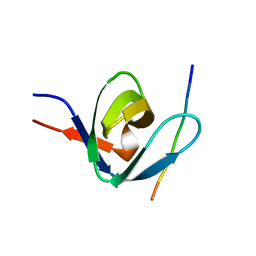
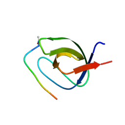
1GCP
DownloadVisualize
BU of 1gcp by Molmil
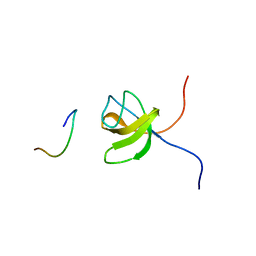
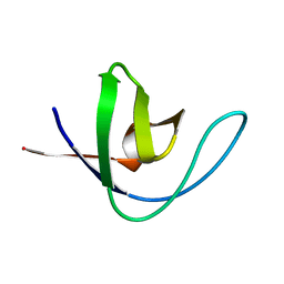
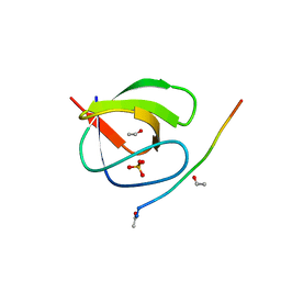
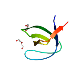
CRYSTAL STRUCTURE OF VAV SH3 DOMAIN
Descriptor: VAV PROTO-ONCOGENE
Authors:Nishida, M, Nagata, K, Hachimori, Y, Ogura, K, Inagaki, F.
Deposit date:2000-08-08
Release date:2001-08-08
Last modified:2023-10-25
Method:X-RAY DIFFRACTION (2.1 Å)
Cite:Novel recognition mode between Vav and Grb2 SH3 domains.
EMBO J., 20, 2001
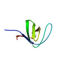
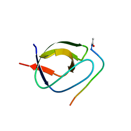
1GBR
DownloadVisualize
BU of 1gbr by Molmil
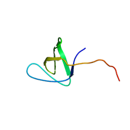
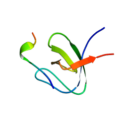
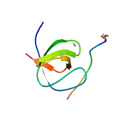
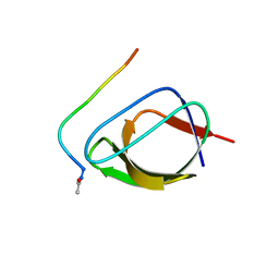
ORIENTATION OF PEPTIDE FRAGMENTS FROM SOS PROTEINS BOUND TO THE N-TERMINAL SH3 DOMAIN OF GRB2 DETERMINED BY NMR SPECTROSCOPY
Descriptor: GROWTH FACTOR RECEPTOR-BOUND PROTEIN 2, SOS-A PEPTIDE
Authors:Wittekind, M, Mapelli, C, Farmer, B.T, Suen, K.-L, Goldfarb, V, Tsao, J, Lavoie, T, Barbacid, M, Meyers, C.A, Mueller, L.
Deposit date:1994-08-12
Release date:1995-01-26
Last modified:2022-02-23
Method:SOLUTION NMR
Cite:Orientation of peptide fragments from Sos proteins bound to the N-terminal SH3 domain of Grb2 determined by NMR spectroscopy.
Biochemistry, 33, 1994
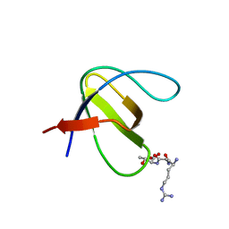
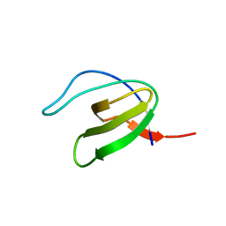
1GL5
DownloadVisualize
BU of 1gl5 by Molmil
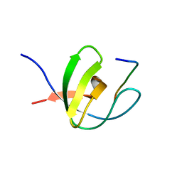
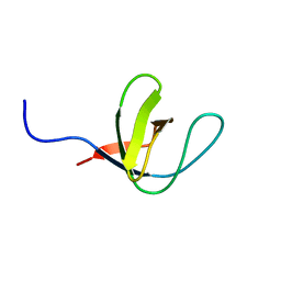
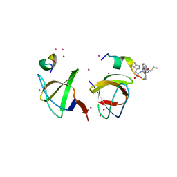
NMR structure of the SH3 domain from the Tec protein tyrosine kinase
Descriptor: TYROSINE-PROTEIN KINASE TEC
Authors:Mulhern, T.D, Pursglove, S.E, Booker, G.W.
Deposit date:2001-08-28
Release date:2001-11-28
Last modified:2024-05-15
Method:SOLUTION NMR
Cite:The solution structure and intramolecular associations of the Tec kinase SRC homology 3 domain.
J. Biol. Chem., 277, 2002
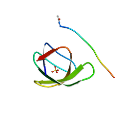
1FYN
DownloadVisualize
BU of 1fyn by Molmil
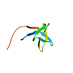
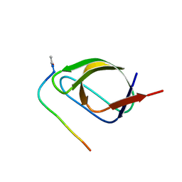
PHOSPHOTRANSFERASE
Descriptor: 3BP-2, PHOSPHOTRANSFERASE FYN
Authors:Musacchio, A, Saraste, M, Wilmanns, M.
Deposit date:1995-05-17
Release date:1996-11-08
Last modified:2024-02-07
Method:X-RAY DIFFRACTION (2.3 Å)
Cite:High-resolution crystal structures of tyrosine kinase SH3 domains complexed with proline-rich peptides.
Nat.Struct.Biol., 1, 1994
1HD3
DownloadVisualize
BU of 1hd3 by Molmil
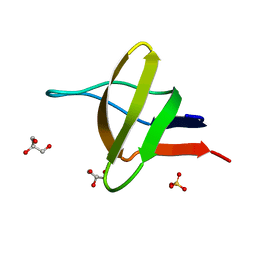
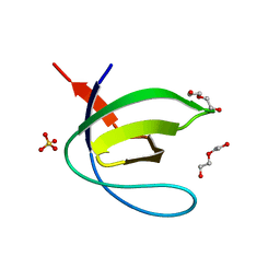
A-SPECTRIN SH3 DOMAIN F52Y MUTANT
Descriptor: GLYCEROL, SPECTRIN ALPHA CHAIN, SULFATE ION
Authors:Vega, M.C, Viguera, A.R, Serrano, L.
Deposit date:2000-11-06
Release date:2001-11-01
Last modified:2023-12-13
Method:X-RAY DIFFRACTION (1.98 Å)
Cite:Unspecific Hydrophobic Stabilization of Folding Transition States
Proc.Natl.Acad.Sci.USA, 99, 2002
1H8K
DownloadVisualize
BU of 1h8k by Molmil
A-SPECTRIN SH3 DOMAIN A11V, V23L, M25V, V53I, V58L MUTANT
Descriptor: SPECTRIN ALPHA CHAIN
Authors:Vega, M.C, Serrano, L.
Deposit date:2001-02-09
Release date:2002-05-23
Last modified:2023-12-13
Method:X-RAY DIFFRACTION (2.7 Å)
Cite:Conformational Strain in the Hydrophobic Core and its Implications for Protein Folding and Design
Nat.Struct.Biol., 9, 2002
1H3H
DownloadVisualize
BU of 1h3h by Molmil
Structural Basis for Specific Recognition of an RxxK-containing SLP-76 peptide by the Gads C-terminal SH3 domain
Descriptor: GRB2-RELATED ADAPTOR PROTEIN 2, LYMPHOCYTE CYTOSOLIC PROTEIN 2
Authors:Liu, Q, Berry, D, Nash, P, Pawson, T, McGlade, C.J, Li, S.S.
Deposit date:2002-09-03
Release date:2003-03-06
Last modified:2024-05-15
Method:SOLUTION NMR
Cite:Structural Basis for Specific Binding of the Gads SH3 Domain to an Rxxk Motif-Containing Slp-76 Peptide: A Novel Mode of Peptide Recognition
Mol.Cell, 11, 2003
1H92
DownloadVisualize
BU of 1h92 by Molmil
SH3 domain of human Lck tyrosine kinase
Descriptor: PROTO-ONCOGENE TYROSINE-PROTEIN KINASE LCK
Authors:Schweimer, K, Hoffmann, S, Friedrich, U, Biesinger, B, Roesch, P, Sticht, H.
Deposit date:2001-02-22
Release date:2001-10-22
Last modified:2024-05-15
Method:SOLUTION NMR
Cite:Structural Investigation of the Binding of a Herpesviral Protein to the SH3 Domain of Tyrosine Kinase Lck
Biochemistry, 41, 2002
1HSQ
DownloadVisualize
BU of 1hsq by Molmil
SOLUTION STRUCTURE OF THE SH3 DOMAIN OF PHOSPHOLIPASE CGAMMA
Descriptor: PHOSPHOLIPASE C-GAMMA (SH3 DOMAIN)
Authors:Kohda, D, Hatanaka, H, Odaka, M, Inagaki, F.
Deposit date:1994-06-13
Release date:1994-08-31
Last modified:2024-05-01
Method:SOLUTION NMR
Cite:Solution structure of the SH3 domain of phospholipase C-gamma.
Cell(Cambridge,Mass.), 72, 1993
1I07
DownloadVisualize
BU of 1i07 by Molmil
EPS8 SH3 DOMAIN INTERTWINED DIMER
Descriptor: EPIDERMAL GROWTH FACTOR RECEPTOR KINASE SUBSTRATE EPS8
Authors:Kishan, K.V.R, Newcomer, M.E.
Deposit date:2001-01-29
Release date:2001-05-09
Last modified:2023-08-09
Method:X-RAY DIFFRACTION (1.8 Å)
Cite:Effect of pH and salt bridges on structural assembly: molecular structures of the monomer and intertwined dimer of the Eps8 SH3 domain.
Protein Sci., 10, 2001
1I0C
DownloadVisualize
BU of 1i0c by Molmil
EPS8 SH3 CLOSED MONOMER
Descriptor: EPIDERMAL GROWTH FACTOR RECEPTOR KINASE SUBSTRATE EPS8
Authors:Kishan, K.V.R, Newcomer, M.E.
Deposit date:2001-01-29
Release date:2001-05-09
Last modified:2023-08-09
Method:X-RAY DIFFRACTION (2 Å)
Cite:Effect of pH and salt bridges on structural assembly: molecular structures of the monomer and intertwined dimer of the Eps8 SH3 domain.
Protein Sci., 10, 2001
1IO6
DownloadVisualize
BU of 1io6 by Molmil
GROWTH FACTOR RECEPTOR-BOUND PROTEIN 2 (GRB2) C-TERMINAL SH3 DOMAIN COMPLEXED WITH A LIGAND PEPTIDE (NMR, MINIMIZED MEAN STRUCTURE)
Descriptor: A LIGAND PEPTIDE, GROWTH FACTOR RECEPTOR-BOUND PROTEIN 2
Authors:Kawasaki, M, Ogura, K, Hatanaka, H, Inagaki, F.
Deposit date:2001-01-25
Release date:2001-02-14
Last modified:2023-12-27
Method:SOLUTION NMR
Cite:Solution Structure of the C-Terminal SH3 Domain of Grb2 Complexed with a Ligand Peptide: A Ligand Exchange Model of the SH3 Domain
To be Published
4HCK
DownloadVisualize
BU of 4hck by Molmil
HUMAN HCK SH3 DOMAIN, NMR, 25 STRUCTURES
Descriptor: HEMATOPOIETIC CELL KINASE
Authors:Horita, D.A, Baldisseri, D.M, Zhang, W, Altieri, A.S, Smithgall, T.E, Gmeiner, W.H, Byrd, R.A.
Deposit date:1998-03-09
Release date:1998-06-17
Last modified:2024-05-01
Method:SOLUTION NMR
Cite:Solution structure of the human Hck SH3 domain and identification of its ligand binding site.
J.Mol.Biol., 278, 1998
4HXJ
DownloadVisualize
BU of 4hxj by Molmil
Crystal structure of SH3:RGT complex
Descriptor: C-terminal 3-mer peptide from Integrin beta-3, Proto-oncogene tyrosine-protein kinase Src
Authors:Xiao, R, Meng, G.
Deposit date:2012-11-11
Release date:2012-11-28
Last modified:2023-11-08
Method:X-RAY DIFFRACTION (2 Å)
Cite:Structural framework of c-Src activation by integrin beta 3
Blood, 121, 2013
4HVV
DownloadVisualize
BU of 4hvv by Molmil
Crystal structure of the T98E c-Src-SH3 domain mutant in complex with the high affinity peptide APP12
Descriptor: Proto-oncogene tyrosine-protein kinase Src, SULFATE ION, SYNTHETIC PEPTIDE Acetyl-APPLPPRNRP
Authors:Camara-Artigas, A.
Deposit date:2012-11-07
Release date:2013-05-01
Last modified:2023-09-20
Method:X-RAY DIFFRACTION (1.1 Å)
Cite:Atomic resolution structures of the c-Src SH3 domain in complex with two high-affinity peptides from classes I and II.
Acta Crystallogr.,Sect.D, 69, 2013
4HVU
DownloadVisualize
BU of 4hvu by Molmil
Crystal structure of the T98D c-Src-SH3 domain mutant in complex with the high affinity peptide APP12
Descriptor: ACETYL GROUP, Proto-oncogene tyrosine-protein kinase Src, SULFATE ION, ...
Authors:Camara-Artigas, A.
Deposit date:2012-11-07
Release date:2013-05-01
Last modified:2023-09-20
Method:X-RAY DIFFRACTION (0.98 Å)
Cite:Atomic resolution structures of the c-Src SH3 domain in complex with two high-affinity peptides from classes I and II.
Acta Crystallogr.,Sect.D, 69, 2013
4HVW
DownloadVisualize
BU of 4hvw by Molmil
Crystal structure of the T98E c-Src-SH3 domain mutant in complex with the high affinity peptide VSL12
Descriptor: ACETYL GROUP, Proto-oncogene tyrosine-protein kinase Src, SULFATE ION, ...
Authors:Camara-Artigas, A.
Deposit date:2012-11-07
Release date:2013-05-01
Last modified:2023-09-20
Method:X-RAY DIFFRACTION (0.98 Å)
Cite:Atomic resolution structures of the c-Src SH3 domain in complex with two high-affinity peptides from classes I and II.
Acta Crystallogr.,Sect.D, 69, 2013
4IIM
DownloadVisualize
BU of 4iim by Molmil
Crystal structure of the Second SH3 Domain of ITSN1 bound with a synthetic peptide
Descriptor: Intersectin-1, UNKNOWN ATOM OR ION, peptide ligand
Authors:Dong, A, Guan, X, Huang, H, Wernimont, A, Gu, J, Sidhu, S, Bountra, C, Arrowsmith, C.H, Edwards, A.M, Tong, Y, Structural Genomics Consortium (SGC)
Deposit date:2012-12-20
Release date:2013-01-23
Last modified:2023-09-20
Method:X-RAY DIFFRACTION (1.8 Å)
Cite:Crystal structure of the Second SH3 Domain of ITSN1 bound with a synthetic peptide
To be Published
4J9E
DownloadVisualize
BU of 4j9e by Molmil
Crystal structure of the N114A mutant of the Abl-SH3 domain complexed with the high affinity peptide P17
Descriptor: P17, SULFATE ION, Tyrosine-protein kinase ABL1
Authors:Camara-Artigas, A.
Deposit date:2013-02-16
Release date:2014-01-29
Last modified:2023-09-20
Method:X-RAY DIFFRACTION (1.4 Å)
Cite:Crystal structure of the N114A mutant of the Abl-SH3 domain complexed with the high affinity peptide P17
To be Published
4J9B
DownloadVisualize
BU of 4j9b by Molmil
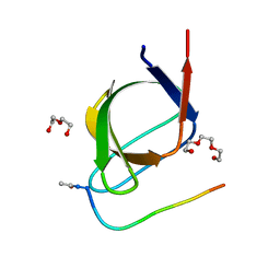
Crystal structure of the Abl-SH3 domain H59Q-N96T mutant
Descriptor: DI(HYDROXYETHYL)ETHER, Tyrosine-protein kinase ABL1
Authors:Camara-Artigas, A.
Deposit date:2013-02-16
Release date:2014-01-29
Last modified:2023-09-20
Method:X-RAY DIFFRACTION (1.702 Å)
Cite:Crystal structure of the Abl-SH3 domain H59Q-N96T mutant
To be Published
4JJB
DownloadVisualize
BU of 4jjb by Molmil
Crystal structure of the Abl-SH3 domain at pH3
Descriptor: DI(HYDROXYETHYL)ETHER, SULFATE ION, Tyrosine-protein kinase ABL1
Authors:Camara-Artigas, A, Martin-Garcia, J.M.
Deposit date:2013-03-07
Release date:2014-03-12
Last modified:2023-11-08
Method:X-RAY DIFFRACTION (1.65 Å)
Cite:Crystal structure of the Abl-SH3 domain at pH3
To be Published
4J9D
DownloadVisualize
BU of 4j9d by Molmil
Crystal structure of the N114A mutant of the Abl-SH3 domain complexed with the high affinity peptide P0
Descriptor: P0, SULFATE ION, Tyrosine-protein kinase ABL1
Authors:Camara-Artigas, A.
Deposit date:2013-02-16
Release date:2014-01-29
Last modified:2023-09-20
Method:X-RAY DIFFRACTION (1.5 Å)
Cite:Crystal structure of the N114A mutant of the Abl-SH3 domain complexed with the high affinity peptide P0
To be Published
4J9C
DownloadVisualize
BU of 4j9c by Molmil
Crystal structure of the Abl-SH3 domain H59Q-N96T mutant complexed with the designed high-affinity peptide ligand P17
Descriptor: DI(HYDROXYETHYL)ETHER, P17, TRIETHYLENE GLYCOL, ...
Authors:Camara-Artigas, A.
Deposit date:2013-02-16
Release date:2014-01-29
Last modified:2023-09-20
Method:X-RAY DIFFRACTION (1.051 Å)
Cite:Crystal structure of the Abl-SH3 domain H59Q-N96T mutant complexed with the designed high-affinity peptide ligand P17
To be Published
4J9H
DownloadVisualize
BU of 4j9h by Molmil
Crystal structure of the ABL-SH3 domain complexed with the designed high-affinity peptide ligand P7 at pH 8
Descriptor: P7, SULFATE ION, Tyrosine-protein kinase ABL1
Authors:Camara-Artigas, A.
Deposit date:2013-02-16
Release date:2014-01-29
Last modified:2023-09-20
Method:X-RAY DIFFRACTION (1.7 Å)
Cite:Crystal structure of the ABL-SH3 domain complexed with the designed high-affinity peptide ligand P7
To be Published
4J9G
DownloadVisualize
BU of 4j9g by Molmil
Crystal structure of the ABL-SH3 domain complexed with the designed high-affinity peptide ligand P7 at pH7
Descriptor: GLYCEROL, P7, SULFATE ION, ...
Authors:Camara-Artigas, A.
Deposit date:2013-02-16
Release date:2014-01-29
Last modified:2023-09-20
Method:X-RAY DIFFRACTION (1.8 Å)
Cite:Crystal structure of the ABL-SH3 domain complexed with the designed high-affinity peptide ligand P7
To be Published

219869

건을2024-05-15부터공개중

PDB statisticsPDBj update infoContact PDBjnumon