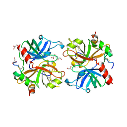
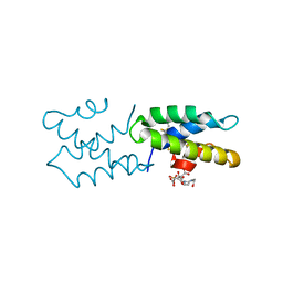
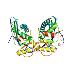
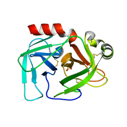
1GUU
 
 | | CRYSTAL STRUCTURE OF C-MYB R1 | | 分子名称: | MYB PROTO-ONCOGENE PROTEIN, SODIUM ION | | 著者 | Tahirov, T.H, Ogata, K. | | 登録日 | 2002-01-30 | | 公開日 | 2003-06-26 | | 最終更新日 | 2023-12-13 | | 実験手法 | X-RAY DIFFRACTION (1.6 Å) | | 主引用文献 | Crystal Structure of C-Myb DNA-Binding Domain: Specific Na+ Binding and Correlation with NMR Structure
To be Published
|
|
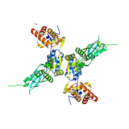
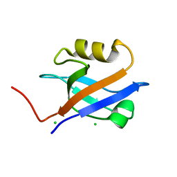
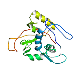
1GV5
 | | CRYSTAL STRUCTURE OF C-MYB R2 | | 分子名称: | MYB PROTO-ONCOGENE PROTEIN, SODIUM ION | | 著者 | Tahirov, T.H, Ogata, K. | | 登録日 | 2002-02-06 | | 公開日 | 2003-07-03 | | 最終更新日 | 2023-12-13 | | 実験手法 | X-RAY DIFFRACTION (1.58 Å) | | 主引用文献 | Crystal Structure of C-Myb DNA-Binding Domain: Specific Na+ Binding and Correlation with NMR Structure
To be Published
|
|
2AFQ
 | |
5F1G
 | | Crystal structure of AmpC BER adenylylated in the cytoplasm | | 分子名称: | 1,2-ETHANEDIOL, ADENOSINE MONOPHOSPHATE, Beta-lactamase, ... | | 著者 | An, Y.J, Kim, M.K, Na, J.H, Cha, S.S. | | 登録日 | 2015-11-30 | | 公開日 | 2016-12-07 | | 最終更新日 | 2023-11-08 | | 実験手法 | X-RAY DIFFRACTION (1.76 Å) | | 主引用文献 | Structural and mechanistic insights into the inhibition of class C beta-lactamases through the adenylylation of the nucleophilic serine.
J.Antimicrob.Chemother., 72, 2017
|
|
4ZPX
 | |
1GQ5
 | |
2WIT
 | | CRYSTAL STRUCTURE OF THE SODIUM-COUPLED GLYCINE BETAINE SYMPORTER BETP FROM CORYNEBACTERIUM GLUTAMICUM WITH BOUND SUBSTRATE | | 分子名称: | GLYCINE BETAINE TRANSPORTER BETP, TRIMETHYL GLYCINE | | 著者 | Ressl, S, Terwisscha Van Scheltinga, A.C, Vonrhein, C, Ott, V, Ziegler, C. | | 登録日 | 2009-05-17 | | 公開日 | 2009-05-26 | | 最終更新日 | 2023-11-15 | | 実験手法 | X-RAY DIFFRACTION (3.35 Å) | | 主引用文献 | Molecular Basis of Transport and Regulation in the Na(+)/Betaine Symporter Betp.
Nature, 458, 2009
|
|
2YGG
 | | Complex of CaMBR and CaM | | 分子名称: | (4R)-2-METHYLPENTANE-2,4-DIOL, (4S)-2-METHYL-2,4-PENTANEDIOL, CALCIUM ION, ... | | 著者 | Koester, S, Yildiz, O. | | 登録日 | 2011-04-15 | | 公開日 | 2011-09-28 | | 最終更新日 | 2024-05-08 | | 実験手法 | X-RAY DIFFRACTION (2.227 Å) | | 主引用文献 | Structure of Human Na+/H+ Exchanger Nhe1 Regulatory Region in Complex with Cam and Ca2+
J.Biol.Chem., 286, 2011
|
|
7X9R
 | | Crystal structure of the antirepressor GmaR | | 分子名称: | Glycosyl transferase family 2 | | 著者 | Cho, S.Y, Na, H.W, Oh, H.B, Kwak, Y.M, Song, W.S, Park, S.C, Yoon, S.I. | | 登録日 | 2022-03-16 | | 公開日 | 2022-11-09 | | 最終更新日 | 2022-11-16 | | 実験手法 | X-RAY DIFFRACTION (2.25 Å) | | 主引用文献 | Structural basis of flagellar motility regulation by the MogR repressor and the GmaR antirepressor in Listeria monocytogenes.
Nucleic Acids Res., 50, 2022
|
|
7X9S
 | | Crystal structure of a complex between the antirepressor GmaR and the transcriptional repressor MogR | | 分子名称: | GmaR, Motility gene repressor MogR | | 著者 | Cho, S.Y, Na, H.W, Oh, H.B, Kwak, Y.M, Song, W.S, Park, S.C, Yoon, S.I. | | 登録日 | 2022-03-16 | | 公開日 | 2022-11-23 | | 最終更新日 | 2023-11-29 | | 実験手法 | X-RAY DIFFRACTION (3.11 Å) | | 主引用文献 | Structural basis of flagellar motility regulation by the MogR repressor and the GmaR antirepressor in Listeria monocytogenes.
Nucleic Acids Res., 50, 2022
|
|
3S64
 | | Saposin-like protein Ac-SLP-1 | | 分子名称: | 4-(2-HYDROXYETHYL)-1-PIPERAZINE ETHANESULFONIC ACID, CITRIC ACID, Saposin-like protein 1 | | 著者 | Willis, C, Wang, C.K, Osman, A, Simon, A, Mulvenna, J, Pickering, D, Riboldi-Tunicliffe, A, Jones, M.K, Loukas, A, Hofmann, A. | | 登録日 | 2011-05-25 | | 公開日 | 2012-01-18 | | 実験手法 | X-RAY DIFFRACTION (2.301 Å) | | 主引用文献 | Insights into the membrane interactions of the saposin-like proteins Na-SLP-1 and Ac-SLP-1 from human and dog hookworm.
Plos One, 6, 2011
|
|
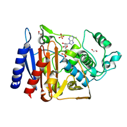
1TTQ
 | | TRYPTOPHAN SYNTHASE (E.C.4.2.1.20) IN THE PRESENCE OF POTASSIUM AT ROOM TEMPERATURE | | 分子名称: | POTASSIUM ION, PYRIDOXAL-5'-PHOSPHATE, TRYPTOPHAN SYNTHASE | | 著者 | Rhee, S, Parris, K, Ahmed, S, Miles, E.W, Davies, D.R. | | 登録日 | 1995-10-11 | | 公開日 | 1996-03-08 | | 最終更新日 | 2011-07-13 | | 実験手法 | X-RAY DIFFRACTION (2 Å) | | 主引用文献 | Exchange of K+ or Cs+ for Na+ induces local and long-range changes in the three-dimensional structure of the tryptophan synthase alpha2beta2 complex.
Biochemistry, 35, 1996
|
|
1TTP
 | | TRYPTOPHAN SYNTHASE (E.C.4.2.1.20) IN THE PRESENCE OF CESIUM, ROOM TEMPERATURE | | 分子名称: | CESIUM ION, PYRIDOXAL-5'-PHOSPHATE, TRYPTOPHAN SYNTHASE | | 著者 | Rhee, S, Parris, K, Ahmed, S, Miles, E.W, Davies, D.R. | | 登録日 | 1995-10-11 | | 公開日 | 1996-03-08 | | 最終更新日 | 2011-07-13 | | 実験手法 | X-RAY DIFFRACTION (2.3 Å) | | 主引用文献 | Exchange of K+ or Cs+ for Na+ induces local and long-range changes in the three-dimensional structure of the tryptophan synthase alpha2beta2 complex.
Biochemistry, 35, 1996
|
|
1L4J
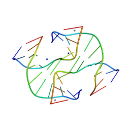 | | Holliday Junction TCGGTACCGA with Na and Ca Binding Sites. | | 分子名称: | 5'-D(*TP*CP*GP*GP*TP*AP*CP*CP*GP*A)-3', CALCIUM ION, SODIUM ION | | 著者 | Thorpe, J.H, Gale, B.C, Teixeira, S.C.M, Cardin, C.J. | | 登録日 | 2002-03-05 | | 公開日 | 2003-03-04 | | 最終更新日 | 2024-02-14 | | 実験手法 | X-RAY DIFFRACTION (1.85 Å) | | 主引用文献 | Conformational and Hydration Effects of Site-selective Sodium, Calcium and
Strontium Ion Binding to the DNA Holliday Junction Structure
d(TCGGTACCGA)4
J.Mol.Biol., 327, 2003
|
|
1XHK
 | | Crystal structure of M. jannaschii Lon proteolytic domain | | 分子名称: | 2-(N-MORPHOLINO)-ETHANESULFONIC ACID, Putative protease La homolog, SULFATE ION | | 著者 | Im, Y.J, Na, Y, Kang, G.B, Rho, S.-H, Kim, M.-K, Lee, J.H, Chung, C.H, Eom, S.H. | | 登録日 | 2004-09-20 | | 公開日 | 2004-10-05 | | 最終更新日 | 2011-07-13 | | 実験手法 | X-RAY DIFFRACTION (1.9 Å) | | 主引用文献 | The active site of a lon protease from Methanococcus jannaschii distinctly differs from the canonical catalytic Dyad of Lon proteases.
J.Biol.Chem., 279, 2004
|
|
1GQ4
 | |
1I0Q
 | | 1.3 A STRUCTURE OF THE A-DECAMER GCGTATACGC WITH A SINGLE 2'-O-METHYL-[TRI(OXYETHYL)] THYMINE IN PLACE OF T6, MEDIUM NA-SALT | | 分子名称: | 5'-D(*GP*CP*GP*TP*AP*(126)P*AP*CP*GP*C)-3', SODIUM ION | | 著者 | Tereshko, V, Wilds, C.J, Minasov, G, Prakash, T.P, Maier, M.A, Howard, A, Wawrzak, Z, Manoharan, M, Egli, M. | | 登録日 | 2001-01-29 | | 公開日 | 2001-04-04 | | 最終更新日 | 2024-02-07 | | 実験手法 | X-RAY DIFFRACTION (1.3 Å) | | 主引用文献 | Detection of alkali metal ions in DNA crystals using state-of-the-art X-ray diffraction experiments.
Nucleic Acids Res., 29, 2001
|
|
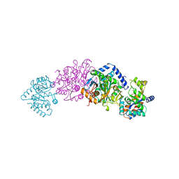
8P82
 | | Cryo-EM structure of dimeric UBR5 | | 分子名称: | E3 ubiquitin-protein ligase UBR5, ZINC ION | | 著者 | Aguirre, J.D, Kater, L, Kempf, G, Cavadini, S, Thoma, N.H. | | 登録日 | 2023-05-31 | | 公開日 | 2023-06-14 | | 実験手法 | ELECTRON MICROSCOPY (3.36 Å) | | 主引用文献 | UBR5 forms ligand-dependent complexes on chromatin to regulate nuclear hormone receptor stability
To Be Published
|
|
2FMJ
 | |
1I0G
 | | 1.45 A STRUCTURE OF THE A-DECAMER GCGTATACGC WITH A SINGLE 2'-O-FLUOROETHYL THYMINE IN PLACE OF T6, MEDIUM NA-SALT | | 分子名称: | 5'-D(*GP*CP*GP*TP*AP*(125)P*AP*CP*GP*C)-3', SODIUM ION | | 著者 | Tereshko, V, Wilds, C.J, Minasov, G, Prakash, T.P, Maier, M.A, Howard, A, Wawrzak, Z, Manoharan, M, Egli, M. | | 登録日 | 2001-01-29 | | 公開日 | 2001-04-04 | | 最終更新日 | 2024-02-07 | | 実験手法 | X-RAY DIFFRACTION (1.45 Å) | | 主引用文献 | Detection of alkali metal ions in DNA crystals using state-of-the-art X-ray diffraction experiments.
Nucleic Acids Res., 29, 2001
|
|
3GIN
 | |
1MO7
 | | ATPase | | 分子名称: | Sodium/Potassium-transporting ATPase alpha-1 chain | | 著者 | Hilge, M, Siegal, G, Vuister, G.W, Guentert, P, Gloor, S.M, Abrahams, J.P. | | 登録日 | 2002-09-08 | | 公開日 | 2003-06-03 | | 最終更新日 | 2022-02-23 | | 実験手法 | SOLUTION NMR | | 主引用文献 | ATP-induced conformational changes of the nucleotide-binding domain of Na,K-ATPase
Nat.Struct.Biol., 10, 2003
|
|
1MO8
 | | ATPase | | 分子名称: | ADENOSINE-5'-TRIPHOSPHATE, Sodium/Potassium-Transporting ATPase alpha-1 | | 著者 | Hilge, M, Siegal, G, Vuister, G.W, Guentert, P, Gloor, S.M, Abrahams, J.P. | | 登録日 | 2002-09-08 | | 公開日 | 2003-06-10 | | 最終更新日 | 2022-02-23 | | 実験手法 | SOLUTION NMR | | 主引用文献 | ATP-induced conformational changes of the nucleotide-binding domain of Na,K-ATPase
Nat.Struct.Biol., 10, 2003
|
|
1U8W
 | | Crystal structure of Arabidopsis thaliana nucleoside diphosphate kinase 1 | | 分子名称: | Nucleoside diphosphate kinase I | | 著者 | Im, Y.J, Kim, J.-I, Shen, Y, Na, Y, Han, Y.-J, Kim, S.-H, Song, P.-S, Eom, S.H. | | 登録日 | 2004-08-07 | | 公開日 | 2004-11-23 | | 最終更新日 | 2024-03-13 | | 実験手法 | X-RAY DIFFRACTION (2.4 Å) | | 主引用文献 | Structural analysis of Arabidopsis thaliana nucleoside diphosphate kinase-2 for phytochrome-mediated light signaling
J.Mol.Biol., 343, 2004
|
|
5X33
 | | Leukotriene B4 receptor BLT1 in complex with BIIL260 | | 分子名称: | 4-[[3-[[4-[2-(4-hydroxyphenyl)propan-2-yl]phenoxy]methyl]phenyl]methoxy]benzenecarboximidamide, LTB4 receptor,Lysozyme,LTB4 receptor | | 著者 | Hori, T, Hirata, K, Yamashita, K, Kawano, Y, Yamamoto, M, Yokoyama, S. | | 登録日 | 2017-02-03 | | 公開日 | 2018-01-03 | | 最終更新日 | 2023-11-22 | | 実験手法 | X-RAY DIFFRACTION (3.7 Å) | | 主引用文献 | Na+-mimicking ligands stabilize the inactive state of leukotriene B4receptor BLT1.
Nat. Chem. Biol., 14, 2018
|
|


