Loading
PDBj
MenuPDBj@FacebookPDBj@TwitterPDBj@YouTubewwPDB FoundationwwPDB
RCSB PDBPDBeBMRBAdv. SearchSearch help

5F9G
DownloadVisualize
BU of 5f9g by Molmil
pnGFP1.5-Y.Cro: circularly permuted green fluorescent protein (with a tyrosine-derived chromophore)
Descriptor: pnGFP1.5-Y.Cro,Green fluorescent protein
Authors:Remington, S.J.
Deposit date:2015-12-09
Release date:2016-02-03
Last modified:2023-11-15
Method:X-RAY DIFFRACTION (2.772 Å)
Cite:pnGFP: circularly permuted green fluorescent protein
To Be Published
3OGO
DownloadVisualize
BU of 3ogo by Molmil
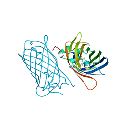
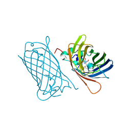
Structure of the GFP:GFP-nanobody complex at 2.8 A resolution in spacegroup P21212
Descriptor: GFP-nanobody, Green fluorescent protein, ISOPROPYL ALCOHOL
Authors:Kubala, M.H, Kovtun, O, Alexandrov, K, Collins, B.M.
Deposit date:2010-08-17
Release date:2010-08-25
Last modified:2024-02-21
Method:X-RAY DIFFRACTION (2.8 Å)
Cite:Structural and thermodynamic analysis of the GFP:GFP-nanobody complex.
Protein Sci., 19, 2010
4XVP
DownloadVisualize
BU of 4xvp by Molmil
X-ray structure of bGFP-C / EGFP complex
Descriptor: BGFP-C, Green fluorescent protein
Authors:Chevrel, A, Urvoas, A, Li de la Sierra-Gallay, I, Van Tilbeurgh, H, Minard, P, Valerio-Lepiniec, M.
Deposit date:2015-01-27
Release date:2015-08-19
Last modified:2024-01-10
Method:X-RAY DIFFRACTION (3.4 Å)
Cite:Specific GFP-binding artificial proteins ( alpha Rep): a new tool for in vitro to live cell applications.
Biosci.Rep., 35, 2015
4XL5
DownloadVisualize
BU of 4xl5 by Molmil
X-ray structure of bGFP-A / EGFP complex
Descriptor: Green fluorescent protein, bGFP-A
Authors:Chevrel, A, Urvoas, A, Li de la Sierra-Gallay, I, Van Tilbeurgh, H, Minard, P, Valerio-Lepiniec, M.
Deposit date:2015-01-13
Release date:2015-08-19
Last modified:2024-01-10
Method:X-RAY DIFFRACTION (2 Å)
Cite:Specific GFP-binding artificial proteins ( alpha Rep): a new tool for in vitro to live cell applications.
Biosci.Rep., 35, 2015
8BXP
DownloadVisualize
BU of 8bxp by Molmil
SfGFP C148 F206 mutant
Descriptor: Green fluorescent protein
Authors:Ahmed, R.D, Rizkallah, P.J, Jones, D.D.
Deposit date:2022-12-09
Release date:2023-12-20
Method:X-RAY DIFFRACTION (1.79 Å)
Cite:SfGFP C148 F206 mutant
To Be Published
8C1X
DownloadVisualize
BU of 8c1x by Molmil
sfGFP C148 F206 mutant
Descriptor: Green fluorescent protein
Authors:Ahmed, R.D, Rizkallah, P.J, Jones, D.D.
Deposit date:2022-12-21
Release date:2024-01-10
Method:X-RAY DIFFRACTION (1.89 Å)
Cite:sfGFP C148 F206 mutant
To Be Published
8AHB
DownloadVisualize
BU of 8ahb by Molmil
rsEGFP2 photoswitched to its off-state at room temperature and back-switched to its on-state at 100K
Descriptor: Green fluorescent protein, SULFATE ION
Authors:Mantovanelli, A, Adam, V.
Deposit date:2022-07-21
Release date:2023-07-19
Last modified:2024-02-07
Method:X-RAY DIFFRACTION (1.79 Å)
Cite:Photophysical Studies at Cryogenic Temperature Reveal a Novel Photoswitching Mechanism of rsEGFP2.
J.Am.Chem.Soc., 145, 2023
8AHA
DownloadVisualize
BU of 8aha by Molmil
rsEGFP2 photoswitched to its off-state at 100K
Descriptor: Green fluorescent protein, SULFATE ION
Authors:Mantovanelli, A, Adam, V.
Deposit date:2022-07-21
Release date:2023-07-19
Last modified:2024-02-07
Method:X-RAY DIFFRACTION (2.38 Å)
Cite:Photophysical Studies at Cryogenic Temperature Reveal a Novel Photoswitching Mechanism of rsEGFP2.
J.Am.Chem.Soc., 145, 2023
2Q57
DownloadVisualize
BU of 2q57 by Molmil
X-ray structure of Cerulean GFP: A tryptophan-based chromophore useful for fluorescence lifetime imaging
Descriptor: Cerulean Green fluorescent protein
Authors:Malo, G.D.
Deposit date:2007-05-31
Release date:2007-11-27
Last modified:2023-11-15
Method:X-RAY DIFFRACTION (2 Å)
Cite:X-ray structure of Cerulean GFP: a tryptophan-based chromophore useful for fluorescence lifetime imaging.
Biochemistry, 46, 2007
4XGY
DownloadVisualize
BU of 4xgy by Molmil
GFP based antibody (fluorobody)
Descriptor: 3,6,9,12,15,18,21,24-OCTAOXAHEXACOSAN-1-OL, Green fluorescent protein, mAb LCDR3
Authors:Shi, N, Chen, Y.G, Wang, S.H.
Deposit date:2015-01-03
Release date:2015-07-01
Last modified:2023-11-15
Method:X-RAY DIFFRACTION (1.494 Å)
Cite:The structure of a GFP-based antibody (fluorobody) to TLH, a toxin from Vibrio parahaemolyticus.
Acta Crystallogr.,Sect.F, 71, 2015
4OGS
DownloadVisualize
BU of 4ogs by Molmil
Crystal structure of GFP S205A/T203V at 2.2 A resolution
Descriptor: CHLORIDE ION, Green fluorescent protein
Authors:Remington, S.J, Saif, M.
Deposit date:2014-01-16
Release date:2014-05-14
Last modified:2014-06-18
Method:X-RAY DIFFRACTION (2.21 Å)
Cite:Insight into the structure and the mechanism of the slow proton transfer in the GFP double mutant T203V/S205A.
Phys Chem Chem Phys, 16, 2014
1YHH
DownloadVisualize
BU of 1yhh by Molmil
Uncyclized precursor structure of S65A Y66S G67A GFP variant
Descriptor: green fluorescent protein
Authors:Barondeau, D.P, Kassmann, C.J, Tainer, J.A, Getzoff, E.D.
Deposit date:2005-01-07
Release date:2005-02-15
Last modified:2024-02-14
Method:X-RAY DIFFRACTION (1.5 Å)
Cite:Understanding GFP Chromophore Biosynthesis: Controlling Backbone Cyclization and Modifying Post-translational Chemistry.
Biochemistry, 44, 2005
1YHI
DownloadVisualize
BU of 1yhi by Molmil
Uncyclized precursor structure of S65A Y66S R96A GFP variant
Descriptor: Green Fluorescent Protein
Authors:Barondeau, D.P, Kassmann, C.J, Tainer, J.A, Getzoff, E.D.
Deposit date:2005-01-08
Release date:2005-02-15
Last modified:2024-02-14
Method:X-RAY DIFFRACTION (1.9 Å)
Cite:Understanding GFP Chromophore Biosynthesis: Controlling Backbone Cyclization and Modifying Post-translational Chemistry(,).
Biochemistry, 44, 2005
1YHG
DownloadVisualize
BU of 1yhg by Molmil
Uncyclized precursor structure of S65G Y66S V68G GFP variant
Descriptor: green fluorescent protein
Authors:Barondeau, D.P, Kassmann, C.J, Tainer, J.A, Getzoff, E.D.
Deposit date:2005-01-07
Release date:2005-02-15
Last modified:2024-02-14
Method:X-RAY DIFFRACTION (2.5 Å)
Cite:Understanding GFP Chromophore Biosynthesis: Controlling Backbone Cyclization and Modifying Post-translational Chemistry.
Biochemistry, 44, 2005
2O2B
DownloadVisualize
BU of 2o2b by Molmil
Spectroscopic and Structural Study of the Heterotropic Linkage between Halide and Proton Ion Binding to Gfp Proteins: E2(GFP)-I Complex
Descriptor: Green fluorescent protein, IODIDE ION
Authors:Garau, G.
Deposit date:2006-11-29
Release date:2007-05-15
Last modified:2023-11-15
Method:X-RAY DIFFRACTION (1.94 Å)
Cite:Spectroscopic and Structural Study of Proton and Halide Ion Cooperative Binding to GFP.
Biophys.J., 93, 2007
2O24
DownloadVisualize
BU of 2o24 by Molmil
Spectroscopic and Structural Study of the Heterotropic Linkage between Halide and Proton Ion Binding to Gfp Proteins: E2(GFP)-Cl Complex
Descriptor: CHLORIDE ION, Green fluorescent protein
Authors:Garau, G.
Deposit date:2006-11-29
Release date:2007-05-15
Last modified:2023-11-15
Method:X-RAY DIFFRACTION (1.45 Å)
Cite:Spectroscopic and Structural Study of Proton and Halide Ion Cooperative Binding to GFP.
Biophys.J., 93, 2007
2O29
DownloadVisualize
BU of 2o29 by Molmil
Spectroscopic and Structural Study of the Heterotropic Linkage between Halide and Proton Ion Binding to Gfp Proteins: E2(GFP)-BR Complex
Descriptor: BROMIDE ION, Green fluorescent protein
Authors:Garau, G.
Deposit date:2006-11-29
Release date:2007-05-15
Last modified:2023-11-15
Method:X-RAY DIFFRACTION (1.8 Å)
Cite:Spectroscopic and Structural Study of Proton and Halide Ion Cooperative Binding to GFP.
Biophys.J., 93, 2007
6LR7
DownloadVisualize
BU of 6lr7 by Molmil
Crystal structure of GFPuv complexed with the nanobody LaG16 at 1.67 Angstron resolution
Descriptor: Green fluorescent protein, Nanobody LaG16
Authors:Zhang, Z.Y, Ding, Y, Hattori, M.
Deposit date:2020-01-15
Release date:2020-04-29
Last modified:2023-04-05
Method:X-RAY DIFFRACTION (1.67 Å)
Cite:Structure-based engineering of anti-GFP nanobody tandems as ultra-high-affinity reagents for purification.
Sci Rep, 10, 2020
2G6E
DownloadVisualize
BU of 2g6e by Molmil
Structure of cyclized F64L S65A Y66S GFP variant
Descriptor: Green fluorescent protein, MAGNESIUM ION
Authors:Barondeau, D.P.
Deposit date:2006-02-24
Release date:2006-04-18
Last modified:2023-11-15
Method:X-RAY DIFFRACTION (1.3 Å)
Cite:Understanding GFP Posttranslational Chemistry: Structures of Designed Variants that Achieve Backbone Fragmentation, Hydrolysis, and Decarboxylation.
J.Am.Chem.Soc., 128, 2006
2H6V
DownloadVisualize
BU of 2h6v by Molmil
Spectroscopic and structural study of the heterotropic linkage between halide and proton ion binding to GFP proteins- E2(GFP) APO FORM
Descriptor: Green fluorescent protein, SODIUM ION
Authors:Garau, G.
Deposit date:2006-06-01
Release date:2007-05-15
Last modified:2021-10-20
Method:X-RAY DIFFRACTION (1.47 Å)
Cite:Spectroscopic and Structural Study of Proton and Halide Ion Cooperative Binding to GFP
Biophys.J., 93, 2007
2G5Z
DownloadVisualize
BU of 2g5z by Molmil
Structure of S65G Y66S GFP variant after spontaneous peptide hydrolysis and decarboxylation
Descriptor: Green fluorescent protein, MAGNESIUM ION
Authors:Barondeau, D.P, Kassmann, C.J, Tainer, J.A, Getzoff, E.D.
Deposit date:2006-02-23
Release date:2006-04-18
Last modified:2023-08-30
Method:X-RAY DIFFRACTION (1.8 Å)
Cite:Understanding GFP Posttranslational Chemistry: Structures of Designed Variants that Achieve Backbone Fragmentation, Hydrolysis, and Decarboxylation.
J.Am.Chem.Soc., 128, 2006
2HGD
DownloadVisualize
BU of 2hgd by Molmil
Structure of S65A Y66F GFP variant with an oxidized chromophore
Descriptor: Green fluorescent protein
Authors:Barondeau, D.P, Kassmann, C.J, Tainer, J.A, Getzoff, E.D.
Deposit date:2006-06-26
Release date:2007-03-27
Last modified:2023-11-15
Method:X-RAY DIFFRACTION (1.6 Å)
Cite:The Case of the Missing Ring: Radical Cleavage of a Carbon-Carbon Bond and Implications for GFP Chromophore Biosynthesis
J.Am.Chem.Soc., 129, 2007
2HGY
DownloadVisualize
BU of 2hgy by Molmil
Structure of S65A Y66F E222A GFP variant after cyclization and carbon-carbon bond cleavage
Descriptor: Green fluorescent protein
Authors:Barondeau, D.P, Kassmann, C.J, Tainer, J.A, Getzoff, E.D.
Deposit date:2006-06-27
Release date:2007-03-27
Last modified:2023-11-15
Method:X-RAY DIFFRACTION (2.05 Å)
Cite:The Case of the Missing Ring: Radical Cleavage of a Carbon-Carbon Bond and Implications for GFP Chromophore Biosynthesis
J.Am.Chem.Soc., 129, 2007
2G3D
DownloadVisualize
BU of 2g3d by Molmil
Structure of S65G Y66A GFP variant after spontaneous peptide hydrolysis
Descriptor: Green fluorescent protein, MAGNESIUM ION
Authors:Barondeau, D.P.
Deposit date:2006-02-17
Release date:2006-04-18
Last modified:2023-08-30
Method:X-RAY DIFFRACTION (1.35 Å)
Cite:Understanding GFP Posttranslational Chemistry: Structures of Designed Variants that Achieve Backbone Fragmentation, Hydrolysis, and Decarboxylation.
J.Am.Chem.Soc., 128, 2006
2G2S
DownloadVisualize
BU of 2g2s by Molmil
Structure of S65G Y66S GFP variant after spontaneous peptide hydrolysis
Descriptor: Green fluorescent protein, MAGNESIUM ION
Authors:Barondeau, D.P.
Deposit date:2006-02-16
Release date:2006-04-18
Last modified:2023-08-30
Method:X-RAY DIFFRACTION (1.2 Å)
Cite:Understanding GFP Posttranslational Chemistry: Structures of Designed Variants that Achieve Backbone Fragmentation, Hydrolysis, and Decarboxylation.
J.Am.Chem.Soc., 128, 2006

219140

數據於2024-05-01公開中

PDB statisticsPDBj update infoContact PDBjnumon