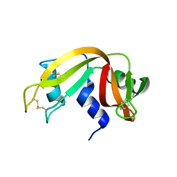
1AWZ
 
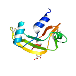
 | | 3D SOLUTION STRUCTURE OF HUMAN ANGIOGENIN DETERMINED BY 1H, 15N NMR SPECTROSCOPY, 30 STRUCTURES | | 分子名称: | ANGIOGENIN | | 著者 | Lequin, O, Thuring, H, Robin, M, Lallemand, J.-Y. | | 登録日 | 1997-10-07 | | 公開日 | 1998-02-25 | | 最終更新日 | 2022-02-16 | | 実験手法 | SOLUTION NMR | | 主引用文献 | Three-dimensional solution structure of human angiogenin determined by 1H,15N-NMR spectroscopy--characterization of histidine protonation states and pKa values.
Eur.J.Biochem., 250, 1997
|
|
4G8Y
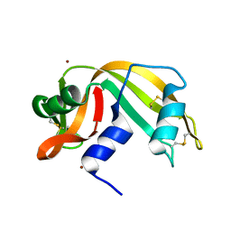
 | | Crystal structure of Ribonuclease A in complex with 5b | | 分子名称: | 1-{[1-(alpha-L-arabinofuranosyl)-1H-1,2,3-triazol-4-yl]methyl}-5-methyl-2,4-dioxo-1,2,3,4-tetrahydropyrimidine, Ribonuclease pancreatic | | 著者 | Chatzileontiadou, D.S.M, Kantsadi, A.L, Leonidas, D.D. | | 登録日 | 2012-07-23 | | 公開日 | 2012-11-21 | | 最終更新日 | 2023-09-13 | | 実験手法 | X-RAY DIFFRACTION (1.8 Å) | | 主引用文献 | Triazole pyrimidine nucleosides as inhibitors of Ribonuclease A. Synthesis, biochemical, and structural evaluation.
Bioorg.Med.Chem., 20, 2012
|
|
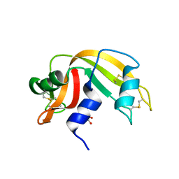
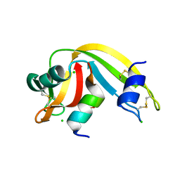
1B1J
 | | CRYSTAL STRUCTURE OF HUMAN ANGIOGENIN VARIANT H13A. | | 分子名称: | HYDROLASE ANGIOGENIN | | 著者 | Leonidas, D.D, Acharya, K.R. | | 登録日 | 1998-11-20 | | 公開日 | 1999-04-02 | | 最終更新日 | 2023-08-09 | | 実験手法 | X-RAY DIFFRACTION (2 Å) | | 主引用文献 | Refined crystal structures of native human angiogenin and two active site variants: implications for the unique functional properties of an enzyme involved in neovascularisation during tumour growth.
J.Mol.Biol., 285, 1999
|
|
1B6V
 | | CRYSTAL STRUCTURE OF A HYBRID BETWEEN RIBONUCLEASE A AND BOVINE SEMINAL RIBONUCLEASE | | 分子名称: | RIBONUCLEASE | | 著者 | Vatzaki, E.H, Allen, S.C, Leonidas, D.D, Acharya, K.R. | | 登録日 | 1999-01-18 | | 公開日 | 1999-06-15 | | 最終更新日 | 2023-08-02 | | 実験手法 | X-RAY DIFFRACTION (2 Å) | | 主引用文献 | Crystal structure of a hybrid between ribonuclease A and bovine seminal ribonuclease--the basic surface, at 2.0 A resolution.
Eur.J.Biochem., 260, 1999
|
|
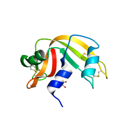
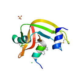
1B1E
 | | CRYSTAL STRUCTURE OF HUMAN ANGIOGENIN VARIANT K40Q | | 分子名称: | CITRIC ACID, HYDROLASE ANGIOGENIN | | 著者 | Leonidas, D.D, Acharya, K.R. | | 登録日 | 1998-11-20 | | 公開日 | 1999-04-02 | | 最終更新日 | 2023-08-09 | | 実験手法 | X-RAY DIFFRACTION (2 Å) | | 主引用文献 | Refined crystal structures of native human angiogenin and two active site variants: implications for the unique functional properties of an enzyme involved in neovascularisation during tumour growth.
J.Mol.Biol., 285, 1999
|
|
1AQP
 | | RIBONUCLEASE A COPPER COMPLEX | | 分子名称: | COPPER (II) ION, RIBONUCLEASE A | | 著者 | Ramasubbu, N. | | 登録日 | 1997-07-31 | | 公開日 | 1998-05-27 | | 最終更新日 | 2023-08-02 | | 実験手法 | X-RAY DIFFRACTION (2 Å) | | 主引用文献 | Crystal structures of the copper and nickel complexes of RNase A: metal-induced interprotein interactions and identification of a novel copper binding motif.
Proc.Natl.Acad.Sci.USA, 94, 1997
|
|
4G90
 | | Crystal structure of Ribonuclease A in complex with 5e | | 分子名称: | 1-{[1-(alpha-L-arabinofuranosyl)-1H-1,2,3-triazol-4-yl]methyl}-5-fluoro-2,4-dioxo-1,2,3,4-tetrahydropyrimidine, Ribonuclease pancreatic | | 著者 | Chatzileontiadou, D.S.M, Kantsadi, A.L, Leonidas, D.D. | | 登録日 | 2012-07-23 | | 公開日 | 2012-11-21 | | 最終更新日 | 2023-09-13 | | 実験手法 | X-RAY DIFFRACTION (1.9 Å) | | 主引用文献 | Triazole pyrimidine nucleosides as inhibitors of Ribonuclease A. Synthesis, biochemical, and structural evaluation.
Bioorg.Med.Chem., 20, 2012
|
|
4G8V
 | | Crystal structure of Ribonuclease A in complex with 5a | | 分子名称: | 1-{[1-(alpha-L-arabinofuranosyl)-1H-1,2,3-triazol-4-yl]methyl}-2,4-dioxo-1,2,3,4-tetrahydropyrimidine, Ribonuclease pancreatic | | 著者 | Chatzileontiadou, D.S.M, Kantsadi, A.L, Leonidas, D.D. | | 登録日 | 2012-07-23 | | 公開日 | 2012-11-21 | | 最終更新日 | 2023-09-13 | | 実験手法 | X-RAY DIFFRACTION (1.7 Å) | | 主引用文献 | Triazole pyrimidine nucleosides as inhibitors of Ribonuclease A. Synthesis, biochemical, and structural evaluation.
Bioorg.Med.Chem., 20, 2012
|
|
4MXF
 | |
4N4C
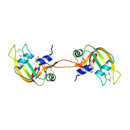 | | Crystal structure of the C-terminal swapped dimer of a Bovine seminal ribonuclease mutant | | 分子名称: | PHOSPHATE ION, Seminal ribonuclease | | 著者 | Pica, A, Russo Krauss, I, Merlino, A, Sica, F. | | 登録日 | 2013-10-08 | | 公開日 | 2013-11-06 | | 最終更新日 | 2023-09-20 | | 実験手法 | X-RAY DIFFRACTION (2.48 Å) | | 主引用文献 | The multiple forms of bovine seminal ribonuclease: Structure and stability of a C-terminal swapped dimer.
Febs Lett., 587, 2013
|
|
4O37
 | | seminsynthetic RNase S1-15-3Pl-7/11 | | 分子名称: | CHLORIDE ION, Ribonuclease pancreatic, S-peptide, ... | | 著者 | Genz, M, Strater, N. | | 登録日 | 2013-12-18 | | 公開日 | 2014-05-07 | | 最終更新日 | 2023-12-06 | | 実験手法 | X-RAY DIFFRACTION (1.4 Å) | | 主引用文献 | An Artificial Imine Reductase based on the Ribonuclease S Scaffold
Chem.Cat.Chem, 2014
|
|
4O36
 | | Semisynthetic RNase S1-15-H7/11-Q10 | | 分子名称: | CHLORIDE ION, Ribonuclease pancreatic, S-peptide, ... | | 著者 | Genz, M, Strater, N. | | 登録日 | 2013-12-18 | | 公開日 | 2014-05-07 | | 最終更新日 | 2023-09-20 | | 実験手法 | X-RAY DIFFRACTION (1.22 Å) | | 主引用文献 | An Artificial Imine Reductase based on the Ribonuclease S scaffold
Chem.Cat.Chem, 2014
|
|
4OXF
 | |
4OKF
 | |
4OOH
 | |
4OWZ
 | |
4OXB
 | |
4OT4
 | |
4J63
 | |
4J67
 | | Crystal structure of Ribonuclease A soaked in 50% 1,6-Hexanediol: One of twelve in MSCS set | | 分子名称: | HEXANE-1,6-DIOL, Ribonuclease pancreatic, SULFATE ION | | 著者 | Kearney, B.M, Dechene, M, Swartz, P.D, Mattos, C. | | 登録日 | 2013-02-11 | | 公開日 | 2014-01-22 | | 最終更新日 | 2023-09-20 | | 実験手法 | X-RAY DIFFRACTION (1.86 Å) | | 主引用文献 | DRoP: A program for analysis of water structure on protein surfaces
to be published
|
|
4J60
 | |
4J64
 | | Crystal structure of Ribonuclease A soaked in 40% Dioxane: One of twelve in MSCS set | | 分子名称: | 1,4-DIETHYLENE DIOXIDE, Ribonuclease pancreatic, SULFATE ION | | 著者 | Kearney, B.M, Dechene, M, Swartz, P.D, Mattos, C. | | 登録日 | 2013-02-11 | | 公開日 | 2014-01-22 | | 最終更新日 | 2023-09-20 | | 実験手法 | X-RAY DIFFRACTION (1.781 Å) | | 主引用文献 | DRoP: A program for analysis of water structure on protein surfaces
to be published
|
|
4J62
 | |
4J69
 | | Crystal structure of Ribonuclease A soaked in 50% S,R,S-bisfuranol: One of twelve in MSCS set | | 分子名称: | (3S,3aR,6aS)-hexahydrofuro[2,3-b]furan-3-ol, Ribonuclease pancreatic, SULFATE ION | | 著者 | Kearney, B.M, Dechene, M, Swartz, P.D, Mattos, C. | | 登録日 | 2013-02-11 | | 公開日 | 2014-01-22 | | 最終更新日 | 2023-09-20 | | 実験手法 | X-RAY DIFFRACTION (1.892 Å) | | 主引用文献 | DRoP: A program for analysis of water structure on protein surfaces
to be published
|
|
4J6A
 | | Crystal structure of Ribonuclease A soaked in 40% 2,2,2-Trifluoroethanol: One of twelve in MSCS set | | 分子名称: | Ribonuclease pancreatic, SULFATE ION, TRIFLUOROETHANOL | | 著者 | Kearney, B.M, Dechene, M, Swartz, P.D, Mattos, C. | | 登録日 | 2013-02-11 | | 公開日 | 2014-01-22 | | 最終更新日 | 2023-09-20 | | 実験手法 | X-RAY DIFFRACTION (2.04 Å) | | 主引用文献 | DRoP: A program for analysis of water structure on protein surfaces
to be published
|
|


