Loading
PDBj
MenuPDBj@FacebookPDBj@X(formerly Twitter)PDBj@BlueSkyPDBj@YouTubewwPDB FoundationwwPDB
RCSB PDBPDBeBMRBAdv. SearchSearch help

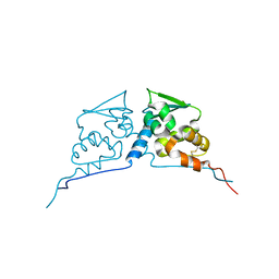
2Z8H
DownloadVisualize
BU of 2z8h by Molmil
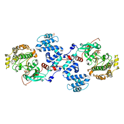
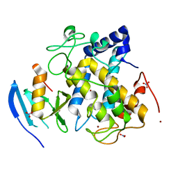
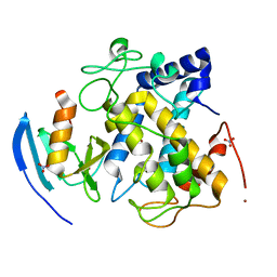
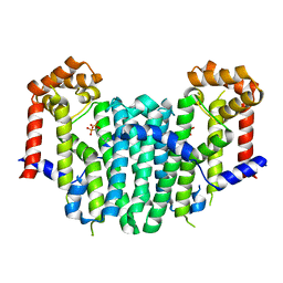
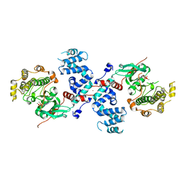
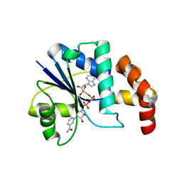
Structure of mouse Bach1 BTB domain
Descriptor: Transcription regulator protein BACH1
Authors:Ito, N, Murayama, K.
Deposit date:2007-09-05
Release date:2008-09-09
Last modified:2024-11-06
Method:X-RAY DIFFRACTION (2.5 Å)
Cite:Structure of mouse Bach1 BTB domain
To be Published
3C50
DownloadVisualize
BU of 3c50 by Molmil
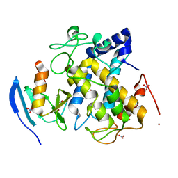
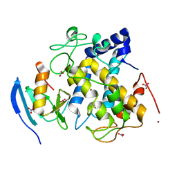
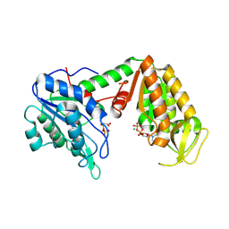
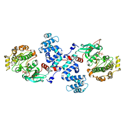
Crystal Structure of G protein coupled receptor kinase 1 bound to ADP and magnesium chloride at 2.6A
Descriptor: 2-(N-MORPHOLINO)-ETHANESULFONIC ACID, ADENOSINE-5'-DIPHOSPHATE, CHLORIDE ION, ...
Authors:Singh, P, Tesmer, J.J.G.
Deposit date:2008-01-30
Release date:2008-03-11
Last modified:2023-08-30
Method:X-RAY DIFFRACTION (2.6 Å)
Cite:Structures of rhodopsin kinase in different ligand states reveal key elements involved in G protein-coupled receptor kinase activation.
J.Biol.Chem., 283, 2008
3C1O
DownloadVisualize
BU of 3c1o by Molmil
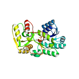
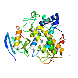
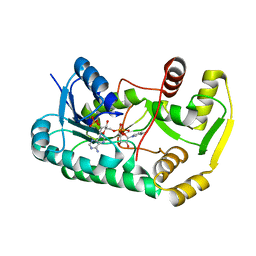
The multiple phenylpropene synthases in both Clarkia breweri and Petunia hybrida represent two distinct lineages
Descriptor: NADP NICOTINAMIDE-ADENINE-DINUCLEOTIDE PHOSPHATE, eugenol synthase
Authors:Louie, G.V, Noel, J.P, Bowman, M.E.
Deposit date:2008-01-23
Release date:2008-05-06
Last modified:2024-02-21
Method:X-RAY DIFFRACTION (1.8 Å)
Cite:The multiple phenylpropene synthases in both Clarkia breweri and Petunia hybrida represent two distinct protein lineages.
Plant J., 54, 2008
3C4Y
DownloadVisualize
BU of 3c4y by Molmil
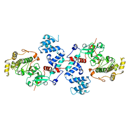
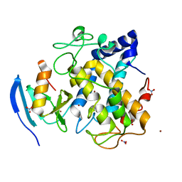
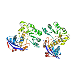
Crystal Structure of Apo form of G protein coupled receptor kinase 1 at 7.51A
Descriptor: Rhodopsin kinase
Authors:Singh, P, Tesmer, J.J.G.
Deposit date:2008-01-30
Release date:2008-03-11
Last modified:2023-08-30
Method:X-RAY DIFFRACTION (7.509 Å)
Cite:Structures of rhodopsin kinase in different ligand states reveal key elements involved in G protein-coupled receptor kinase activation.
J.Biol.Chem., 283, 2008
3AWT
DownloadVisualize
BU of 3awt by Molmil
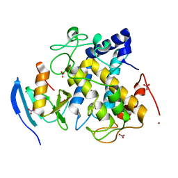
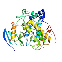
Crystal structure of Streptomyces tyrosinase in a complex with caddie soaked in a Cu(II)-containing solution for 20 hr: occupancy of Cu(II) is high
Descriptor: COPPER (II) ION, MelC, NITRATE ION, ...
Authors:Matoba, Y, Sugiyama, M.
Deposit date:2011-03-26
Release date:2011-06-29
Last modified:2023-11-01
Method:X-RAY DIFFRACTION (1.35 Å)
Cite:A molecular mechanism for copper transportation to tyrosinase that is assisted by a metallochaperone, caddie protein
J.Biol.Chem., 286, 2011
3AWY
DownloadVisualize
BU of 3awy by Molmil
Crystal structure of Streptomyces tyrosinase in a complex with caddie M84L mutant soaked in a Cu(II)-containing solution for 80 hr
Descriptor: COPPER (II) ION, MelC, NITRATE ION, ...
Authors:Matoba, Y, Sugiyama, M.
Deposit date:2011-03-26
Release date:2011-06-29
Last modified:2023-11-01
Method:X-RAY DIFFRACTION (1.58 Å)
Cite:A molecular mechanism for copper transportation to tyrosinase that is assisted by a metallochaperone, caddie protein
J.Biol.Chem., 286, 2011
3AWW
DownloadVisualize
BU of 3aww by Molmil
Crystal structure of Streptomyces tyrosinase in a complex with caddie soaked in a Cu(II)-containing solution for 80 hr: occupancy of CuA is high
Descriptor: COPPER (II) ION, MelC, NITRATE ION, ...
Authors:Matoba, Y, Sugiyama, M.
Deposit date:2011-03-26
Release date:2011-06-29
Last modified:2023-11-01
Method:X-RAY DIFFRACTION (1.35 Å)
Cite:A molecular mechanism for copper transportation to tyrosinase that is assisted by a metallochaperone, caddie protein
J.Biol.Chem., 286, 2011
3AWS
DownloadVisualize
BU of 3aws by Molmil
Crystal structure of Streptomyces tyrosinase in a complex with caddie soaked in a Cu(II)-containing solution for 20 hr: occupancy of Cu(II) is low
Descriptor: COPPER (II) ION, MelC, NITRATE ION, ...
Authors:Matoba, Y, Sugiyama, M.
Deposit date:2011-03-26
Release date:2011-06-29
Last modified:2023-11-01
Method:X-RAY DIFFRACTION (1.24 Å)
Cite:A molecular mechanism for copper transportation to tyrosinase that is assisted by a metallochaperone, caddie protein
J.Biol.Chem., 286, 2011
3AX0
DownloadVisualize
BU of 3ax0 by Molmil
Crystal structure of Streptomyces tyrosinase in a complex with caddie Y98F mutant soaked in a Cu(II)-containing solution for 80 hr
Descriptor: COPPER (II) ION, MelC, NITRATE ION, ...
Authors:Matoba, Y, Sugiyama, M.
Deposit date:2011-03-26
Release date:2011-06-29
Last modified:2023-11-01
Method:X-RAY DIFFRACTION (1.4 Å)
Cite:A molecular mechanism for copper transportation to tyrosinase that is assisted by a metallochaperone, caddie protein
J.Biol.Chem., 286, 2011
3AWZ
DownloadVisualize
BU of 3awz by Molmil
Crystal structure of Streptomyces tyrosinase in a complex with caddie H97Q mutant soaked in a Cu(II)-containing solution for 80 hr
Descriptor: COPPER (II) ION, MelC, NITRATE ION, ...
Authors:Matoba, Y, Sugiyama, M.
Deposit date:2011-03-26
Release date:2011-06-29
Last modified:2023-11-01
Method:X-RAY DIFFRACTION (1.43 Å)
Cite:A molecular mechanism for copper transportation to tyrosinase that is assisted by a metallochaperone, caddie protein
J.Biol.Chem., 286, 2011
3AWU
DownloadVisualize
BU of 3awu by Molmil
Crystal structure of Streptomyces tyrosinase in a complex with caddie soaked in a Cu(II)-containing solution for 40 h
Descriptor: COPPER (II) ION, MelC, NITRATE ION, ...
Authors:Matoba, Y, Sugiyama, M.
Deposit date:2011-03-26
Release date:2011-06-29
Last modified:2023-11-01
Method:X-RAY DIFFRACTION (1.16 Å)
Cite:A molecular mechanism for copper transportation to tyrosinase that is assisted by a metallochaperone, caddie protein
J.Biol.Chem., 286, 2011
3AWX
DownloadVisualize
BU of 3awx by Molmil
Crystal structure of Streptomyces tyrosinase in a complex with caddie H82Q mutant soaked in a Cu(II)-containing solution for 80 hr
Descriptor: COPPER (II) ION, MelC, NITRATE ION, ...
Authors:Matoba, Y, Sugiyama, M.
Deposit date:2011-03-26
Release date:2011-06-29
Last modified:2023-11-01
Method:X-RAY DIFFRACTION (1.25 Å)
Cite:A molecular mechanism for copper transportation to tyrosinase that is assisted by a metallochaperone, caddie protein
J.Biol.Chem., 286, 2011
2Z4Z
DownloadVisualize
BU of 2z4z by Molmil
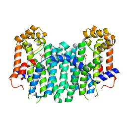
S. cerevisiae geranylgeranyl pyrophosphate synthase in complex with magnesium and BPH-sc01
Descriptor: (6E,11E)-HEPTADECA-6,11-DIENE-9,9-DIYLBIS(PHOSPHONIC ACID), Geranylgeranyl pyrophosphate synthetase, MAGNESIUM ION
Authors:Chen, C.K.-M, Guo, R.T, Hudock, M, Cao, R, Oldfield, E, Wang, A.H.-J.
Deposit date:2007-06-26
Release date:2008-07-01
Last modified:2023-11-01
Method:X-RAY DIFFRACTION (2.09 Å)
Cite:Inhibition of geranylgeranyl diphosphate synthase by bisphosphonates: a crystallographic and computational investigation
J.Med.Chem., 51, 2008
2Z50
DownloadVisualize
BU of 2z50 by Molmil
S. cerevisiae geranylgeranyl pyrophosphate synthase in complex with BPH-28
Descriptor: (1-HYDROXYHEPTANE-1,1-DIYL)BIS(PHOSPHONIC ACID), Geranylgeranyl pyrophosphate synthetase
Authors:Guo, R.T, Chen, C.K.-M, Hudock, M, Cao, R, Oldfield, E, Wang, A.H.-J.
Deposit date:2007-06-26
Release date:2008-07-01
Last modified:2023-11-01
Method:X-RAY DIFFRACTION (2.01 Å)
Cite:Inhibition of geranylgeranyl diphosphate synthase by bisphosphonates: a crystallographic and computational investigation
J.Med.Chem., 51, 2008
3C3X
DownloadVisualize
BU of 3c3x by Molmil
The multiple phenylpropene synthases in both Clarkia breweri and Petunia hybrida represent two distinct lineages
Descriptor: Eugenol synthase 1, NADP NICOTINAMIDE-ADENINE-DINUCLEOTIDE PHOSPHATE
Authors:Louie, G.V, Noel, J.P, Bowman, M.E.
Deposit date:2008-01-29
Release date:2008-05-06
Last modified:2023-08-30
Method:X-RAY DIFFRACTION (2.15 Å)
Cite:The multiple phenylpropene synthases in both Clarkia breweri and Petunia hybrida represent two distinct protein lineages.
Plant J., 54, 2008
3C3C
DownloadVisualize
BU of 3c3c by Molmil
Crystal Structure of human phosphoglycerate kinase bound to 3-phosphoglycerate and L-CDP
Descriptor: 3-PHOSPHOGLYCERIC ACID, CYTIDINE-5'-DIPHOSPHATE, MAGNESIUM ION, ...
Authors:Arold, S.T, Gondeau, C, Lionne, C, Chaloin, L.
Deposit date:2008-01-28
Release date:2008-07-01
Last modified:2024-10-16
Method:X-RAY DIFFRACTION (2.4 Å)
Cite:Molecular basis for the lack of enantioselectivity of human 3-phosphoglycerate kinase
Nucleic Acids Res., 36, 2008
3CAW
DownloadVisualize
BU of 3caw by Molmil
Crystal structure of o-succinylbenzoate synthase from Bdellovibrio bacteriovorus liganded with Mg
Descriptor: MAGNESIUM ION, o-succinylbenzoate synthase
Authors:Fedorov, A.A, Fedorov, E.V, Sakai, A, Burley, S.K, Gerlt, J.A, Almo, S.C, New York SGX Research Center for Structural Genomics (NYSGXRC)
Deposit date:2008-02-20
Release date:2008-03-04
Last modified:2024-02-21
Method:X-RAY DIFFRACTION (1.87 Å)
Cite:Loss of quaternary structure is associated with rapid sequence divergence in the OSBS family.
Proc.Natl.Acad.Sci.USA, 111, 2014
3C51
DownloadVisualize
BU of 3c51 by Molmil
Crystal structure of G protein coupled receptor kinase 1 bound to ADP and magnesium chloride at 3.55A
Descriptor: ADENOSINE-5'-DIPHOSPHATE, BROMIDE ION, MAGNESIUM ION, ...
Authors:Singh, P, Tesmer, J.J.G.
Deposit date:2008-01-30
Release date:2008-03-11
Last modified:2023-08-30
Method:X-RAY DIFFRACTION (3.55 Å)
Cite:Structures of rhodopsin kinase in different ligand states reveal key elements involved in G protein-coupled receptor kinase activation.
J.Biol.Chem., 283, 2008
3C4X
DownloadVisualize
BU of 3c4x by Molmil
Crystal Structure of G protein coupled receptor kinase 1 bound to ATP and magnesium chloride at 2.9A
Descriptor: ADENOSINE-5'-TRIPHOSPHATE, CHLORIDE ION, MAGNESIUM ION, ...
Authors:Singh, P, Tesmer, J.J.G.
Deposit date:2008-01-30
Release date:2008-03-11
Last modified:2023-08-30
Method:X-RAY DIFFRACTION (2.9 Å)
Cite:Structures of rhodopsin kinase in different ligand states reveal key elements involved in G protein-coupled receptor kinase activation.
J.Biol.Chem., 283, 2008
3CCD
DownloadVisualize
BU of 3ccd by Molmil
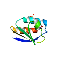
1.0 A Structure of Post-Succinimide His15Asp HPr
Descriptor: Phosphocarrier protein HPr, SULFATE ION
Authors:Napper, S, Prasad, L, Delbaere, L.T.J.
Deposit date:2008-02-25
Release date:2008-10-21
Last modified:2023-08-30
Method:X-RAY DIFFRACTION (1 Å)
Cite:Structural investigation of a phosphorylation-catalyzed, isoaspartate-free, protein succinimide: crystallographic structure of post-succinimide His15Asp histidine-containing protein
Biochemistry, 47, 2008
1EGX
DownloadVisualize
BU of 1egx by Molmil
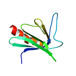
SOLUTION STRUCTURE OF THE ENA-VASP HOMOLOGY 1 (EVH1) DOMAIN OF HUMAN VASODILATOR-STIMULATED PHOSPHOPROTEIN (VASP)
Descriptor: VASODILATOR-STIMULATED PHOSPHOPROTEIN
Authors:Ball, L, Kuhne, R, Hoffmann, B, Hafner, A, Schmieder, P, Volkmer-Engert, R, Hof, M, Wahl, M, Schneider-Mergener, J, Walter, U, Oschkinat, H, Jarchau, T.
Deposit date:2000-02-17
Release date:2000-09-20
Last modified:2024-05-01
Method:SOLUTION NMR
Cite:Dual epitope recognition by the VASP EVH1 domain modulates polyproline ligand specificity and binding affinity.
EMBO J., 19, 2000
1EJ2
DownloadVisualize
BU of 1ej2 by Molmil
Crystal structure of methanobacterium thermoautotrophicum nicotinamide mononucleotide adenylyltransferase with bound NAD+
Descriptor: NICOTINAMIDE MONONUCLEOTIDE ADENYLYLTRANSFERASE, NICOTINAMIDE-ADENINE-DINUCLEOTIDE, SODIUM ION, ...
Authors:Saridakis, V, Christendat, D, Kimber, M.S, Edwards, A.M, Pai, E.F, Midwest Center for Structural Genomics (MCSG), Northeast Structural Genomics Consortium (NESG)
Deposit date:2000-02-29
Release date:2001-03-14
Last modified:2024-02-07
Method:X-RAY DIFFRACTION (1.9 Å)
Cite:Insights into ligand binding and catalysis of a central step in NAD+ synthesis: structures of Methanobacterium thermoautotrophicum NMN adenylyltransferase complexes.
J.Biol.Chem., 276, 2001
1E8K
DownloadVisualize
BU of 1e8k by Molmil
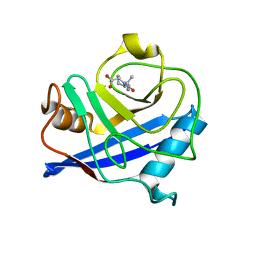
Cyclophilin 3 Complexed With Dipeptide Ala-Pro
Descriptor: ALANINE, PEPTIDYL-PROLYL CIS-TRANS ISOMERASE 3, PROLINE
Authors:Wu, S.Y, Dornan, J, Kontopidis, G, Taylor, P, Walkinshaw, M.D.
Deposit date:2000-09-25
Release date:2001-09-20
Last modified:2023-12-13
Method:X-RAY DIFFRACTION (1.9 Å)
Cite:The First Direct Determination of a Ligand Binding Constant in Protein Crystals
Angew.Chem.Int.Ed.Engl., 40, 2001
1E3X
DownloadVisualize
BU of 1e3x by Molmil
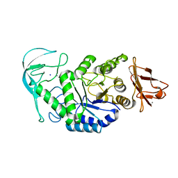
Native structure of chimaeric amylase from B. amyloliquefaciens and B. licheniformis at 1.92A
Descriptor: ALPHA-AMYLASE, CALCIUM ION, SODIUM ION
Authors:Brzozowski, A.M, Lawson, D.M, Turkenburg, J.P, Bisgaard-Frantzen, H, Svendsen, A, Borchert, T.V, Dauter, Z, Wilson, K.S, Davies, G.J.
Deposit date:2000-06-26
Release date:2001-06-21
Last modified:2024-05-08
Method:X-RAY DIFFRACTION (1.9 Å)
Cite:Structural Analysis of a Chimeric Bacterial Alpha-Amylase. High Resolution Analysis of Native and Ligand Complexes
Biochemistry, 39, 2000
1HH9
DownloadVisualize
BU of 1hh9 by Molmil
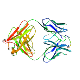
ANTI-P24 (HIV-1) FAB FRAGMENT CB41 COMPLEXED WITH A PEPTIDE
Descriptor: IGG2A KAPPA ANTIBODY CB41 (HEAVY CHAIN), IGG2A KAPPA ANTIBODY CB41 (LIGHT CHAIN), PEP-2
Authors:Hahn, M, Wessner, H, Schneider-Mergener, J, Hohne, W.
Deposit date:2000-12-21
Release date:2001-01-12
Last modified:2024-11-06
Method:X-RAY DIFFRACTION (2.7 Å)
Cite:Evolutionary Transition Pathways for Changing Peptide Ligand Specificity and Structure
Embo J., 19, 2000

227344

PDB entries from 2024-11-13

PDB statisticsPDBj update infoContact PDBjnumon