2CRJ
 
 | |
2CSJ
 | |
2COE
 | |
2CR6
 | |
2CSH
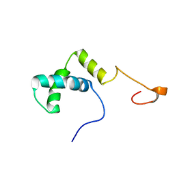
 | | Solution structure of tandem repeat of the zf-C2H2 domains of human zinc finger protein 297B | | Descriptor: | ZINC ION, Zinc finger protein 297B | | Authors: | Inoue, K, Saitoh, K, Hayashi, F, Kigawa, T, Yokoyama, S, RIKEN Structural Genomics/Proteomics Initiative (RSGI) | | Deposit date: | 2005-05-21 | | Release date: | 2005-11-21 | | Last modified: | 2024-05-29 | | Method: | SOLUTION NMR | | Cite: | Solution structure of tandem repeat of the zf-C2H2 domains of human zinc finger protein 297B
To be Published
|
|
2CQQ
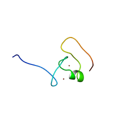
 | | Solution Structure of RSGI RUH-037, a myb DNA-binding domain in human cDNA | | Descriptor: | DnaJ homolog subfamily C member 1 | | Authors: | Doi-Katayama, Y, Hirota, H, Hayashi, F, Yokoyama, S, RIKEN Structural Genomics/Proteomics Initiative (RSGI) | | Deposit date: | 2005-05-20 | | Release date: | 2005-11-20 | | Last modified: | 2024-05-29 | | Method: | SOLUTION NMR | | Cite: | Solution Structure of RSGI RUH-037, a myb DNA-binding domain in human cDNA
To be Published
|
|
2CRP
 | |
2CRG
 | |
2CRW
 | |
2CSK
 | |
2CSO
 | |
2CU1
 | |
2D9X
 | | Solution structure of the PH domain of Oxysterol binding protein-related protein 11 from human | | Descriptor: | Oxysterol binding protein-related protein 11 | | Authors: | Li, H, Sato, M, Koshiba, S, Inoue, M, Kigawa, T, Yokoyama, S, RIKEN Structural Genomics/Proteomics Initiative (RSGI) | | Deposit date: | 2005-12-13 | | Release date: | 2006-06-13 | | Last modified: | 2024-05-29 | | Method: | SOLUTION NMR | | Cite: | Solution structure of the PH domain of Oxysterol binding protein-related protein 11 from human
To be Published
|
|
2DAG
 | | Solution Structure of the First UBA Domain in the Human Ubiquitin Specific Protease 5 (Isopeptidase 5) | | Descriptor: | Ubiquitin carboxyl-terminal hydrolase 5 | | Authors: | Zhao, C, Kigawa, T, Tomizawa, T, Koshiba, S, Inoue, M, Yokoyama, S, RIKEN Structural Genomics/Proteomics Initiative (RSGI) | | Deposit date: | 2005-12-14 | | Release date: | 2006-06-14 | | Last modified: | 2024-05-29 | | Method: | SOLUTION NMR | | Cite: | Solution Structure of the First UBA Domain in the Human Ubiquitin Specific Protease 5 (Isopeptidase 5)
To be Published
|
|
2D88
 | | Solution structure of the CH domain from human MICAL-3 protein | | Descriptor: | Protein MICAL-3 | | Authors: | Tomizawa, T, Kigawa, T, Koshiba, S, Inoue, M, Yokoyama, S, RIKEN Structural Genomics/Proteomics Initiative (RSGI) | | Deposit date: | 2005-12-02 | | Release date: | 2006-06-02 | | Last modified: | 2024-05-29 | | Method: | SOLUTION NMR | | Cite: | Solution structure of the CH domain from human MICAL-3 protein
To be Published
|
|
2D93
 | |
2D7L
 | | Solution structure of the HMG box domain from human WD repeat and HMG-box DNA binding protein 1 | | Descriptor: | WD repeat and HMG-box DNA binding protein 1 | | Authors: | Tomizawa, T, Kigawa, T, Saito, K, Koshiba, S, Inoue, M, Kamatari, Y.O, Yokoyama, S, RIKEN Structural Genomics/Proteomics Initiative (RSGI) | | Deposit date: | 2005-11-24 | | Release date: | 2006-05-24 | | Last modified: | 2024-05-29 | | Method: | SOLUTION NMR | | Cite: | Solution structure of the HMG box domain from human WD repeat and HMG-box DNA binding protein 1
To be Published
|
|
2D8U
 | | Solution structure of the B-box domain of the human tripartite motif-containing 63 protein | | Descriptor: | Ubiquitin ligase TRIM63, ZINC ION | | Authors: | Miyamoto, K, Kigawa, T, Tomizawa, T, Koshiba, S, Inoue, M, Yokoyama, S, RIKEN Structural Genomics/Proteomics Initiative (RSGI) | | Deposit date: | 2005-12-08 | | Release date: | 2006-06-08 | | Last modified: | 2024-05-29 | | Method: | SOLUTION NMR | | Cite: | Solution structure of the B-box domain of the human tripartite motif-containing 63 protein
To be Published
|
|
2D9W
 | | Solution structure of the PH domain of Docking protein 2 from human | | Descriptor: | Docking protein 2 | | Authors: | Li, H, Tomizawa, T, Koshiba, S, Inoue, M, Kigawa, T, Yokoyama, S, RIKEN Structural Genomics/Proteomics Initiative (RSGI) | | Deposit date: | 2005-12-13 | | Release date: | 2006-06-13 | | Last modified: | 2024-05-29 | | Method: | SOLUTION NMR | | Cite: | Solution structure of the PH domain of Docking protein 2 from human
To be Published
|
|
2DAF
 | | Solution Structure of the Novel Identified Ubiquitin-like Domain in the Human Hypothetical Protein FLJ35834 | | Descriptor: | FLJ35834 protein | | Authors: | Zhao, C, Kigawa, T, Saito, K, Koshiba, S, Inoue, M, Yokoyama, S, RIKEN Structural Genomics/Proteomics Initiative (RSGI) | | Deposit date: | 2005-12-14 | | Release date: | 2006-06-14 | | Last modified: | 2024-05-29 | | Method: | SOLUTION NMR | | Cite: | Solution Structure of the Novel Identified Ubiquitin-like Domain in the Human Hypothetical Protein FLJ35834
To be Published
|
|
2DA3
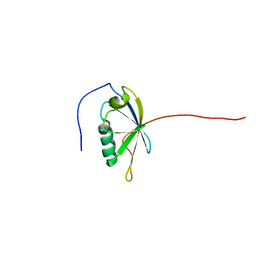
 | | Solution structure of the third homeobox domain of AT-binding transcription factor 1 (ATBF1) | | Descriptor: | Alpha-fetoprotein enhancer binding protein | | Authors: | Ohnishi, S, Kigawa, T, Tomizawa, T, Koshiba, S, Inoue, M, Yokoyama, S, RIKEN Structural Genomics/Proteomics Initiative (RSGI) | | Deposit date: | 2005-12-13 | | Release date: | 2006-06-13 | | Last modified: | 2024-05-29 | | Method: | SOLUTION NMR | | Cite: | Solution structure of the third homeobox domain of AT-binding transcription factor 1 (ATBF1)
To be Published
|
|
2DAI
 | | Solution Structure of the First UBA Domain in the Human Ubiquitin Associated Domain Containing 1 (UBADC1) | | Descriptor: | Ubiquitin associated domain containing 1 | | Authors: | Zhao, C, Kigawa, T, Sato, M, Koshiba, S, Inoue, M, Yokoyama, S, RIKEN Structural Genomics/Proteomics Initiative (RSGI) | | Deposit date: | 2005-12-14 | | Release date: | 2006-06-14 | | Last modified: | 2024-05-29 | | Method: | SOLUTION NMR | | Cite: | Solution Structure of the First UBA Domain in the Human Ubiquitin Associated Domain Containing 1 (UBADC1)
To be Published
|
|
2D8C
 | | Solution structure of the sam-domain of mouse phosphatidyl ceramidecholinephosphotransferase 1 | | Descriptor: | Phosphatidylcholine:ceramide cholinephosphotransferase 1 | | Authors: | Goroncy, A.K, Kigawa, T, Koshiba, S, Tomizawa, T, Kobayashi, N, Tochio, N, Inoue, M, Yokoyama, S, RIKEN Structural Genomics/Proteomics Initiative (RSGI) | | Deposit date: | 2005-12-02 | | Release date: | 2006-06-02 | | Last modified: | 2024-05-29 | | Method: | SOLUTION NMR | | Cite: | Solution structure of the sam-domain of mouse phosphatidyl ceramidecholinephosphotransferase 1
To be Published
|
|
2DA9
 | | Solution structure of the third SH3 domain of SH3-domain kinase binding protein 1 (Regulator of ubiquitous kinase, Ruk) | | Descriptor: | SH3-domain kinase binding protein 1 | | Authors: | Ohnishi, S, Kigawa, T, Saito, K, Koshiba, S, Inoue, M, Yokoyama, S, RIKEN Structural Genomics/Proteomics Initiative (RSGI) | | Deposit date: | 2005-12-13 | | Release date: | 2006-06-13 | | Last modified: | 2024-05-29 | | Method: | SOLUTION NMR | | Cite: | Solution structure of the third SH3 domain of SH3-domain kinase binding protein 1 (Regulator of ubiquitous kinase, Ruk)
To be Published
|
|
2D9M
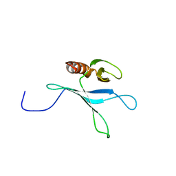
 | | Solution structure of CCCH type zinc-finger domain 3 in zinc finger CCCH-type domain containing 7A | | Descriptor: | ZINC ION, Zinc finger CCCH-type domain containing protein 7A | | Authors: | Abe, C, Muto, Y, Inoue, M, Kigawa, T, Terada, T, Shirouzu, M, Yokoyama, S, RIKEN Structural Genomics/Proteomics Initiative (RSGI) | | Deposit date: | 2005-12-12 | | Release date: | 2006-06-12 | | Last modified: | 2024-05-29 | | Method: | SOLUTION NMR | | Cite: | Solution structure of CCCH type zinc-finger domain 3 in zinc finger CCCH-type domain containing 7A
To be Published
|
|


