Loading
PDBj
MenuPDBj@FacebookPDBj@TwitterPDBj@YouTubewwPDB FoundationwwPDB
RCSB PDBPDBeBMRBAdv. SearchSearch help

2XAX
DownloadVisualize
BU of 2xax by Molmil
Ribonucleotide reductase Y730NO2Y and Y731A modified R1 subunit of E. coli
Descriptor: RIBONUCLEOSIDE-DIPHOSPHATE REDUCTASE 1 SUBUNIT ALPHA, RIBONUCLEOSIDE-DIPHOSPHATE REDUCTASE 1 SUBUNIT BETA
Authors:Yokoyama, K, Uhlin, U, Stubbe, J.
Deposit date:2010-04-01
Release date:2010-04-14
Last modified:2023-12-20
Method:X-RAY DIFFRACTION (2.75 Å)
Cite:Site-Specific Incorporation of 3-Nitrotyrosine as a Probe of Pk(A) Perturbation of Redox-Active Tyrosines in Ribonucleotide Reductase.
J.Am.Chem.Soc., 132, 2010
1VBE
DownloadVisualize
BU of 1vbe by Molmil
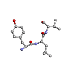
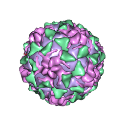
POLIOVIRUS (TYPE 3, SABIN STRAIN, MUTANT 242-H2) COMPLEXED WITH R78206
Descriptor: (METHYLPYRIDAZINE PIPERIDINE PROPYLOXYPHENYL)ETHYLACETATE, MYRISTIC ACID, POLIOVIRUS TYPE 3
Authors:Grant, R.A, Hiremath, C.N, Filman, D.J, Syed, R, Andries, K, Hogle, J.M.
Deposit date:1996-01-02
Release date:1996-07-11
Last modified:2021-11-03
Method:X-RAY DIFFRACTION (2.8 Å)
Cite:Structures of poliovirus complexes with anti-viral drugs: implications for viral stability and drug design.
Curr.Biol., 4, 1994
2XAK
DownloadVisualize
BU of 2xak by Molmil
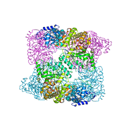
Ribonucleotide reductase Y730NO2Y modified R1 subunit of E. coli
Descriptor: RIBONUCLEOSIDE-DIPHOSPHATE REDUCTASE 1 SUBUNIT ALPHA, RIBONUCLEOSIDE-DIPHOSPHATE REDUCTASE 1 SUBUNIT BETA
Authors:Yokoyama, K, Uhlin, U, Stubbe, J.
Deposit date:2010-03-31
Release date:2010-04-14
Last modified:2023-12-20
Method:X-RAY DIFFRACTION (2.8 Å)
Cite:Site-Specific Incorporation of 3-Nitrotyrosine as a Probe of Pk(A) Perturbation of Redox-Active Tyrosines in Ribonucleotide Reductase.
J.Am.Chem.Soc., 132, 2010
1VBA
DownloadVisualize
BU of 1vba by Molmil
POLIOVIRUS (TYPE 3, SABIN STRAIN) (P3/SABIN, P3/LEON/12A(1)B) COMPLEXED WITH R78206
Descriptor: (METHYLPYRIDAZINE PIPERIDINE PROPYLOXYPHENYL)ETHYLACETATE, MYRISTIC ACID, POLIOVIRUS TYPE 3
Authors:Grant, R.A, Hiremath, C.N, Filman, D.J, Syed, R, Andries, K, Hogle, J.M.
Deposit date:1996-01-02
Release date:1996-07-11
Last modified:2024-06-05
Method:X-RAY DIFFRACTION (2.9 Å)
Cite:Structures of poliovirus complexes with anti-viral drugs: implications for viral stability and drug design.
Curr.Biol., 4, 1994
3GU4
DownloadVisualize
BU of 3gu4 by Molmil
Crystal structure of DAPKQ23V-AMPPNP
Descriptor: Death-associated protein kinase 1, PHOSPHOAMINOPHOSPHONIC ACID-ADENYLATE ESTER
Authors:McNamara, L.K, Schavocky, J.S, Watterson, D.M, Brunzelle, J.S.
Deposit date:2009-03-28
Release date:2010-03-09
Last modified:2023-09-06
Method:X-RAY DIFFRACTION (1.35 Å)
Cite:Enzymatic activity and crystallgoraphic analyses of a glycine-rich loop mutant of DAPK
To be Published
3GU6
DownloadVisualize
BU of 3gu6 by Molmil
Crystal structure of DAPKQ23V-ADP
Descriptor: ADENOSINE-5'-DIPHOSPHATE, Death-associated protein kinase 1
Authors:McNamara, L.K, Schavocky, J.S, Watterson, D.M, Brunzelle, J.S.
Deposit date:2009-03-28
Release date:2010-03-09
Last modified:2023-09-06
Method:X-RAY DIFFRACTION (1.49 Å)
Cite:Enzymatic activity and crystallographic analyses of a glycine-rich loop mutant of DAPK
To be Published
3GUB
DownloadVisualize
BU of 3gub by Molmil
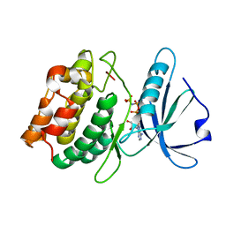
Crystal structure of DAPKL93G complexed with N6-(2-Phenylethyl)adenosine
Descriptor: 9-alpha-L-lyxofuranosyl-N-(2-phenylethyl)-9H-purin-6-amine, Death-associated protein kinase 1, SULFATE ION
Authors:McNamara, L.K, Schumacher, A.M, Schavocky, J.S, Watterson, D.M, Brunzelle, J.S.
Deposit date:2009-03-29
Release date:2010-03-09
Last modified:2023-09-06
Method:X-RAY DIFFRACTION (1.71 Å)
Cite:Crystal structures of the DAPK gatekeeper mutant complexed with N6-modified adenosine analogs.
To be Published
3GU7
DownloadVisualize
BU of 3gu7 by Molmil
Crystal structure of DAPKQ23V-ADP-Mg2+
Descriptor: ADENOSINE-5'-DIPHOSPHATE, Death-associated protein kinase 1, MAGNESIUM ION
Authors:McNamara, L.K, Schavocky, J.S, Watterson, D.M, Brunzelle, J.S.
Deposit date:2009-03-28
Release date:2010-03-09
Last modified:2023-09-06
Method:X-RAY DIFFRACTION (1.9 Å)
Cite:Enzyme activity and crystallographic analyses of a glycine-rich loop mutant of DAPK
To be Published
3GU8
DownloadVisualize
BU of 3gu8 by Molmil
Crystal structure of DAPKL93G with N6-cyclopentyladenosine
Descriptor: Death-associated protein kinase 1, N6-cyclopentyladenosine
Authors:McNamara, L.K, Schumacher, A.M, Schavocky, J.S, Watterson, D.M, Brunzelle, J.S.
Deposit date:2009-03-28
Release date:2010-03-09
Last modified:2023-09-06
Method:X-RAY DIFFRACTION (1.6 Å)
Cite:Crystal structures of the DAPK gatekeeper mutant with N6-modified adenosine analogs.
To be Published
3GVX
DownloadVisualize
BU of 3gvx by Molmil
Crystal structure of Glycerate dehydrogenase related protein from Thermoplasma acidophilum
Descriptor: Glycerate dehydrogenase related protein, POTASSIUM ION
Authors:Syed Ibrahim, B, Kumaran, D, Burley, S.K, Swaminathan, S, New York SGX Research Center for Structural Genomics (NYSGXRC)
Deposit date:2009-03-31
Release date:2009-04-28
Last modified:2021-02-10
Method:X-RAY DIFFRACTION (2.2 Å)
Cite:Crystal Structure of Glycerate dehydrogenase related protein from Thermoplasma acidophilum
To be Published
3H49
DownloadVisualize
BU of 3h49 by Molmil
Crystal structure of a putative Ribokinase (Apo Form) from E.coli at 1.8A resolution
Descriptor: Ribokinase
Authors:Satyanarayana, L, Eswaramoorthy, S, Burley, S.K, Swaminathan, S, New York SGX Research Center for Structural Genomics (NYSGXRC)
Deposit date:2009-04-18
Release date:2009-04-28
Last modified:2021-02-10
Method:X-RAY DIFFRACTION (1.8 Å)
Cite:Crystal structure of a putative Ribokinase (Apo Form) from E.coli at 1.8A resolution.
To be Published
3HIC
DownloadVisualize
BU of 3hic by Molmil
THE CRYSTAL STRUCTURE OF PHOSPHOFRUCTOKINASE(Lin2199)FROM Listeria innocua
Descriptor: Lin2199 protein, MAGNESIUM ION
Authors:Satyanarayana, L, Burley, S.K, Swaminathan, S, New York SGX Research Center for Structural Genomics (NYSGXRC)
Deposit date:2009-05-19
Release date:2009-07-07
Last modified:2021-02-10
Method:X-RAY DIFFRACTION (2.02 Å)
Cite:THE CRYSTAL STRUCTURE OF PHOSPHOFRUCTOKINASE(Lin2199)FROM Listeria innocua
To be Published
3IN1
DownloadVisualize
BU of 3in1 by Molmil
Crystal structure of a putative Ribokinase in complex with ADP from E.coli
Descriptor: ADENOSINE-5'-DIPHOSPHATE, SODIUM ION, Uncharacterized sugar kinase ydjH
Authors:Satyanarayana, L, Burley, S.K, Swaminathan, S, New York SGX Research Center for Structural Genomics (NYSGXRC)
Deposit date:2009-08-11
Release date:2009-09-08
Last modified:2023-11-22
Method:X-RAY DIFFRACTION (2.15 Å)
Cite:Crystal structure of a putative Ribokinase in complex with ADP from E.coli
To be Published
3IH0
DownloadVisualize
BU of 3ih0 by Molmil
Crystal structure of an uncharacterized sugar kinase PH1459 from Pyrococcus horikoshii in complex with AMP-PNP
Descriptor: GLYCEROL, PHOSPHOAMINOPHOSPHONIC ACID-ADENYLATE ESTER, Uncharacterized sugar kinase PH1459
Authors:Kumar, G, Eswaramoorthy, S, Burley, S.K, Swaminathan, S, New York SGX Research Center for Structural Genomics (NYSGXRC)
Deposit date:2009-07-29
Release date:2009-09-08
Last modified:2023-11-22
Method:X-RAY DIFFRACTION (1.9 Å)
Cite:Crystal structure of an uncharacterized sugar kinase PH1459 from Pyrococcus horikoshii in complex with AMP-PNP
To be Published
3IKH
DownloadVisualize
BU of 3ikh by Molmil
Crystal structure of Ribokinase in Complex with ATP and glycerol in the active site from Klebsiella pneumoniae
Descriptor: ADENOSINE-5'-TRIPHOSPHATE, Carbohydrate kinase, GLYCEROL
Authors:Satyanarayana, L, Burley, S.K, Swaminathan, S, New York SGX Research Center for Structural Genomics (NYSGXRC)
Deposit date:2009-08-05
Release date:2009-08-18
Last modified:2023-11-22
Method:X-RAY DIFFRACTION (1.88 Å)
Cite:Crystal structure of Ribokinase in Complex with ATP and glycerol in the active site from Klebsiella pneumoniae
To be Published
3IE7
DownloadVisualize
BU of 3ie7 by Molmil
The crystal structure of phosphofructokinase (lin2199) from Listeria innocua in complex with ATP at 1.6A
Descriptor: ADENOSINE-5'-TRIPHOSPHATE, GLYCEROL, Lin2199 protein, ...
Authors:Satyanarayana, L, Burley, S.K, Swaminathan, S, New York SGX Research Center for Structural Genomics (NYSGXRC)
Deposit date:2009-07-22
Release date:2009-08-04
Last modified:2023-11-22
Method:X-RAY DIFFRACTION (1.6 Å)
Cite:The crystal structure of phosphofructokinase (lin2199) from Listeria innocua in complex with ATP at 1.6A
To be Published
3ILH
DownloadVisualize
BU of 3ilh by Molmil
Crystal structure of Two component response regulator from Cytophaga hutchinsonii
Descriptor: Two component response regulator
Authors:Syed Ibrahim, B, Burley, S.K, Swaminathan, S, New York SGX Research Center for Structural Genomics (NYSGXRC)
Deposit date:2009-08-07
Release date:2009-08-18
Last modified:2024-02-21
Method:X-RAY DIFFRACTION (2.59 Å)
Cite:Crystal structure of Two component response regulator from Cytophaga hutchinsonii
To be Published
3HZ4
DownloadVisualize
BU of 3hz4 by Molmil
Crystal Structure of Thioredoxin from Methanosarcina mazei
Descriptor: 1,2-ETHANEDIOL, Thioredoxin
Authors:Syed Ibrahim, B, Burley, S.K, Swaminathan, S, New York SGX Research Center for Structural Genomics (NYSGXRC)
Deposit date:2009-06-23
Release date:2009-06-30
Last modified:2021-02-10
Method:X-RAY DIFFRACTION (2.3 Å)
Cite:Crystal Structure of Thioredoxin from Methanosarcina mazei
To be Published
3IAO
DownloadVisualize
BU of 3iao by Molmil
Conformational plasticity of the coiled coil domain of BmrR is required for bmr promoter binding-the unliganded structure of BmrR
Descriptor: Multidrug-efflux transporter 1 regulator
Authors:Kumaraswami, M, Newberry, K.J, Brennan, R.G.
Deposit date:2009-07-14
Release date:2010-04-14
Last modified:2023-09-06
Method:X-RAY DIFFRACTION (2.8 Å)
Cite:Conformational plasticity of the coiled-coil domain of BmrR is required for bmr operator binding: the structure of unliganded BmrR.
J.Mol.Biol., 398, 2010
3IQ0
DownloadVisualize
BU of 3iq0 by Molmil
Crystal structure of a putative Ribokinase II in complex with ATP and Mg+2 from E.coli
Descriptor: ADENOSINE-5'-TRIPHOSPHATE, MAGNESIUM ION, putative Ribokinase II
Authors:Satyanarayana, L, Burley, S.K, Swaminathan, S, New York SGX Research Center for Structural Genomics (NYSGXRC)
Deposit date:2009-08-18
Release date:2009-09-08
Last modified:2021-02-10
Method:X-RAY DIFFRACTION (1.79 Å)
Cite:Crystal structure of a putative Ribokinase (II)in complex with ATP and Mg+2 from E.coli
To be Published
3HP0
DownloadVisualize
BU of 3hp0 by Molmil
Crystal structure of a Putative polyketide biosynthesis enoyl-CoA hydratase (pksH) from Bacillus subtilis
Descriptor: Putative polyketide biosynthesis enoyl-CoA hydratase homolog pksH
Authors:Satyanarayana, L, Eswaramoorthy, S, Burley, S.K, Swaminathan, S, New York SGX Research Center for Structural Genomics (NYSGXRC)
Deposit date:2009-06-03
Release date:2009-06-23
Last modified:2021-02-10
Method:X-RAY DIFFRACTION (2.32 Å)
Cite:Crystal structure of a Putative polyketide biosynthesis enoyl-CoA hydratase (pksH) from Bacillus subtilis
To be Published
3GBV
DownloadVisualize
BU of 3gbv by Molmil
Crystal structure of a putative LacI transcriptional regulator from Bacteroides fragilis
Descriptor: 1,2-ETHANEDIOL, Putative LacI-family transcriptional regulator, SODIUM ION
Authors:Syed Ibrahim, B, Kumaran, D, Burley, S.K, Swaminathan, S, New York SGX Research Center for Structural Genomics (NYSGXRC)
Deposit date:2009-02-20
Release date:2009-03-10
Last modified:2021-02-10
Method:X-RAY DIFFRACTION (2.2 Å)
Cite:Crystal structure of a putative LacI transcriptional regulator from Bacteroides fragilis
To be Published
3GU5
DownloadVisualize
BU of 3gu5 by Molmil
Crystal structure of DAPKQ23V-AMPPNP-Mg2+
Descriptor: Death-associated protein kinase 1, MAGNESIUM ION, PHOSPHOAMINOPHOSPHONIC ACID-ADENYLATE ESTER
Authors:McNamara, L.K, Schavocky, J.S, Watterson, D.M, Brunzelle, J.S.
Deposit date:2009-03-28
Release date:2010-03-09
Last modified:2023-09-06
Method:X-RAY DIFFRACTION (1.65 Å)
Cite:Enzyme activity and crystallographic analyses of a glycine-rich loop mutant of DAPK
To be Published
3JUL
DownloadVisualize
BU of 3jul by Molmil
Crystal structure of Listeria innocua D-Tagatose-6-Phosphate Kinase bound with substrate
Descriptor: 6-O-phosphono-beta-D-tagatofuranose, Lin2199 protein, MAGNESIUM ION
Authors:Satyanarayana, L, Burley, S.K, Swaminathan, S, New York SGX Research Center for Structural Genomics (NYSGXRC)
Deposit date:2009-09-15
Release date:2009-10-06
Last modified:2023-11-22
Method:X-RAY DIFFRACTION (2.4 Å)
Cite:Crystal structure of Listeria innocua D-Tagatose-6-Phosphate Kinase bound with substrate
To be Published
3HEB
DownloadVisualize
BU of 3heb by Molmil
Crystal Structure of Response regulator receiver domain from Rhodospirillum rubrum
Descriptor: PHOSPHATE ION, Response regulator receiver domain protein (CheY)
Authors:Syed Ibrahim, B, Burley, S.K, Swaminathan, S, New York SGX Research Center for Structural Genomics (NYSGXRC)
Deposit date:2009-05-08
Release date:2009-05-19
Last modified:2021-02-10
Method:X-RAY DIFFRACTION (2.4 Å)
Cite:Crystal Structure of Response regulator receiver domain from Rhodospirillum rubrum
To be Published

225158

PDB entries from 2024-09-18

PDB statisticsPDBj update infoContact PDBjnumon