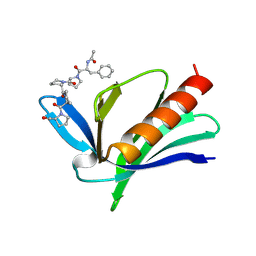
9OJW
 
 | |
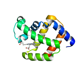
1EVH
 | |
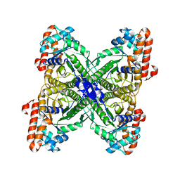
9R0L
 | | Three dimensional structure of human carbonic anhydrase XII in complex with sulfonamide | | Descriptor: | 3-(cyclooctylamino)-2,6-difluoro-4-((3-hydroxypropyl)sulfonyl)-5-(piperidin-1-yl)benzenesulfonamide, Carbonic anhydrase 12, ZINC ION | | Authors: | Eimonta, V, Leitans, J, Tars, K. | | Deposit date: | 2025-04-24 | | Release date: | 2025-09-17 | | Last modified: | 2025-09-24 | | Method: | X-RAY DIFFRACTION (1.45 Å) | | Cite: | Di- meta -Substituted Fluorinated Benzenesulfonamides as Potent and Selective Anticancer Inhibitors of Carbonic Anhydrase IX and XII.
J.Med.Chem., 68, 2025
|
|
9R40
 | | HaloTag bound to compound MRC71 | | Descriptor: | (2~{S},4~{S})-1-[(3~{S})-3-azanyl-3-(2-methoxyphenyl)propanoyl]-~{N}-[(2~{R})-1-[2-[2-[2-[2-(6-chloranylhexoxy)ethoxy]ethoxy]ethoxy]ethylamino]-1-oxidanylidene-3-pyridin-4-yl-propan-2-yl]-4-cyclohexyl-pyrrolidine-2-carboxamide, CHLORIDE ION, Haloalkane dehalogenase | | Authors: | Luptak, J. | | Deposit date: | 2025-05-06 | | Release date: | 2025-10-08 | | Method: | X-RAY DIFFRACTION (2.043 Å) | | Cite: | Development of TRIM21 PROTAC molecules
To Be Published
|
|
9NXL
 | | Crystal structure of steroid aldehyde dehydrogenase (Sad) from Caldimonas tepidiphilia | | Descriptor: | (4S)-2-METHYL-2,4-PENTANEDIOL, 2-AMINO-2-HYDROXYMETHYL-PROPANE-1,3-DIOL, Steroid aldehyde dehydrogenase | | Authors: | Rolfe, N, Myskiw, D, Patton, M.T, Forrester, T.J.B, Kimber, M.S, Seah, S.Y.K. | | Deposit date: | 2025-03-25 | | Release date: | 2025-10-08 | | Method: | X-RAY DIFFRACTION (1.8 Å) | | Cite: | Sad from Proteobacteria is a Structurally Distinct ALDH3 Enzyme Specialized for the Oxidation of Steroidal Aldehydes.
Biochemistry, 64, 2025
|
|
9OLZ
 | | Crystal structure of E. coli ApaH in complex with Ap4A | | Descriptor: | 4-(2-HYDROXYETHYL)-1-PIPERAZINE ETHANESULFONIC ACID, BIS(ADENOSINE)-5'-TETRAPHOSPHATE, Bis(5'-nucleosyl)-tetraphosphatase [symmetrical], ... | | Authors: | Nuthanakanti, A, Serganov, A. | | Deposit date: | 2025-05-13 | | Release date: | 2025-09-03 | | Method: | X-RAY DIFFRACTION (1.71 Å) | | Cite: | ApaH decaps Np 4 N-capped RNAs in two alternative orientations.
Nat.Chem.Biol., 2025
|
|
1ECA
 | |
9R8O
 | |
9QCT
 | | Crystal structure of Rhizobium etli L-asparaginase ReAV R47A mutant | | Descriptor: | 1,2-ETHANEDIOL, CHLORIDE ION, L-asparaginase II, ... | | Authors: | Pokrywka, K, Grzechowiak, M, Loch, J.I, Ruszkowski, M, Gilski, M, Jaskolski, M. | | Deposit date: | 2025-03-05 | | Release date: | 2025-07-16 | | Last modified: | 2025-08-06 | | Method: | X-RAY DIFFRACTION (1.75 Å) | | Cite: | Probing the Active Site of Class 3 L-Asparaginase by Mutagenesis: Mutations of the Ser-Lys Tandems of ReAV.
Biomolecules, 15, 2025
|
|
9ONG
 | |
9QCU
 | | Crystal structure of Rhizobium etli L-asparaginase ReAV S48A mutant | | Descriptor: | 1,2-ETHANEDIOL, CHLORIDE ION, DI(HYDROXYETHYL)ETHER, ... | | Authors: | Pokrywka, K, Grzechowiak, M, Loch, J.I, Ruszkowski, M, Gilski, M, Jaskolski, M. | | Deposit date: | 2025-03-05 | | Release date: | 2025-07-16 | | Last modified: | 2025-08-06 | | Method: | X-RAY DIFFRACTION (1.4 Å) | | Cite: | Probing the Active Site of Class 3 L-Asparaginase by Mutagenesis: Mutations of the Ser-Lys Tandems of ReAV.
Biomolecules, 15, 2025
|
|
9QZ1
 | | MINPP1 from Bacteroides thetaiotaomicron E325N mutant complex with myo-inositol(1,2,4,5,6)pentaphosphate | | Descriptor: | (1R,2R,3R,4S,5R,6R)-6-hydroxycyclohexane-1,2,3,4,5-pentayl pentakis[dihydrogen (phosphate)], multiple inositol polyphosphate histidine phosphatase 1 | | Authors: | Li, A.W.H, Shang, X.Y, Hemmings, A.M. | | Deposit date: | 2025-04-22 | | Release date: | 2025-06-11 | | Method: | X-RAY DIFFRACTION (2.45 Å) | | Cite: | A Structural Basis for the Stereospecificity of Multiple Inositol Polyphosphate Phosphatases
To Be Published
|
|
9QC3
 | | CRYSTAL STRUCTURE OF LYSYL-TRNA SYNTHETASE FROM Mycobacterium tuberculosis COMPLEXED WITH L-LYSINE AND INHIBITOR DDD01993593 | | Descriptor: | 2-azanyl-4-methoxy-6-[(1~{R},2~{S})-2-oxidanylcycloheptyl]-7~{H}-pyrrolo[3,4-d]pyrimidin-5-one, LYSINE, Lysine--tRNA ligase 1 | | Authors: | Dawson, A, Cleghorn, L.A.T, Davis, S.H. | | Deposit date: | 2025-03-04 | | Release date: | 2025-08-13 | | Last modified: | 2025-08-27 | | Method: | X-RAY DIFFRACTION (2.28 Å) | | Cite: | Design and Development of Lysyl tRNA Synthetase Inhibitors, for the Treatment of Tuberculosis.
J.Med.Chem., 68, 2025
|
|
9SG3
 | | X-ray structure of acetylcholine binding protein (AChBP) in complex with IOTA739 | | Descriptor: | 1,10-PHENANTHROLINE, 2-acetamido-2-deoxy-beta-D-glucopyranose, Acetylcholine-binding protein, ... | | Authors: | Cederfelt, D, Lund, B.A, Boronat, P, Hennig, S, Dobritzsch, D, Danielson, U.H. | | Deposit date: | 2025-08-21 | | Release date: | 2025-09-03 | | Last modified: | 2025-10-15 | | Method: | X-RAY DIFFRACTION (3 Å) | | Cite: | Detection and characterisation of ligand-induced conformational changes in acetylcholine binding proteins using biosensors and X-ray crystallography.
Rsc Chem Biol, 6, 2025
|
|
9QEI
 | | CRYSTAL STRUCTURE OF LYSYL-TRNA SYNTHETASE FROM Mycobacterium tuberculosis COMPLEXED WITH L-LYSINE AND INHIBITOR DDD01866774 | | Descriptor: | 2-azanyl-4-ethoxy-6-[(1~{R},2~{S})-2-oxidanylcyclohexyl]-7~{H}-pyrrolo[3,4-d]pyrimidin-5-one, LYSINE, Lysine--tRNA ligase 1 | | Authors: | Dawson, A, Cleghorn, L.A.T, Davis, S.H. | | Deposit date: | 2025-03-10 | | Release date: | 2025-08-13 | | Last modified: | 2025-08-27 | | Method: | X-RAY DIFFRACTION (2.4 Å) | | Cite: | Design and Development of Lysyl tRNA Synthetase Inhibitors, for the Treatment of Tuberculosis.
J.Med.Chem., 68, 2025
|
|
9OLY
 | |
9QC4
 | | CRYSTAL STRUCTURE OF LYSYL-TRNA SYNTHETASE FROM Mycobacterium tuberculosis COMPLEXED WITH L-LYSINE AND INHIBITOR DDD01869767 | | Descriptor: | 6-azanyl-1-[2-(azetidin-3-yl)ethyl]-2-cycloheptyl-4-ethoxy-pyrazolo[3,4-d]pyrimidin-3-one, LYSINE, Lysine--tRNA ligase 1 | | Authors: | Dawson, A, Cleghorn, L.A.T, Davis, S.H. | | Deposit date: | 2025-03-04 | | Release date: | 2025-08-13 | | Last modified: | 2025-08-27 | | Method: | X-RAY DIFFRACTION (2.601 Å) | | Cite: | Design and Development of Lysyl tRNA Synthetase Inhibitors, for the Treatment of Tuberculosis.
J.Med.Chem., 68, 2025
|
|
9QFY
 | | Structure of CHIP E3 ubiquitin ligase TPR domain in complex with compound 9. | | Descriptor: | E3 ubiquitin-protein ligase CHIP, SULFATE ION, ~{N},~{N}-dimethyl-1-[(3~{S})-1-[[4,5,6,7-tetrakis(fluoranyl)-1~{H}-indol-3-yl]carbonyl]piperidin-3-yl]methanesulfonamide | | Authors: | Breed, J. | | Deposit date: | 2025-03-12 | | Release date: | 2025-08-27 | | Method: | X-RAY DIFFRACTION (1.064 Å) | | Cite: | Discovery of Small-Molecule Ligands for the E3 Ligase STUB1/CHIP from a DNA-Encoded Library Screen.
Acs Med.Chem.Lett., 16, 2025
|
|
1EPX
 | | CRYSTAL STRUCTURE ANALYSIS OF ALDOLASE FROM L. MEXICANA | | Descriptor: | FRUCTOSE-1,6-BISPHOSPHATE ALDOLASE | | Authors: | Chudzik, D.M, Michels, P.A, de Walque, S, Hol, W.G.J. | | Deposit date: | 2000-03-29 | | Release date: | 2000-07-13 | | Last modified: | 2024-04-03 | | Method: | X-RAY DIFFRACTION (1.8 Å) | | Cite: | Structures of type 2 peroxisomal targeting signals in two trypanosomatid aldolases.
J.Mol.Biol., 300, 2000
|
|
9QDJ
 | | CRYSTAL STRUCTURE OF LYSYL-TRNA SYNTHETASE FROM Mycobacterium tuberculosis COMPLEXED WITH L-LYSINE AND INHIBITOR DDD018870911 | | Descriptor: | 2-azanyl-6-[(1~{S})-2,2-bis(fluoranyl)cyclohexyl]-4-methoxy-7~{H}-pyrrolo[3,4-d]pyrimidin-5-one, LYSINE, Lysine--tRNA ligase 1 | | Authors: | Dawson, A, Cleghorn, L.A.T, Davis, S.H. | | Deposit date: | 2025-03-06 | | Release date: | 2025-08-13 | | Last modified: | 2025-08-27 | | Method: | X-RAY DIFFRACTION (2.597 Å) | | Cite: | Design and Development of Lysyl tRNA Synthetase Inhibitors, for the Treatment of Tuberculosis.
J.Med.Chem., 68, 2025
|
|
1EXG
 | | SOLUTION STRUCTURE OF A CELLULOSE BINDING DOMAIN FROM CELLULOMONAS FIMI BY NUCLEAR MAGNETIC RESONANCE SPECTROSCOPY | | Descriptor: | EXO-1,4-BETA-D-GLYCANASE | | Authors: | Xu, G.-Y, Ong, E, Gilkes, N.R, Kilburn, D.G, Muhandiram, D.R, Harris-Brandts, M, Carver, J.P, Kay, L.E, Harvey, T.S. | | Deposit date: | 1995-03-14 | | Release date: | 1995-06-03 | | Last modified: | 2024-10-23 | | Method: | SOLUTION NMR | | Cite: | Solution structure of a cellulose-binding domain from Cellulomonas fimi by nuclear magnetic resonance spectroscopy.
Biochemistry, 34, 1995
|
|
1EEF
 | | HEAT-LABILE ENTEROTOXIN B-PENTAMER COMPLEXED WITH BOUND LIGAND PEPG | | Descriptor: | 2-PHENETHYL-2,3-DIHYDRO-PHTHALAZINE-1,4-DIONE, PROTEIN (HEAT-LABILE ENTEROTOXIN B CHAIN), alpha-D-galactopyranose | | Authors: | Merritt, E.A, Hol, W.G.J. | | Deposit date: | 2000-01-31 | | Release date: | 2000-02-16 | | Last modified: | 2024-10-16 | | Method: | X-RAY DIFFRACTION (1.8 Å) | | Cite: | Exploration of the GM1 receptor-binding site of heat-labile enterotoxin and cholera toxin by phenyl-ring-containing galactose derivatives.
Acta Crystallogr.,Sect.D, 57, 2001
|
|
8Y54
 | |
1E9H
 | | Thr 160 phosphorylated CDK2 - Human cyclin A3 complex with the inhibitor indirubin-5-sulphonate bound | | Descriptor: | 2',3-DIOXO-1,1',2',3-TETRAHYDRO-2,3'-BIINDOLE-5'-SULFONIC ACID, CELL DIVISION PROTEIN KINASE 2, CYCLIN A3 | | Authors: | Davies, T.G, Tunnah, P, Noble, M.E.M, Endicott, J.A. | | Deposit date: | 2000-10-16 | | Release date: | 2001-10-11 | | Last modified: | 2024-11-13 | | Method: | X-RAY DIFFRACTION (2.5 Å) | | Cite: | Inhibitor Binding to Active and Inactive Cdk2: The Crystal Structure of Cdk2-Cyclin A/Indirubin-5-Sulphonate
Structure, 9, 2001
|
|
1ED9
 | | STRUCTURE OF E. COLI ALKALINE PHOSPHATASE WITHOUT THE INORGANIC PHOSPHATE AT 1.75A RESOLUTION | | Descriptor: | ALKALINE PHOSPHATASE, MAGNESIUM ION, SULFATE ION, ... | | Authors: | Stec, B, Holtz, K.M, Kantrowitz, E.R. | | Deposit date: | 2000-01-27 | | Release date: | 2000-09-20 | | Last modified: | 2024-11-20 | | Method: | X-RAY DIFFRACTION (1.75 Å) | | Cite: | A revised mechanism for the alkaline phosphatase reaction involving three metal ions.
J.Mol.Biol., 299, 2000
|
|


