Loading
PDBj
MenuPDBj@FacebookPDBj@TwitterPDBj@YouTubewwPDB FoundationwwPDB
RCSB PDBPDBeBMRBAdv. SearchSearch help

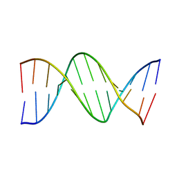
1D16
DownloadVisualize
BU of 1d16 by Molmil
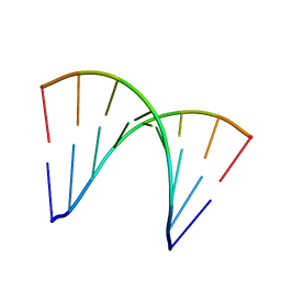
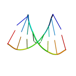
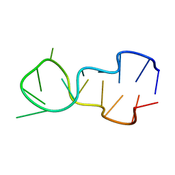
STRUCTURE OF A T4 HAIRPIN LOOP ON A Z-DNA STEM AND COMPARISON WITH A-RNA AND B-DNA LOOPS
Descriptor: DNA (5'-D(*CP*GP*CP*GP*CP*GP*TP*TP*TP*TP*CP*GP*CP*GP*CP*G)-3')
Authors:Chattopadhyaya, R, Grzeskowiak, K, Dickerson, R.E.
Deposit date:1988-04-12
Release date:1989-01-09
Last modified:2024-02-07
Method:X-RAY DIFFRACTION (2.1 Å)
Cite:Structure of a T4 hairpin loop on a Z-DNA stem and comparison with A-RNA and B-DNA loops.
J.Mol.Biol., 211, 1990
3PFK
DownloadVisualize
BU of 3pfk by Molmil
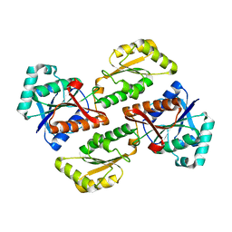
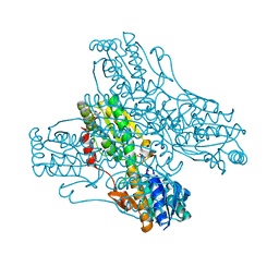
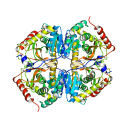
PHOSPHOFRUCTOKINASE. STRUCTURE AND CONTROL
Descriptor: PHOSPHATE ION, PHOSPHOFRUCTOKINASE
Authors:Evans, P.R, Hudson, P.J.
Deposit date:1988-01-25
Release date:1989-01-09
Last modified:2024-02-21
Method:X-RAY DIFFRACTION (2.4 Å)
Cite:Phosphofructokinase: structure and control.
Philos.Trans.R.Soc.London,Ser.B, 293, 1981
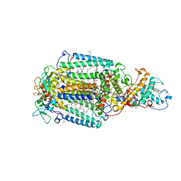
1PRC
DownloadVisualize
BU of 1prc by Molmil
CRYSTALLOGRAPHIC REFINEMENT AT 2.3 ANGSTROMS RESOLUTION AND REFINED MODEL OF THE PHOTOSYNTHETIC REACTION CENTER FROM RHODOPSEUDOMONAS VIRIDIS
Descriptor: 15-trans-1,2-dihydroneurosporene, BACTERIOCHLOROPHYLL B, BACTERIOPHEOPHYTIN B, ...
Authors:Deisenhofer, J, Epp, O, Miki, K, Huber, R, Michel, H.
Deposit date:1988-02-04
Release date:1989-01-09
Last modified:2021-03-10
Method:X-RAY DIFFRACTION (2.3 Å)
Cite:Crystallographic refinement at 2.3 A resolution and refined model of the photosynthetic reaction centre from Rhodopseudomonas viridis.
J.Mol.Biol., 246, 1995
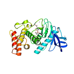
3TMN
DownloadVisualize
BU of 3tmn by Molmil
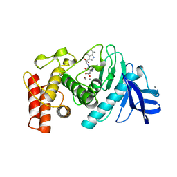
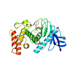
THE BINDING OF L-VALYL-L-TRYPTOPHAN TO CRYSTALLINE THERMOLYSIN ILLUSTRATES THE MODE OF INTERACTION OF A PRODUCT OF PEPTIDE HYDROLYSIS
Descriptor: CALCIUM ION, THERMOLYSIN, TRYPTOPHAN, ...
Authors:Holden, H.M, Matthews, B.W.
Deposit date:1987-06-29
Release date:1989-01-09
Last modified:2024-05-22
Method:X-RAY DIFFRACTION (1.7 Å)
Cite:The binding of L-valyl-L-tryptophan to crystalline thermolysin illustrates the mode of interaction of a product of peptide hydrolysis.
J.Biol.Chem., 263, 1988
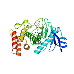
4TMN
DownloadVisualize
BU of 4tmn by Molmil
SLOW-AND FAST-BINDING INHIBITORS OF THERMOLYSIN DISPLAY DIFFERENT MODES OF BINDING. CRYSTALLOGRAPHIC ANALYSIS OF EXTENDED PHOSPHONAMIDATE TRANSITION-STATE ANALOGUES
Descriptor: CALCIUM ION, N-[(S)-[(1R)-1-{[(benzyloxy)carbonyl]amino}-2-phenylethyl](hydroxy)phosphoryl]-L-leucyl-L-alanine, THERMOLYSIN, ...
Authors:Holden, H.M, Tronrud, D.E, Monzingo, A.F, Weaver, L.H, Matthews, B.W.
Deposit date:1987-06-29
Release date:1989-01-09
Last modified:2024-05-22
Method:X-RAY DIFFRACTION (1.7 Å)
Cite:Slow- and fast-binding inhibitors of thermolysin display different modes of binding: crystallographic analysis of extended phosphonamidate transition-state analogues.
Biochemistry, 26, 1987
4DNB
DownloadVisualize
BU of 4dnb by Molmil
METHYLATION OF THE ECORI RECOGNITION SITE DOES NOT ALTER DNA CONFORMATION. THE CRYSTAL STRUCTURE OF D(CGCGAM6ATTCGCG) AT 2.0 ANGSTROMS RESOLUTION
Descriptor: DNA (5'-D(*CP*GP*CP*GP*AP*(6MA)P*TP*TP*CP*GP*CP*G)-3')
Authors:Frederick, C.A, Quigley, G.J, Van Der Marel, G.A, Van Boom, J.H, Wang, A.H.-J, Rich, A.
Deposit date:1988-08-29
Release date:1989-01-09
Last modified:2024-02-28
Method:X-RAY DIFFRACTION (2 Å)
Cite:Methylation of the EcoRI recognition site does not alter DNA conformation: the crystal structure of d(CGCGAm6ATTCGCG) at 2.0-A resolution.
J.Biol.Chem., 263, 1988
3ANA
DownloadVisualize
BU of 3ana by Molmil
THREE-DIMENSIONAL STRUCTURE OF D(GGGATCCC) IN THE CRYSTALLINE STATE
Descriptor: DNA (5'-D(*GP*GP*GP*AP*TP*CP*CP*C)-3')
Authors:Heinemann, U, Lauble, H.
Deposit date:1988-07-06
Release date:1989-01-09
Last modified:2024-02-21
Method:X-RAY DIFFRACTION (2.5 Å)
Cite:Three-dimensional structure of d(GGGATCCC) in the crystalline state.
Nucleic Acids Res., 16, 1988
2PFK
DownloadVisualize
BU of 2pfk by Molmil
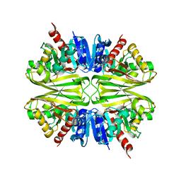
THE CRYSTAL STRUCTURE OF UNLIGANDED PHOSPHOFRUCTOKINASE FROM ESCHERICHIA COLI
Descriptor: 6-PHOSPHOFRUCTOKINASE ISOZYME I
Authors:Rypniewski, W.R, Evans, P.R.
Deposit date:1988-01-25
Release date:1989-01-09
Last modified:2024-02-21
Method:X-RAY DIFFRACTION (2.4 Å)
Cite:Crystal structure of unliganded phosphofructokinase from Escherichia coli.
J.Mol.Biol., 207, 1989
1LRP
DownloadVisualize
BU of 1lrp by Molmil
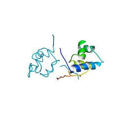
COMPARISON OF THE STRUCTURES OF CRO AND LAMBDA REPRESSOR PROTEINS FROM BACTERIOPHAGE LAMBDA
Descriptor: LAMBDA REPRESSOR
Authors:Pabo, C, Lewis, M.
Deposit date:1987-12-04
Release date:1989-01-09
Last modified:2023-09-27
Method:X-RAY DIFFRACTION (3.2 Å)
Cite:Comparison of the structures of cro and lambda repressor proteins from bacteriophage lambda.
J.Mol.Biol., 169, 1983
1TMN
DownloadVisualize
BU of 1tmn by Molmil
Binding of n-carboxymethyl dipeptide inhibitors to thermolysin determined by x-ray crystallography. a novel class of transition-state analogues for zinc peptidases
Descriptor: CALCIUM ION, N-[(1R)-1-carboxy-3-phenylpropyl]-L-leucyl-L-tryptophan, THERMOLYSIN, ...
Authors:Monzingo, A.F, Matthews, B.W.
Deposit date:1987-06-29
Release date:1989-01-09
Last modified:2024-05-22
Method:X-RAY DIFFRACTION (1.9 Å)
Cite:Binding of N-carboxymethyl dipeptide inhibitors to thermolysin determined by X-ray crystallography: a novel class of transition-state analogues for zinc peptidases
Biochemistry, 23, 1984
1DNE
DownloadVisualize
BU of 1dne by Molmil
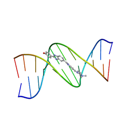
MOLECULAR STRUCTURE OF THE NETROPSIN-D(CGCGATATCGCG) COMPLEX: DNA CONFORMATION IN AN ALTERNATING AT SEGMENT; CONFORMATION 2
Descriptor: DNA (5'-D(*CP*GP*CP*GP*AP*TP*AP*TP*CP*GP*CP*G)-3'), NETROPSIN
Authors:Coll, M, Aymami, J, Van Der Marel, G.A, Van Boom, J.H, Rich, A, Wang, A.H.-J.
Deposit date:1988-09-14
Release date:1989-01-09
Last modified:2024-02-07
Method:X-RAY DIFFRACTION (2.4 Å)
Cite:Molecular structure of the netropsin-d(CGCGATATCGCG) complex: DNA conformation in an alternating AT segment.
Biochemistry, 28, 1989
1DCG
DownloadVisualize
BU of 1dcg by Molmil
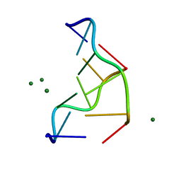
THE MOLECULAR STRUCTURE OF THE LEFT-HANDED Z-DNA DOUBLE HELIX AT 1.0 ANGSTROM ATOMIC RESOLUTION. GEOMETRY, CONFORMATION, AND IONIC INTERACTIONS OF D(CGCGCG)
Descriptor: DNA (5'-D(*CP*GP*CP*GP*CP*G)-3'), MAGNESIUM ION
Authors:Gessner, R.V, Frederick, C.A, Quigley, G.J, Rich, A, Wang, A.H.-J.
Deposit date:1988-08-29
Release date:1989-01-09
Last modified:2024-02-07
Method:X-RAY DIFFRACTION (1 Å)
Cite:The molecular structure of the left-handed Z-DNA double helix at 1.0-A atomic resolution. Geometry, conformation, and ionic interactions of d(CGCGCG).
J.Biol.Chem., 264, 1989
2DND
DownloadVisualize
BU of 2dnd by Molmil
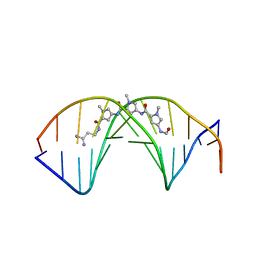
A BIFURCATED HYDROGEN-BONDED CONFORMATION IN THE D(A.T) BASE PAIRS OF THE DNA DODECAMER D(CGCAAATTTGCG) AND ITS COMPLEX WITH DISTAMYCIN
Descriptor: DISTAMYCIN A, DNA (5'-D(*CP*GP*CP*AP*AP*AP*TP*TP*TP*GP*CP*G)-3')
Authors:Coll, M, Frederick, C.A, Wang, A.H.-J, Rich, A.
Deposit date:1988-08-29
Release date:1989-01-09
Last modified:2024-02-14
Method:X-RAY DIFFRACTION (2.2 Å)
Cite:A bifurcated hydrogen-bonded conformation in the d(A.T) base pairs of the DNA dodecamer d(CGCAAATTTGCG) and its complex with distamycin.
Proc.Natl.Acad.Sci.USA, 84, 1987
1DNH
DownloadVisualize
BU of 1dnh by Molmil
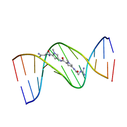
THE MOLECULAR STRUCTURE OF THE COMPLEX OF HOECHST 33258 AND THE DNA DODECAMER D(CGCGAATTCGCG)
Descriptor: 2'-(4-HYDROXYPHENYL)-5-(4-METHYL-1-PIPERAZINYL)-2,5'-BI-BENZIMIDAZOLE, DNA (5'-D(*CP*GP*CP*GP*AP*AP*TP*TP*CP*GP*CP*G)-3')
Authors:Teng, M.-K, Usman, N, Frederick, C.A, Wang, A.H.-J.
Deposit date:1988-02-16
Release date:1989-01-09
Last modified:2024-02-07
Method:X-RAY DIFFRACTION (2.25 Å)
Cite:The molecular structure of the complex of Hoechst 33258 and the DNA dodecamer d(CGCGAATTCGCG).
Nucleic Acids Res., 16, 1988
1BDS
DownloadVisualize
BU of 1bds by Molmil
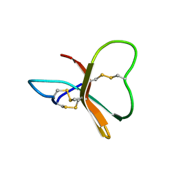
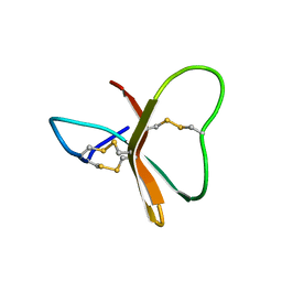
DETERMINATION OF THE THREE-DIMENSIONAL SOLUTION STRUCTURE OF THE ANTIHYPERTENSIVE AND ANTIVIRAL PROTEIN BDS-I FROM THE SEA ANEMONE ANEMONIA SULCATA. A STUDY USING NUCLEAR MAGNETIC RESONANCE AND HYBRID DISTANCE GEOMETRY-DYNAMICAL SIMULATED ANNEALING
Descriptor: BDS-I
Authors:Clore, G.M, Driscoll, P.C, Gronenborn, A.M.
Deposit date:1988-11-14
Release date:1989-01-09
Last modified:2017-11-29
Method:SOLUTION NMR
Cite:Determination of the three-dimensional solution structure of the antihypertensive and antiviral protein BDS-I from the sea anemone Anemonia sulcata: a study using nuclear magnetic resonance and hybrid distance geometry-dynamical simulated annealing.
Biochemistry, 28, 1989
4PFK
DownloadVisualize
BU of 4pfk by Molmil
PHOSPHOFRUCTOKINASE. STRUCTURE AND CONTROL
Descriptor: 6-O-phosphono-beta-D-fructofuranose, ADENOSINE-5'-DIPHOSPHATE, MAGNESIUM ION, ...
Authors:Evans, P.R, Hudson, P.J.
Deposit date:1988-01-25
Release date:1989-01-09
Last modified:2024-02-28
Method:X-RAY DIFFRACTION (2.4 Å)
Cite:Phosphofructokinase: structure and control.
Philos.Trans.R.Soc.London,Ser.B, 293, 1981
1TLP
DownloadVisualize
BU of 1tlp by Molmil
CRYSTALLOGRAPHIC STRUCTURAL ANALYSIS OF PHOSPHORAMIDATES AS INHIBITORS AND TRANSITION-STATE ANALOGS OF THERMOLYSIN
Descriptor: CALCIUM ION, N-ALPHA-L-RHAMNOPYRANOSYLOXY(HYDROXYPHOSPHINYL)-L-LEUCYL-L-TRYPTOPHAN, THERMOLYSIN, ...
Authors:Tronrud, D.E, Monzingo, A.F, Matthews, B.W.
Deposit date:1987-06-29
Release date:1989-01-09
Last modified:2024-03-13
Method:X-RAY DIFFRACTION (2.3 Å)
Cite:Crystallographic structural analysis of phosphoramidates as inhibitors and transition-state analogs of thermolysin.
Eur.J.Biochem., 157, 1986
1VJ4
DownloadVisualize
BU of 1vj4 by Molmil
SEQUENCE-DEPENDENT CONFORMATION OF AN A-DNA DOUBLE HELIX: THE CRYSTAL STRUCTURE OF THE OCTAMER D(G-G-T-A-T-A-C-C)
Descriptor: 5'-D(*GP*GP*TP*AP*TP*AP*CP*C)-3'
Authors:Shakked, Z, Rabinovich, D, Kennard, O, Cruse, W.B, Salisbury, S.A, Viswamitra, M.A.
Deposit date:1989-01-11
Release date:1989-01-11
Last modified:2024-02-14
Method:X-RAY DIFFRACTION (1.8 Å)
Cite:Sequence-dependent conformation of an A-DNA double helix: The crystal structure of the octamer d(G-G-T-A-T-A-C-C)
J.Mol.Biol., 166, 1983
2HVP
DownloadVisualize
BU of 2hvp by Molmil
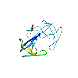
THREE-DIMENSIONAL STRUCTURE OF ASPARTYL PROTEASE FROM HUMAN IMMUNODEFICIENCY VIRUS HIV-1
Descriptor: HIV-1 PROTEASE
Authors:Navia, M.A, Fitzgerald, P.M.D, Mckeever, B.M, Springer, J.P.
Deposit date:1989-04-10
Release date:1989-04-19
Last modified:2024-02-14
Method:X-RAY DIFFRACTION (3 Å)
Cite:Three-dimensional structure of aspartyl protease from human immunodeficiency virus HIV-1.
Nature, 337, 1989
2BDS
DownloadVisualize
BU of 2bds by Molmil
DETERMINATION OF THE THREE-DIMENSIONAL SOLUTION STRUCTURE OF THE ANTIHYPERTENSIVE AND ANTIVIRAL PROTEIN BDS-I FROM THE SEA ANEMONE ANEMONIA SULCATA. A STUDY USING NUCLEAR MAGNETIC RESONANCE AND HYBRID DISTANCE GEOMETRY-DYNAMICAL SIMULATED ANNEALING
Descriptor: BDS-I
Authors:Clore, G.M, Driscoll, P.C, Gronenborn, A.M.
Deposit date:1988-11-14
Release date:1989-04-19
Last modified:2017-11-29
Method:SOLUTION NMR
Cite:Determination of the three-dimensional solution structure of the antihypertensive and antiviral protein BDS-I from the sea anemone Anemonia sulcata: a study using nuclear magnetic resonance and hybrid distance geometry-dynamical simulated annealing.
Biochemistry, 28, 1989
1RDG
DownloadVisualize
BU of 1rdg by Molmil
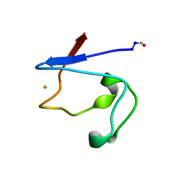
RUBREDOXIN FROM DESULFOVIBRIO GIGAS. A MOLECULAR MODEL OF THE OXIDIZED FORM AT 1.4 ANGSTROMS RESOLUTION
Descriptor: FE (III) ION, FORMYL GROUP, RUBREDOXIN
Authors:Frey, M, Sieker, L.C, Payan, F.
Deposit date:1988-03-17
Release date:1989-04-19
Last modified:2024-06-05
Method:X-RAY DIFFRACTION (1.4 Å)
Cite:Rubredoxin from Desulfovibrio gigas. A molecular model of the oxidized form at 1.4 A resolution.
J.Mol.Biol., 197, 1987
4MDH
DownloadVisualize
BU of 4mdh by Molmil
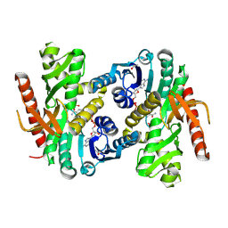
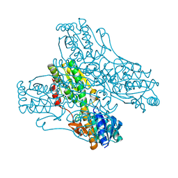
REFINED CRYSTAL STRUCTURE OF CYTOPLASMIC MALATE DEHYDROGENASE AT 2.5-ANGSTROMS RESOLUTION
Descriptor: CYTOPLASMIC MALATE DEHYDROGENASE, NICOTINAMIDE-ADENINE-DINUCLEOTIDE, SULFATE ION
Authors:Birktoft, J.J, Banaszak, L.J.
Deposit date:1989-04-12
Release date:1989-04-19
Last modified:2017-11-29
Method:X-RAY DIFFRACTION (2.5 Å)
Cite:Refined crystal structure of cytoplasmic malate dehydrogenase at 2.5-A resolution.
Biochemistry, 28, 1989
1HOE
DownloadVisualize
BU of 1hoe by Molmil
CRYSTAL STRUCTURE DETERMINATION, REFINEMENT AND THE MOLECULAR MODEL OF THE ALPHA-AMYLASE INHIBITOR HOE-467A
Descriptor: ALPHA-AMYLASE INHIBITOR
Authors:Pflugrath, J.W, Wiegand, G, Huber, R.
Deposit date:1989-01-27
Release date:1989-04-19
Last modified:2017-11-29
Method:X-RAY DIFFRACTION (2 Å)
Cite:Crystal structure determination, refinement and the molecular model of the alpha-amylase inhibitor Hoe-467A.
J.Mol.Biol., 189, 1986
2LDX
DownloadVisualize
BU of 2ldx by Molmil
CHARACTERIZATION OF THE ANTIGENIC SITES ON THE REFINED 3-ANGSTROMS RESOLUTION STRUCTURE OF MOUSE TESTICULAR LACTATE DEHYDROGENASE C4
Descriptor: APO-LACTATE DEHYDROGENASE
Authors:Griffith, J.P, Rossmann, M.G.
Deposit date:1987-11-25
Release date:1989-04-19
Last modified:2023-09-06
Method:X-RAY DIFFRACTION (2.96 Å)
Cite:Characterization of the antigenic sites on the refined 3-A resolution structure of mouse testicular lactate dehydrogenase C4.
J.Biol.Chem., 262, 1987
4GPD
DownloadVisualize
BU of 4gpd by Molmil
THE STRUCTURE OF LOBSTER APO-D-GLYCERALDEHYDE-3-PHOSPHATE DEHYDROGENASE AT 3.0 ANGSTROMS RESOLUTION
Descriptor: APO-D-GLYCERALDEHYDE-3-PHOSPHATE DEHYDROGENASE
Authors:Griffith, J.P, Song, S, Rossmann, M.G.
Deposit date:1988-01-04
Release date:1989-07-12
Last modified:2024-02-28
Method:X-RAY DIFFRACTION (2.8 Å)
Cite:Structure of lobster apo-D-glyceraldehyde-3-phosphate dehydrogenase at 3.0 A resolution.
J.Mol.Biol., 138, 1980

222624

PDB entries from 2024-07-17

PDB statisticsPDBj update infoContact PDBjnumon