Loading
PDBj
MenuPDBj@FacebookPDBj@TwitterPDBj@YouTubewwPDB FoundationwwPDB
RCSB PDBPDBeBMRBAdv. SearchSearch help

3SSP
DownloadVisualize
BU of 3ssp by Molmil
Engineered low-affinity halide-binding protein derived from YFP: halide-free
Descriptor: Green fluorescent protein
Authors:Wang, W, Grimley, J.S, Beese, L.S, Hellinga, H.W.
Deposit date:2011-07-08
Release date:2012-07-11
Last modified:2023-11-15
Method:X-RAY DIFFRACTION (1.628 Å)
Cite:Determination of engineered chloride-binding site structures in fluorescent proteins reveals principles of halide recognition
To be Published
8A6O
DownloadVisualize
BU of 8a6o by Molmil
Room temperature rsEGFP2 with a chlorinated chromophore 600 fs after Photoexcitation
Descriptor: Green fluorescent protein
Authors:Fadini, A, van Thor, J.
Deposit date:2022-06-18
Release date:2023-07-19
Last modified:2024-02-07
Method:X-RAY DIFFRACTION (1.63 Å)
Cite:Serial Femtosecond Crystallography Reveals that Photoactivation in a Fluorescent Protein Proceeds via the Hula Twist Mechanism.
J.Am.Chem.Soc., 145, 2023
8A6P
DownloadVisualize
BU of 8a6p by Molmil
Room temperature rsEGFP2 with a chlorinated chromophore 900 fs after photoexcitation
Descriptor: Green fluorescent protein
Authors:Fadini, A, van Thor, J.
Deposit date:2022-06-18
Release date:2023-07-19
Last modified:2024-02-07
Method:X-RAY DIFFRACTION (1.63 Å)
Cite:Serial Femtosecond Crystallography Reveals that Photoactivation in a Fluorescent Protein Proceeds via the Hula Twist Mechanism.
J.Am.Chem.Soc., 145, 2023
8A6R
DownloadVisualize
BU of 8a6r by Molmil
Room temperature rsEGFP2 with a chlorinated chromophore 100 ps after Photoexcitation
Descriptor: Green fluorescent protein
Authors:Fadini, A, van Thor, J.
Deposit date:2022-06-18
Release date:2023-07-19
Last modified:2024-02-07
Method:X-RAY DIFFRACTION (1.63 Å)
Cite:Serial Femtosecond Crystallography Reveals that Photoactivation in a Fluorescent Protein Proceeds via the Hula Twist Mechanism.
J.Am.Chem.Soc., 145, 2023
8A6G
DownloadVisualize
BU of 8a6g by Molmil
Room temperature rsEGFP2 with a chlorinated chromophore in the non-fluorescent OFF-state
Descriptor: Green fluorescent protein
Authors:Fadini, A, van Thor, J.
Deposit date:2022-06-17
Release date:2023-07-19
Last modified:2024-02-07
Method:X-RAY DIFFRACTION (1.63 Å)
Cite:Serial Femtosecond Crystallography Reveals that Photoactivation in a Fluorescent Protein Proceeds via the Hula Twist Mechanism.
J.Am.Chem.Soc., 145, 2023
8A6N
DownloadVisualize
BU of 8a6n by Molmil
Room temperature rsEGFP2 with a chlorinated chromophore 300 fs after Photoexcitation
Descriptor: Green fluorescent protein
Authors:Fadini, A, van Thor, J.
Deposit date:2022-06-18
Release date:2023-07-19
Last modified:2024-02-07
Method:X-RAY DIFFRACTION (1.63 Å)
Cite:Serial Femtosecond Crystallography Reveals that Photoactivation in a Fluorescent Protein Proceeds via the Hula Twist Mechanism.
J.Am.Chem.Soc., 145, 2023
8A6Q
DownloadVisualize
BU of 8a6q by Molmil
Room temperature rsEGFP2 with a chlorinated chromophore 5 ps after photoexcitation
Descriptor: Green fluorescent protein
Authors:Fadini, A, van Thor, J.
Deposit date:2022-06-18
Release date:2023-07-19
Last modified:2024-02-07
Method:X-RAY DIFFRACTION (1.63 Å)
Cite:Serial Femtosecond Crystallography Reveals that Photoactivation in a Fluorescent Protein Proceeds via the Hula Twist Mechanism.
J.Am.Chem.Soc., 145, 2023
8A6S
DownloadVisualize
BU of 8a6s by Molmil
Room temperature rsEGFP2 with a chlorinated chromophore 1 microsecond after Photoexcitation
Descriptor: Green fluorescent protein
Authors:Fadini, A, van Thor, J.
Deposit date:2022-06-19
Release date:2023-07-19
Last modified:2024-02-07
Method:X-RAY DIFFRACTION (1.63 Å)
Cite:Serial Femtosecond Crystallography Reveals that Photoactivation in a Fluorescent Protein Proceeds via the Hula Twist Mechanism.
J.Am.Chem.Soc., 145, 2023
7AMB
DownloadVisualize
BU of 7amb by Molmil
Crystal structure of rsFolder2 in its fluorescent on-state
Descriptor: GLYCEROL, Green fluorescent protein
Authors:Moreno-Chicano, T, El Khatib, M, Colletier, J.-P.
Deposit date:2020-10-08
Release date:2022-08-24
Last modified:2024-01-31
Method:X-RAY DIFFRACTION (1.63 Å)
Cite:Rational Control of Off-State Heterogeneity in a Photoswitchable Fluorescent Protein Provides Switching Contrast Enhancement.
Chemphyschem, 23, 2022
7AMF
DownloadVisualize
BU of 7amf by Molmil
Crystal structure of rsFolder2 in its non-fluorescent off-state
Descriptor: GLYCEROL, Green fluorescent protein
Authors:Moreno-Chicano, T, El Khatib, M, Colletier, J.-P.
Deposit date:2020-10-08
Release date:2022-08-24
Last modified:2024-01-31
Method:X-RAY DIFFRACTION (1.63 Å)
Cite:Rational Control of Off-State Heterogeneity in a Photoswitchable Fluorescent Protein Provides Switching Contrast Enhancement.
Chemphyschem, 23, 2022
7UGS
DownloadVisualize
BU of 7ugs by Molmil
Crystal structure of monomeric hyperfolder YFP (K206V mutant)
Descriptor: Hyperfolder yellow fluorescent protein
Authors:Campbell, B.C, Liu, C.F, Petsko, G.A.
Deposit date:2022-03-25
Release date:2022-10-26
Last modified:2023-11-15
Method:X-RAY DIFFRACTION (1.63 Å)
Cite:Chemically stable fluorescent proteins for advanced microscopy.
Nat.Methods, 19, 2022
2YE1
DownloadVisualize
BU of 2ye1 by Molmil
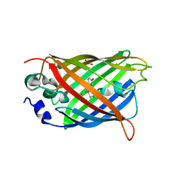
X-ray structure of the cyan fluorescent proteinmTurquoise-GL (K206A mutant)
Descriptor: GREEN FLUORESCENT PROTEIN, MAGNESIUM ION
Authors:von Stetten, D, Noirclerc-Savoye, M, Goedhart, J, Gadella, T.W.J, Royant, A.
Deposit date:2011-03-25
Release date:2012-04-11
Last modified:2023-12-20
Method:X-RAY DIFFRACTION (1.63 Å)
Cite:Structural Characterization of the Cyan Fluorescent Protein Mturquoise-Gl
To be Published
3IR8
DownloadVisualize
BU of 3ir8 by Molmil
Red fluorescent protein mKeima at pH 7.0
Descriptor: Large stokes shift fluorescent protein
Authors:Henderson, J.N, Osborn, M.F, Koon, N, Gepshtein, R, Huppert, D, Remington, S.J.
Deposit date:2009-08-21
Release date:2009-09-08
Last modified:2023-11-15
Method:X-RAY DIFFRACTION (1.63 Å)
Cite:Excited state proton transfer in the red fluorescent protein mKeima.
J.Am.Chem.Soc., 131, 2009
5WJ4
DownloadVisualize
BU of 5wj4 by Molmil
Crystal structure of redox-sensitive green fluorescent protein Clover mutant roClover1
Descriptor: Green fluorescent protein
Authors:Liu, C, Campbell, B.C, Petsko, G.A.
Deposit date:2017-07-21
Release date:2018-01-17
Last modified:2023-11-15
Method:X-RAY DIFFRACTION (1.631 Å)
Cite:Crystal Structure of Green Fluorescent Protein Clover and Design of Clover-Based Redox Sensors.
Structure, 26, 2018
5K4Z
DownloadVisualize
BU of 5k4z by Molmil
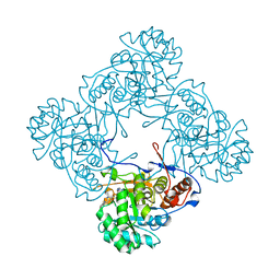
M. thermoresistible IMPDH in complex with IMP and Compound 6
Descriptor: INOSINIC ACID, Inosine-5'-monophosphate dehydrogenase,Inosine-5'-monophosphate dehydrogenase, ~{N}-(4-fluorophenyl)-4-(2~{H}-indazol-6-ylsulfamoyl)-3,5-dimethyl-1~{H}-pyrrole-2-carboxamide
Authors:Pacitto, A, Ascher, D.B, Blundell, T.L.
Deposit date:2016-05-22
Release date:2016-10-19
Last modified:2024-05-01
Method:X-RAY DIFFRACTION (1.64 Å)
Cite:Essential but Not Vulnerable: Indazole Sulfonamides Targeting Inosine Monophosphate Dehydrogenase as Potential Leads against Mycobacterium tuberculosis.
ACS Infect Dis, 3, 2017
2VAE
DownloadVisualize
BU of 2vae by Molmil
Fast maturing red fluorescent protein, DsRed.T4
Descriptor: 1,2-ETHANEDIOL, RED FLUORESCENT PROTEIN
Authors:Strongin, D.E, Bevis, B, Khuong, N, Downing, M.E, Strack, R.L, Sundaram, K, Glick, B.S, Keenan, R.J.
Deposit date:2007-08-31
Release date:2007-11-06
Last modified:2023-12-13
Method:X-RAY DIFFRACTION (1.64 Å)
Cite:Structural Rearrangements Near the Chromophore Influence the Maturation Speed and Brightness of Dsred Variants.
Protein Eng.Des.Sel., 20, 2007
7AMU
DownloadVisualize
BU of 7amu by Molmil
Crystal structure of rsEGFP2 T204A in its fluorescent on-state
Descriptor: GLYCEROL, Green fluorescent protein, SULFATE ION
Authors:Moreno-Chicano, T, Schlichting, I, Hartmann, E, Zala, N, Colletier, J.-P, Weik, M.
Deposit date:2020-10-09
Release date:2022-04-20
Last modified:2024-01-31
Method:X-RAY DIFFRACTION (1.64 Å)
Cite:Crystal structure of rsEGFP2 in its fluorescent on-state at pH 8.0
To Be Published
6IR6
DownloadVisualize
BU of 6ir6 by Molmil
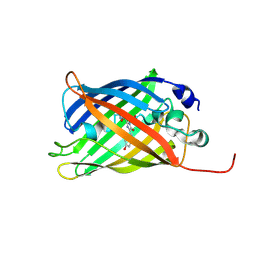
Green fluorescent protein variant GFPuv with the native lysine residue at the C-terminus
Descriptor: Green fluorescent protein, SULFATE ION
Authors:Nakatani, T, Yasui, N, Yamashita, A.
Deposit date:2018-11-12
Release date:2019-04-03
Last modified:2023-11-22
Method:X-RAY DIFFRACTION (1.642 Å)
Cite:Specific modification at the C-terminal lysine residue of the green fluorescent protein variant, GFPuv, expressed in Escherichia coli.
Sci Rep, 9, 2019
6GO8
DownloadVisualize
BU of 6go8 by Molmil
Structure of GFPmut2 crystallized at pH 6
Descriptor: (4R)-2-METHYLPENTANE-2,4-DIOL, (4S)-2-METHYL-2,4-PENTANEDIOL, Green fluorescent protein
Authors:Lolli, G, Raboni, S, Pasqualetto, E, Campanini, B, Mozzarelli, A, Bettati, S, Battistutta, R.
Deposit date:2018-06-01
Release date:2018-12-19
Last modified:2024-01-17
Method:X-RAY DIFFRACTION (1.648 Å)
Cite:Insight into GFPmut2 pH Dependence by Single Crystal Microspectrophotometry and X-ray Crystallography.
J.Phys.Chem.B, 122, 2018
6OFN
DownloadVisualize
BU of 6ofn by Molmil
Crystal structure of green fluorescent protein (GFP); S65T, T203(3-OMeY); ih circular permutant (50-51)
Descriptor: Green fluorescent protein (GFP); S65T, T203(3-OMeY); ih circular permutant (50-51)
Authors:Lin, C.-Y, Romei, M.G, Mathews, I.I, Boxer, S.G.
Deposit date:2019-03-31
Release date:2019-07-10
Last modified:2023-11-15
Method:X-RAY DIFFRACTION (1.649 Å)
Cite:Unified Model for Photophysical and Electro-Optical Properties of Green Fluorescent Proteins.
J.Am.Chem.Soc., 141, 2019
4TPS
DownloadVisualize
BU of 4tps by Molmil
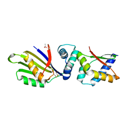
Sporulation Inhibitor of DNA Replication, SirA, in complex with Domain I of DnaA
Descriptor: ACETATE ION, BETA-MERCAPTOETHANOL, Chromosomal replication initiator protein DnaA, ...
Authors:Jameson, K.H, Turkenburg, J.P, Fogg, M.J, Grahl, A, Wilkinson, A.J.
Deposit date:2014-06-09
Release date:2014-07-30
Last modified:2024-04-03
Method:X-RAY DIFFRACTION (1.65 Å)
Cite:Structure and interactions of the Bacillus subtilis sporulation inhibitor of DNA replication, SirA, with domain I of DnaA.
Mol.Microbiol., 93, 2014
7SFA
DownloadVisualize
BU of 7sfa by Molmil
Branchiostoma floridae fluorescent protein LanFP10A2
Descriptor: Fluorescent protein lanFP10A2
Authors:Pletnev, S, Pletneva, N, Pletnev, V.Z, Muslinkina, L.
Deposit date:2021-10-03
Release date:2022-03-30
Last modified:2023-11-15
Method:X-RAY DIFFRACTION (1.65 Å)
Cite:A novel violet fluorescent protein contains a unique oxidized tyrosine as the simplest chromophore ever reported in fluorescent proteins.
Protein Sci., 31, 2022
3U0M
DownloadVisualize
BU of 3u0m by Molmil
Crystal structure of the engineered fluorescent protein mRuby, crystal form 1, pH 8.5
Descriptor: mRuby
Authors:Akerboom, J, Looger, L.L, Schreiter, E.R.
Deposit date:2011-09-28
Release date:2012-10-03
Last modified:2023-12-06
Method:X-RAY DIFFRACTION (1.65 Å)
Cite:Genetically encoded calcium indicators for multi-color neural activity imaging and combination with optogenetics.
Front Mol Neurosci, 6, 2013
7A8D
DownloadVisualize
BU of 7a8d by Molmil
rsGreen0.7-K206A-F165W partially in the green-on state
Descriptor: Green fluorescent protein
Authors:De Zitter, E, Dedecker, P, Van Meervelt, L.
Deposit date:2020-08-30
Release date:2021-02-17
Last modified:2021-05-05
Method:X-RAY DIFFRACTION (1.65 Å)
Cite:Structure-Function Dataset Reveals Environment Effects within a Fluorescent Protein Model System*.
Angew.Chem.Int.Ed.Engl., 60, 2021
2ZMU
DownloadVisualize
BU of 2zmu by Molmil
Crystal Structure of Monomeric Kusabira-Orange (MKO), Orange-Emitting GFP-like Protein, at pH 9.1
Descriptor: Fluorescent protein
Authors:Kikuchi, A, Fukumura, E, Karasawa, S, Mizuno, H, Miyawaki, A, Shiro, Y, RIKEN Structural Genomics/Proteomics Initiative (RSGI)
Deposit date:2008-04-21
Release date:2008-10-21
Last modified:2023-11-15
Method:X-RAY DIFFRACTION (1.65 Å)
Cite:Structural Characterization of a Thiazoline-Containing Chromophore in an Orange Fluorescent Protein, Monomeric Kusabira Orange
Biochemistry, 47, 2008

221051

PDB entries from 2024-06-12

PDB statisticsPDBj update infoContact PDBjnumon