2ODV
 
 | |
2ODU
 | |
4Q57
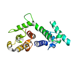
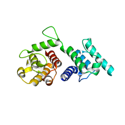
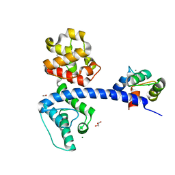 | | Crystal structure of the plectin 1a actin-binding domain/N-terminal domain of calmodulin complex | | Descriptor: | 1,2-ETHANEDIOL, CALCIUM ION, CHLORIDE ION, ... | | Authors: | Song, J.-G, Kostan, J, Grishkovskaya, I, Djinovic-Carugo, K. | | Deposit date: | 2014-04-16 | | Release date: | 2014-07-23 | | Last modified: | 2024-02-28 | | Method: | X-RAY DIFFRACTION (1.8 Å) | | Cite: | Crystal structure of the plectin 1a actin-binding domain/N-terminal domain of calmodulin complex
To be Published
|
|
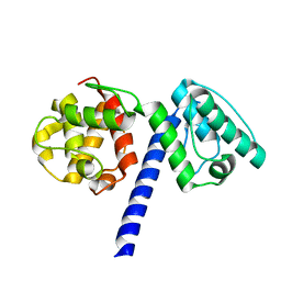
3F7P
 | |
1MB8
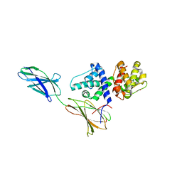
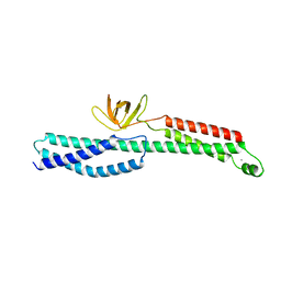
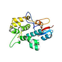
 | | Crystal Structure of the actin binding domain of plectin | | Descriptor: | Plectin | | Authors: | de Pereda, J.M. | | Deposit date: | 2002-08-02 | | Release date: | 2003-06-10 | | Last modified: | 2024-02-14 | | Method: | X-RAY DIFFRACTION (2.15 Å) | | Cite: | Structural and Functional Analysis of the Actin Binding Domain of Plectin
Suggests Alternative Mechanisms for Binding to F-Actin and Integrin Beta4
Structure, 11, 2003
|
|
1SH6
 | |
1SH5
 | |
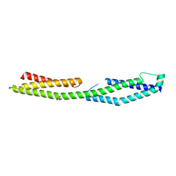
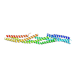
4GDO
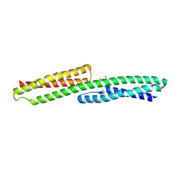
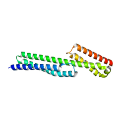
 | | Structure of a fragment of the rod domain of plectin | | Descriptor: | Plectin | | Authors: | De Pereda, J.M, Buey, R.M, Uson, I, Sammito, M.D, De Marino, I. | | Deposit date: | 2012-08-01 | | Release date: | 2013-09-11 | | Last modified: | 2024-02-28 | | Method: | X-RAY DIFFRACTION (1.7 Å) | | Cite: | Exploiting tertiary structure through local folds for crystallographic phasing.
Nat.Methods, 10, 2013
|
|
3PDY
 | |
3PE0
 | |
4Q58
 | |
4Q59
 | |
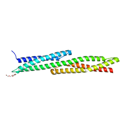
2N03
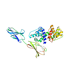
 | | Solution NMR Structure plectin repeat domain 6 (4403-4606) of Plectin from Homo sapiens, Northeast Structural Genomics Consortium (NESG) Target HR6354E | | Descriptor: | Plectin | | Authors: | Pulavarti, S, Eletsky, A, Huang, Y, Janjua, H, Xiao, R, Acton, T, Everett, J, Montelione, G, Szyperski, T, Northeast Structural Genomics Consortium (NESG) | | Deposit date: | 2015-03-04 | | Release date: | 2015-05-13 | | Last modified: | 2024-05-15 | | Method: | SOLUTION NMR | | Cite: | Solution NMR Structure plectin repeat domain 6 (4403-4606) of Plectin from Homo sapiens, Northeast Structural Genomics Consortium (NESG) Target HR6354E
To be Published
|
|
5J1F
 | |
5J1H
 | |
5J1I
 | |
5J1G
 | |


