Loading
PDBj
MenuPDBj@FacebookPDBj@TwitterPDBj@YouTubewwPDB FoundationwwPDB
RCSB PDBPDBeBMRBAdv. SearchSearch help
PDB: 599 results

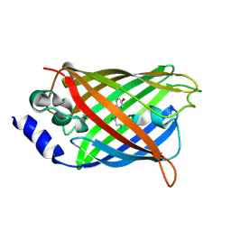
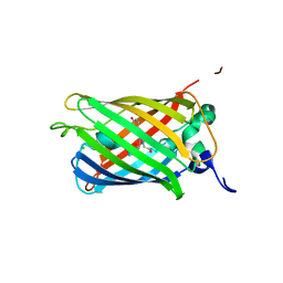
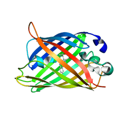
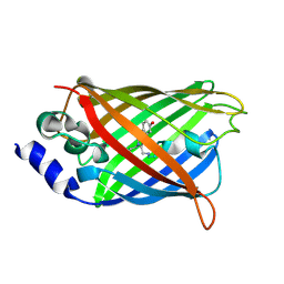
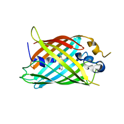
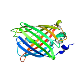
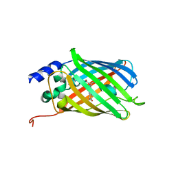
5DTX
DownloadVisualize
BU of 5dtx by Molmil
Crystal structure of rsEGFP2 in the fluorescent on-state
Descriptor: Green fluorescent protein
Authors:Adam, V, Martins, A.
Deposit date:2015-09-18
Release date:2016-01-20
Last modified:2024-01-10
Method:X-RAY DIFFRACTION (1.45 Å)
Cite:Rational design of ultrastable and reversibly photoswitchable fluorescent proteins for super-resolution imaging of the bacterial periplasm.
Sci Rep, 6, 2016
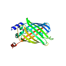
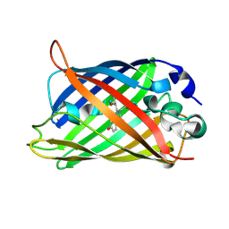
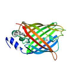
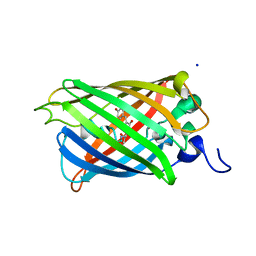
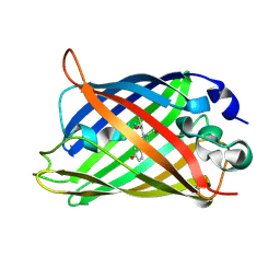
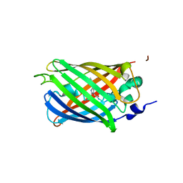
3DQH
DownloadVisualize
BU of 3dqh by Molmil
Structure of the Yellow Fluorescent Protein Citrine Frozen at 1000 Atmospheres Number 5: Structure 9 in a Series of 26 High Pressure Structures
Descriptor: Green fluorescent protein
Authors:Barstow, B, Kim, C.U.
Deposit date:2008-07-09
Release date:2008-09-23
Last modified:2023-11-15
Method:X-RAY DIFFRACTION (1.45 Å)
Cite:Alteration of citrine structure by hydrostatic pressure explains the accompanying spectral shift.
Proc.Natl.Acad.Sci.Usa, 105, 2008
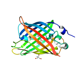
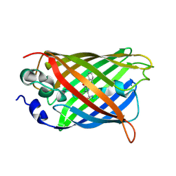
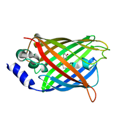
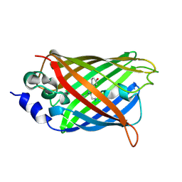
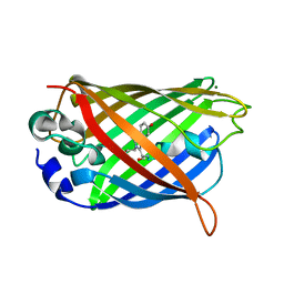
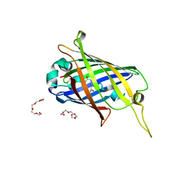
6PFT
DownloadVisualize
BU of 6pft by Molmil
rsEGFP2 with a chlorinated chromophore in the non-fluorescent off-state
Descriptor: Green fluorescent protein, SULFATE ION
Authors:Chang, J, Romei, M.G, Boxer, S.G.
Deposit date:2019-06-22
Release date:2019-08-07
Last modified:2023-11-15
Method:X-RAY DIFFRACTION (1.45 Å)
Cite:Structural Evidence of Photoisomerization Pathways in Fluorescent Proteins.
J.Am.Chem.Soc., 141, 2019
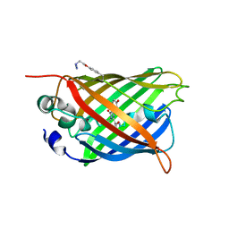
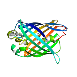
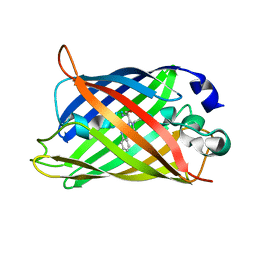
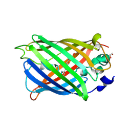
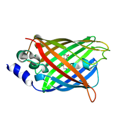
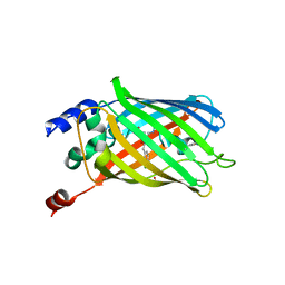
4H48
DownloadVisualize
BU of 4h48 by Molmil
1.45 angstrom CyPet Structure at pH7.0
Descriptor: 2-AMINO-2-HYDROXYMETHYL-PROPANE-1,3-DIOL, Green fluorescent protein
Authors:Hu, X.-J, Liu, R.
Deposit date:2012-09-17
Release date:2013-09-18
Last modified:2023-12-06
Method:X-RAY DIFFRACTION (1.45 Å)
Cite:Structure insight of the fluorescent state of CyPet
To be Published
5EHU
DownloadVisualize
BU of 5ehu by Molmil
sfGFP mutant with unnatural amino acid 4-azidoethoxy-L-phenylalanine incorporated at the 149 site
Descriptor: Green fluorescent protein
Authors:Tookmanian, E.M, Phillips-Piro, C.M, Fenlon, E.E, Brewer, S.B.
Deposit date:2015-10-28
Release date:2015-12-23
Last modified:2023-11-15
Method:X-RAY DIFFRACTION (1.45 Å)
Cite:Azidoethoxyphenylalanine as a Vibrational Reporter and Click Chemistry Partner in Proteins.
Chemistry, 21, 2015
2O24
DownloadVisualize
BU of 2o24 by Molmil
Spectroscopic and Structural Study of the Heterotropic Linkage between Halide and Proton Ion Binding to Gfp Proteins: E2(GFP)-Cl Complex
Descriptor: CHLORIDE ION, Green fluorescent protein
Authors:Garau, G.
Deposit date:2006-11-29
Release date:2007-05-15
Last modified:2023-11-15
Method:X-RAY DIFFRACTION (1.45 Å)
Cite:Spectroscopic and Structural Study of Proton and Halide Ion Cooperative Binding to GFP.
Biophys.J., 93, 2007
1Q4A
DownloadVisualize
BU of 1q4a by Molmil
S65T Q80R Green Fluorescent Protein (GFP) pH 8.5
Descriptor: Green Fluorescent Protein
Authors:Jain, R.K, Ranganathan, R.
Deposit date:2003-08-02
Release date:2004-02-03
Last modified:2023-11-15
Method:X-RAY DIFFRACTION (1.45 Å)
Cite:Local complexity of amino acid interactions in a protein core.
Proc.Natl.Acad.Sci.USA, 101, 2004
4B5Y
DownloadVisualize
BU of 4b5y by Molmil
X-ray structure of the cyan fluorescent protein mTurquoise-GL (K206A mutant) in space group C222(1)
Descriptor: GREEN FLUORESCENT PROTEIN
Authors:von Stetten, D, Lelimousin, M, Oost, K, Noirclerc-Savoye, M, Gadella, T.W.J, Goedhart, J, Royant, A.
Deposit date:2012-08-08
Release date:2013-08-28
Last modified:2023-12-20
Method:X-RAY DIFFRACTION (1.45 Å)
Cite:Influence of the H148G Mutation on Fluorescence Properties of Cyan Fluorescent Proteins
To be Published
3EVP
DownloadVisualize
BU of 3evp by Molmil
crystal structure of circular-permutated EGFP
Descriptor: Green fluorescent protein,Green fluorescent protein
Authors:Wang, Q, Shui, B, Kotlikoff, M.I, Sondermann, H.
Deposit date:2008-10-13
Release date:2008-12-09
Last modified:2023-12-27
Method:X-RAY DIFFRACTION (1.453 Å)
Cite:Structural Basis for Calcium Sensing by GCaMP2.
Structure, 16, 2008
6QQ8
DownloadVisualize
BU of 6qq8 by Molmil
Cryogenic temperature structure of the fluorescent protein Cerulean recorded after an accumulated dose of 290 kGy
Descriptor: Green fluorescent protein
Authors:Gotthard, G, Aumonier, S, Royant, A.
Deposit date:2019-02-18
Release date:2019-06-19
Last modified:2024-01-24
Method:X-RAY DIFFRACTION (1.46 Å)
Cite:Specific radiation damage is a lesser concern at room temperature.
Iucrj, 6, 2019
8A7V
DownloadVisualize
BU of 8a7v by Molmil
Room temperature rsEGFP2 in its OFF-state obtained with SFX
Descriptor: Green fluorescent protein
Authors:Fadini, A, van Thor, J.
Deposit date:2022-06-21
Release date:2023-07-19
Last modified:2024-02-07
Method:X-RAY DIFFRACTION (1.46 Å)
Cite:Serial Femtosecond Crystallography Reveals that Photoactivation in a Fluorescent Protein Proceeds via the Hula Twist Mechanism.
J.Am.Chem.Soc., 145, 2023
3DQF
DownloadVisualize
BU of 3dqf by Molmil
Structure of the Yellow Fluorescent Protein Citrine Frozen at 1000 Atmospheres Number 6: Structure 10 in a Series of 26 High Pressure Structures
Descriptor: Green fluorescent protein
Authors:Barstow, B, Kim, C.U.
Deposit date:2008-07-09
Release date:2008-09-23
Last modified:2023-11-15
Method:X-RAY DIFFRACTION (1.46 Å)
Cite:Alteration of citrine structure by hydrostatic pressure explains the accompanying spectral shift.
Proc.Natl.Acad.Sci.Usa, 105, 2008
2YE0
DownloadVisualize
BU of 2ye0 by Molmil
X-ray structure of the cyan fluorescent protein mTurquoise (K206A mutant)
Descriptor: GREEN FLUORESCENT PROTEIN
Authors:von Stetten, D, Goedhart, J, Noirclerc-Savoye, M, Lelimousin, M, Joosen, L, Hink, M.A, van Weeren, L, Gadella, T.W.J, Royant, A.
Deposit date:2011-03-25
Release date:2012-03-21
Last modified:2023-12-20
Method:X-RAY DIFFRACTION (1.47 Å)
Cite:Structure-Guided Evolution of Cyan Fluorescent Proteins Towards a Quantum Yield of 93%
Nat.Commun, 3, 2012
3DQL
DownloadVisualize
BU of 3dql by Molmil
Structure of the Yellow Fluorescent Protein Citrine Frozen at 1000 Atmospheres Number 1: Structure 5 in a Series of 26 High Pressure Structures
Descriptor: Green fluorescent protein
Authors:Barstow, B, Kim, C.U.
Deposit date:2008-07-09
Release date:2008-09-23
Last modified:2023-11-15
Method:X-RAY DIFFRACTION (1.47 Å)
Cite:Alteration of citrine structure by hydrostatic pressure explains the accompanying spectral shift.
Proc.Natl.Acad.Sci.Usa, 105, 2008
2H6V
DownloadVisualize
BU of 2h6v by Molmil
Spectroscopic and structural study of the heterotropic linkage between halide and proton ion binding to GFP proteins- E2(GFP) APO FORM
Descriptor: Green fluorescent protein, SODIUM ION
Authors:Garau, G.
Deposit date:2006-06-01
Release date:2007-05-15
Last modified:2021-10-20
Method:X-RAY DIFFRACTION (1.47 Å)
Cite:Spectroscopic and Structural Study of Proton and Halide Ion Cooperative Binding to GFP
Biophys.J., 93, 2007
3DQ4
DownloadVisualize
BU of 3dq4 by Molmil
Structure of the Yellow Fluorescent Protein Citrine Frozen at 2000 Atmospheres Number 2: Structure 20 in a Series of 26 High Pressure Structures
Descriptor: Green fluorescent protein
Authors:Barstow, B, Kim, C.U.
Deposit date:2008-07-09
Release date:2008-09-23
Last modified:2023-11-15
Method:X-RAY DIFFRACTION (1.47 Å)
Cite:Alteration of citrine structure by hydrostatic pressure explains the accompanying spectral shift.
Proc.Natl.Acad.Sci.Usa, 105, 2008
7A7P
DownloadVisualize
BU of 7a7p by Molmil
rsGreen0.7-K206A partially in the green-off state
Descriptor: 1,2-ETHANEDIOL, Green fluorescent protein, NITRATE ION
Authors:De Zitter, E, Dedecker, P, Van Meervelt, L.
Deposit date:2020-08-30
Release date:2021-02-17
Last modified:2021-05-05
Method:X-RAY DIFFRACTION (1.48 Å)
Cite:Structure-Function Dataset Reveals Environment Effects within a Fluorescent Protein Model System*.
Angew.Chem.Int.Ed.Engl., 60, 2021
6OFM
DownloadVisualize
BU of 6ofm by Molmil
Crystal structure of green fluorescent protein (GFP); S65T, Y66(3-CH3Y); ih circular permutant (50-51)
Descriptor: Green fluorescent protein (GFP); S65T, Y66(3-CH3Y); ih circular permutant (50-51)
Authors:Lin, C.-Y, Romei, M.G, Mathews, I.I, Boxer, S.G.
Deposit date:2019-03-31
Release date:2019-07-10
Last modified:2023-11-15
Method:X-RAY DIFFRACTION (1.48 Å)
Cite:Unified Model for Photophysical and Electro-Optical Properties of Green Fluorescent Proteins.
J.Am.Chem.Soc., 141, 2019
1Q4B
DownloadVisualize
BU of 1q4b by Molmil
S65T Q80R Green Fluorescent Protein (GFP) pH 5.5
Descriptor: Green Fluorescent Protein
Authors:Jain, R.K, Ranganathan, R.
Deposit date:2003-08-02
Release date:2004-02-03
Last modified:2023-11-15
Method:X-RAY DIFFRACTION (1.48 Å)
Cite:Local complexity of amino acid interactions in a protein core.
Proc.Natl.Acad.Sci.USA, 101, 2004
3CBE
DownloadVisualize
BU of 3cbe by Molmil
Development of a family of redox-sensitive green fluorescent protein indicators for use in relatively oxidizing subcellular environments
Descriptor: Green fluorescent protein, MAGNESIUM ION
Authors:Lohman, J.R.
Deposit date:2008-02-21
Release date:2008-08-19
Last modified:2021-10-20
Method:X-RAY DIFFRACTION (1.488 Å)
Cite:Development of a family of redox-sensitive green fluorescent protein indicators for use in relatively oxidizing subcellular environments
Biochemistry, 47, 2008
3DQC
DownloadVisualize
BU of 3dqc by Molmil
Structure of the Yellow Fluorescent Protein Citrine Frozen at 1250 Atmospheres Number 3: Structure 13 in a Series of 26 High Pressure Structures
Descriptor: Green fluorescent protein
Authors:Barstow, B, Kim, C.U.
Deposit date:2008-07-09
Release date:2008-09-23
Last modified:2023-11-15
Method:X-RAY DIFFRACTION (1.49 Å)
Cite:Alteration of citrine structure by hydrostatic pressure explains the accompanying spectral shift.
Proc.Natl.Acad.Sci.Usa, 105, 2008
3SVE
DownloadVisualize
BU of 3sve by Molmil
Engineered low-affinity halide-binding protein derived from YFP: bromide complex
Descriptor: BROMIDE ION, Green fluorescent protein
Authors:Wang, W, Grimley, J.S, Beese, L.S, Hellinga, H.W.
Deposit date:2011-07-12
Release date:2012-07-18
Last modified:2023-11-15
Method:X-RAY DIFFRACTION (1.492 Å)
Cite:Determination of engineered chloride-binding site structures in fluorescent proteins reveals principles of halide recognition
To be Published
3SS0
DownloadVisualize
BU of 3ss0 by Molmil
Engineered high-affinity halide-binding protein derived from YFP: fluoride complex
Descriptor: Green fluorescent protein
Authors:Wang, W, Grimley, J.S, Beese, L.S, Hellinga, H.W.
Deposit date:2011-07-07
Release date:2012-07-11
Last modified:2023-11-15
Method:X-RAY DIFFRACTION (1.492 Å)
Cite:Determination of engineered chloride-binding site structures in fluorescent proteins reveals principles of halide recognition
To be Published
4XGY
DownloadVisualize
BU of 4xgy by Molmil
GFP based antibody (fluorobody)
Descriptor: 3,6,9,12,15,18,21,24-OCTAOXAHEXACOSAN-1-OL, Green fluorescent protein, mAb LCDR3
Authors:Shi, N, Chen, Y.G, Wang, S.H.
Deposit date:2015-01-03
Release date:2015-07-01
Last modified:2023-11-15
Method:X-RAY DIFFRACTION (1.494 Å)
Cite:The structure of a GFP-based antibody (fluorobody) to TLH, a toxin from Vibrio parahaemolyticus.
Acta Crystallogr.,Sect.F, 71, 2015
6UN4
DownloadVisualize
BU of 6un4 by Molmil
Crystal structure of rsEGFP2, Y67(3-ClY), Y107(3-ClY)
Descriptor: Green fluorescent protein, SULFATE ION
Authors:Lin, C.-Y, Romei, M.G, Boxer, S.G, Chang, J.
Deposit date:2019-10-10
Release date:2020-07-15
Last modified:2023-11-15
Method:X-RAY DIFFRACTION (1.499 Å)
Cite:Structural and spectroscopic characterization of photoactive yellow protein and photoswitchable fluorescent protein constructs containing heavy atoms.
J Photochem Photobiol A Chem, 401, 2020

222415

PDB entries from 2024-07-10

PDB statisticsPDBj update infoContact PDBjnumon