3OM1
 
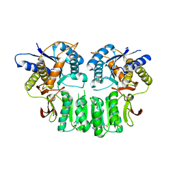 | | Crystal structure of the GluK5 (KA2) ATD dimer at 1.7 Angstrom Resolution | | Descriptor: | 2-acetamido-2-deoxy-beta-D-glucopyranose, 2-acetamido-2-deoxy-beta-D-glucopyranose-(1-4)-2-acetamido-2-deoxy-beta-D-glucopyranose, GLYCEROL, ... | | Authors: | Kumar, J, Mayer, M.L. | | Deposit date: | 2010-08-26 | | Release date: | 2010-11-03 | | Last modified: | 2024-11-20 | | Method: | X-RAY DIFFRACTION (1.677 Å) | | Cite: | Crystal Structures of the Glutamate Receptor Ion Channel GluK3 and GluK5 Amino-Terminal Domains.
J.Mol.Biol., 404, 2010
|
|
3OLZ
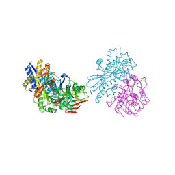 | |
3OM0
 | |


